
Radiotherapy of moving lesions is challenging. Delivery of the therapeutic radiation to a planned target volume may be impacted by organ motion, while anatomical deformations and set-up uncertainties can cause targeting errors. If radiation oncologists had an accurate, real-time 3D radiation dose distribution map, they would be able to alter the level or trajectory of radiation online to achieve more effective and safer treatments.
Ionizing radiation acoustic imaging (iRAI) is a non-invasive technology that could provide this capability. By reconstructing radiation dose using acoustic waves, iRAI can map dose deposition in tumours and adjacent healthy tissues and monitor dose accumulation in real time during radiotherapy, without the need to use additional radiation sources.
A multi-speciality research team at the University of Michigan and Moffitt Cancer Center has now developed a clinical grade iRAI volumetric imaging system. The system, described in Nature Biotechnology, achieved 3D semiquantitative mapping of X-ray beam delivery deep into the body during radiotherapy of a patient with liver metastases.
The iRAI technique works via the thermoacoustic effect. When a high-energy pulsed photon beam generated by a linear accelerator strikes body tissue, it is absorbed. This absorbed energy transfers into heat, which causes localized thermal expansion and generates acoustic waves. These waves are weak, however, and usually undetectable by clinical ultrasound technology.
The new iRAI system detects the acoustic signals with a custom-designed 2D matrix array transducer and a matching multi-channel preamplifier board, driven by a commercial research ultrasound system. The amplified signal is then transferred into an ultrasound device to construct dose-related images in real time.
The researchers explain that their dual-modality system, which combines iRAI with ultrasound imaging, offers “a promising solution to solve the need for real-time monitoring of beam position and online assessment of delivering dose during radiotherapy”. The ultrasound image presents the morphological tissue structures and motion in the body, as well as functional information such as blood flow and vascular density, while the iRAI image can map and quantify the spatially distributed dose deposition in different biological tissues.
“This clinical trial was a pilot study to assess the feasibility of using iRAI in patients receiving abdominal stereotactic body radiation therapy (SBRT),” explains clinical principal investigator Kyle Cuneo from Michigan’s Rogel Cancer Center. “Its findings are enabling us to optimize the iRAI system.”
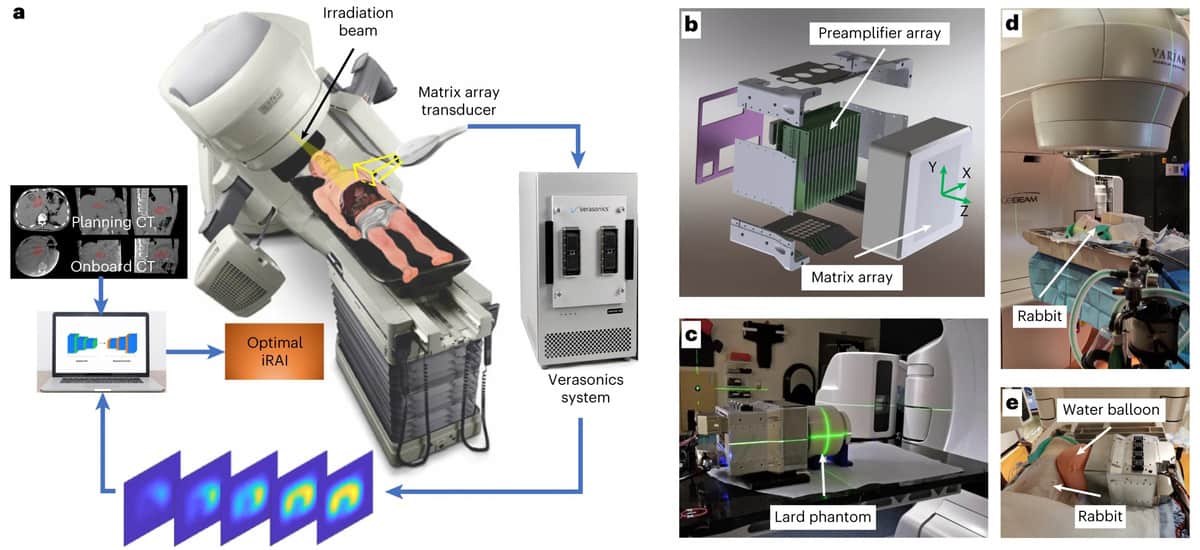
For their proof-of-concept study, the researchers validated the system in a cylindrical lard phantom, a rabbit and then a patient undergoing abdominal SBRT. To enhance the signal-to-noise ratio (SNR) when detecting radiation acoustic signals, they selected a central frequency of 0.35 MHz to match the power spectrum of the acoustic signals generated by the 4 µs X-ray pulse. The SNR was further enhanced by the 1024-channel preamplifier with 46 dB gain integrated with the 2D matrix array, and by displaying the iRAI images with 25 times averaging.
After verifying the system performance using the phantom, the team created and tested a clinical treatment plan to irradiate the liver of a rabbit. The iRAI measurements showed high consistency between the measured dose distribution and that generated by the treatment planning system.
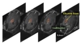
The team then prepared radiotherapy plans for the study participant, with the treatment plan for each fraction divided into two parts. The first part was for iRAI imaging and comprised 2.087 and 0.877 Gy beams delivered in the superior and inferior anterior directions, respectively. This was followed by a volumetric-modulated arc therapy plan (without iRAI imaging) to ensure that the total delivered radiation dose met the clinical requirements.
Both the dose locations and the overall distributions of iRAI measurements matched well with the treatment plan. The iRAI volumetric imaging was able to map the high-dose area with high accuracy. The researchers note that they need to optimize the mapping accuracy for lower dose intensity areas, improve spatial resolution, and develop a comprehensive calibration protocol to provide absolute dose measurement, using advanced reconstruction techniques exploiting artificial intelligence.
Grant principal investigator Issam El Naqa from the Moffitt Cancer Center advises that the current system will be augmented with real-time ultrasound imaging and will also be evaluated in the context of high-risk delivery scenarios such as FLASH radiotherapy.
Acoustic imaging promises real-time dose measurement for FLASH radiotherapy
“One potential application of this technology in the future is real-time adaptive treatment delivery. Current adaptive treatment techniques are based mainly on anatomical changes in the tumour and organs-at-risk (OARs),” explains Cuneo. “With iRAI, we can use both anatomical information, and more importantly, dosimetric information to adapt the radiation plan. This could allow for dose escalation in the target, especially in situations where there is an adjacent OAR, and provide for safer treatments by accurately quantifying the true dose delivered to the target and OARs during each fraction.”
“The system has the unique capability of visualizing radiation deposition while monitoring organ motion, allowing better pinpointing of radiation to targeted tumours while sparing uninvolved tissue in a cost saving manner,” El Naqa adds. “This can be equally applied in both developed and developing countries where financial resources are scarce, leading to improved patient care and better outcomes in these places.”


