Researchers at the Mayo Clinic have combined photon-counting detector (PCD) CT with deep learning-based noise reduction to image patients with multiple myeloma. The new imaging technique demonstrated superior detection of bone disease associated with multiple myeloma compared with conventional low-dose, whole-body CT scans.
PCD CT works by directly converting individual X-ray photons into an electric signal. This allows the use of smaller detector pixels, thereby increasing the image spatial resolution and improving detection of lytic lesions – areas of bone destruction found in around 80% of multiple myeloma patients. This improvement could make a difference in the staging of disease, and potentially impact therapy choice and patient outcomes.
Multiple myeloma is a cancer of plasma cells, a type of white blood cell. Cancerous myeloma cells grow out of control, crowding out normal plasma cells and preventing them from making antibodies to fight infection. Patients may initially be asymptomatic, or develop smouldering multiple myeloma, an intermediary disease phase characterized by a significant increase in plasma cell proliferation, but without any organ damage or symptoms. Active multiple myeloma can cause a range of symptoms, including bone pain, hypercalcaemia, renal insufficiency, anaemia and/or lytic bone lesions.
“When a single lytic lesion larger than 5 mm can be seen on CT, this is a myeloma-defining event that will upstage a patient to active status and require a conversation about initiating treatment,” says principal investigator Francis Baffour.
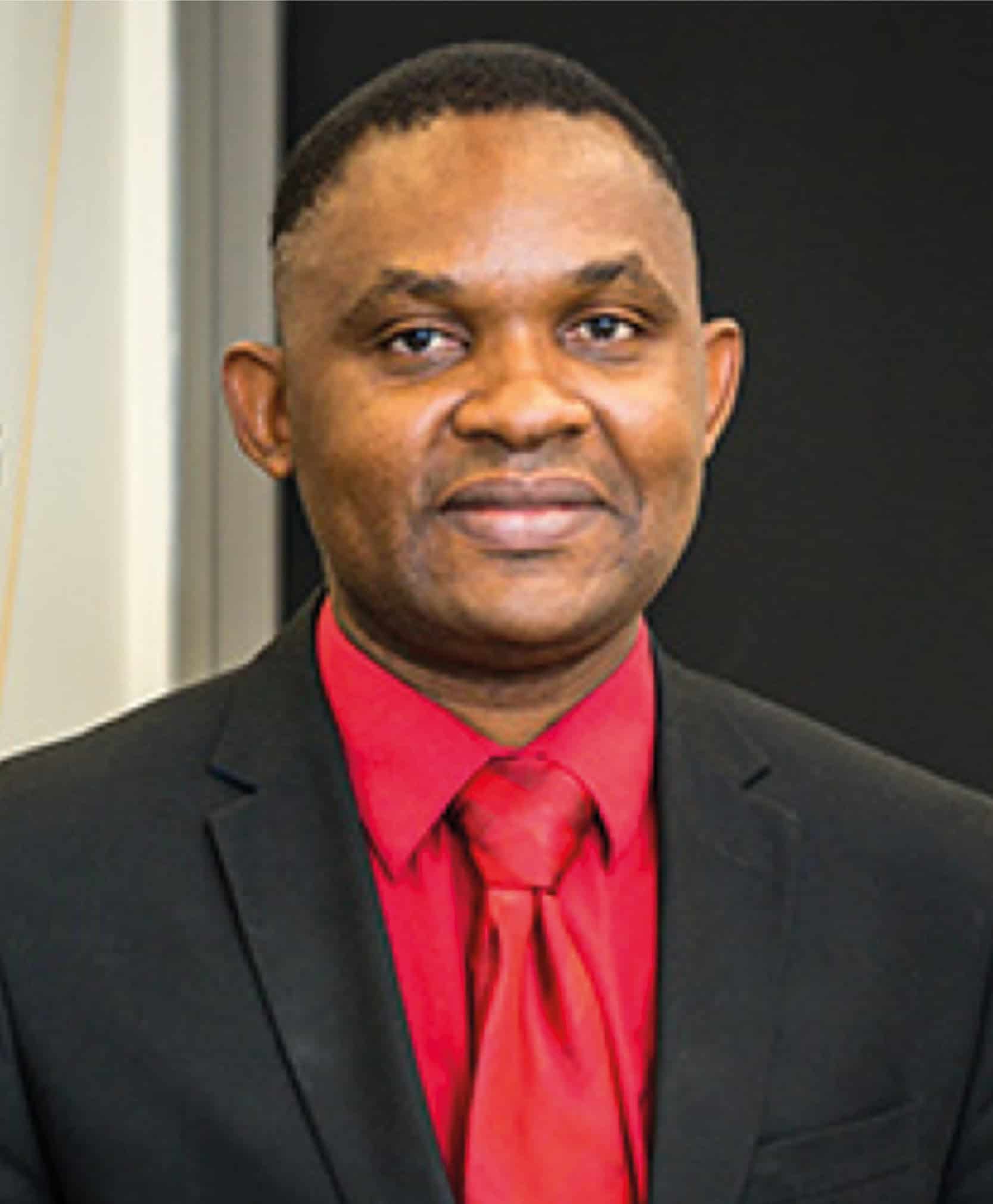
Lead author: Francis Baffour is a diagnostic radiologist at the Mayo Clinic. (Courtesy: Francis Baffour)
“Patients suspected of having multiple myeloma undergo a battery of tests, including bone marrow biopsy. But random biopsy sampling from just one location may not detect the true burden of the disease,” Baffour explains. “Because of its high sensitivity and specificity for lytic bone lesions throughout the body, the International Myeloma Working Group recommends low-dose, whole-body CT as the imaging test to evaluate lytic bone lesions.”
Baffour and colleagues sought to prove that PCD CT, combined with the deep learning denoising software developed by the Mayo team, would increase spatial resolution enough to improve diagnosis using the same radiation dose as a conventional energy-integrating detector (EID) CT scan.
The study, reported in Radiology, included 27 adult patients with different stages of multiple myeloma. Participants underwent both types of scan on the same day: a low-dose, whole-body EID CT scan; and a PCD CT scan at a matched radiation dose (8 mSv for an average adult). The researchers reconstructed EID CT and PCD CT images at 2 mm section thickness. They also reconstructed thinner 0.6 mm PCD CT image sections, which were denoised using a convolutional neural network.
To compare the corresponding 2 mm images, two musculoskeletal radiologists blinded to patient demographics, CT scanner type and scan protocols independently scored the images for features that determined myeloma activity. These included: lytic lesions in the skeleton; hyperdense nodular soft-tissue lesions in the medullary cavity; fat attenuation in myeloma lesions; and pathologic fractures.
The researchers report that PCD CT exhibited superior performance across all visualization assessments, with the strongest differences observed for viewing lytic bone lesions, intramedullary lesions and fat attenuation.
Four weeks later, the radiologists compared the denoised 0.6 mm PCD CT images with the 2.0 mm EID CT images. For each patient, they also recorded the change in the number of detected lytic lesions from the previous set of images. The thin-section PCD CT images again demonstrated improved viewing of all four pathologic abnormalities. Additionally, the readers observed a higher number of lytic lesions on PCD CT relative to EID CT images in 21 of the 27 participants.
“An increase in the number of lytic lesions is highly suggestive of disease progression,” explains Baffour. “When a patient progresses, therapy may be initiated if the patient was previously not being treated or changed if the patient was on therapy. Lytic lesions weaken the bone and, in some locations, put the patient at risk of developing fractures.”
Baffour points out that the improved spatial resolution of PCD CT allows visualization of small structures and more detailed features that may suggest that a lesion is healing or has been treated. “For example, if we see that a lesion has developed a sclerotic rim or has new internal fat density when there was soft tissue previously, we know that the patient has undergone treatment,” he explains.
Baffour tells Physics World that he and his colleagues would like to observe a cohort of patients in various multiple myeloma precursor states, to determine the frequency of new lytic lesions. They also want to investigate PCD CT in other instances in which low-dose protocols are beneficial, such as for paediatric or pregnant patients or screening applications.
“Here at Mayo Clinic, studies are underway to determine how low we can go with scanning doses while still obtaining diagnostic CT images,” says Baffour. “There is much on the horizon and so much potential for photon-counting detector CT in clinical care.”
The gap between the required and the available cancer care widened in the last decade for the majority of cancer patients in the world. Ahmed Elzawawy and Wilfred Ngwa will present the need for global approaches, and campaign for the scientific exploration of avenues to improve the affordability of cancer treatment for millions of patients. They do so while considering the interests and incentives of all stakeholders in the real world, including foremost, cancer patients everywhere, but also governments, professional cancer-care providers, health industries, health businesses and the economy.
Ahmed Elzawawy MD PhD is a professor at Suez Canal University, Ismailia, Egypt, and chairman of the Alsoliman Clinical and Radiation Oncology Centre, Port Said, Egypt. His postgraduate studies and the early years of his medical career took place in Paris, France. He serves as chair of the Global Health Catalyst win–win initiative, and chair of the board of directors at the award-winning Global Oncology University (GO-U). He is a former president of the African Organization for Research and Training in Cancer (AORTIC), and president of International Campaign for Establishment and Development of Oncology Centres (ICEDOC). He is on the board of founders for AFROX–H2 (Africa–Oxford–Harvard/Hopkins), Cancer Research Consortium and ecancerforall (World Premier Comprehensive Cancer Center in the Cloud). He contributed to the World Health Organization’s (WHO) cancer strategy for the new millennium, global strategy for radiotherapy and in scientific, educational, and consultancy missions for ICEDOC and the International Agency for Atomic Energy (IAEA) in Africa, Asia, and East Europe. He contributed to the advisory editorial board of many journals e.g. ecancer, Lancet Oncology, JCO Global Oncology, the World Journal of Surgical Oncology, and Cambridge Scholars Publishing, UK.
Wilfred Ngwa
Wilfred Ngwa is director of the Global Health Catalyst, launched at Harvard in 2015 for catalyzing high-impact international collaborations to reduce health disparities in the USA and globally. He is ICTU distinguished professor of public health, associate professor of radiation oncology at Johns Hopkins University and adjunct professor at University of Massachusetts. He is a co-founder with Prof. Ahmed Elzawawy and others of the Global Oncology University (GO-U), and also founding director of the International Phytomedicines Institute. His work at Harvard and Johns Hopkins Medicine has led to development of award-winning tiny drones to target cancer with potential to increase world-wide access to radiotherapy and immunotherapy. Dr Ngwa has won more than 35 prestigious awards/honours from Harvard, the National Institutes of Health, and national and international professional societies and organizations. He chairs the Africa Health and Infrastructure Committee advising the USA government on global health. He has also been appointed by the director of the USA National Institutes of Health to serve in the prestigious role of a charter member for NIH review section from 2022–2028. His has published three books on global health and serves on the editorial board for four leading journals including ASCO’s Journal of Global Health, and Frontiers in Oncology. He serves as a chair of the Lancet Oncology Commission for Sub-Saharan Africa.
The Editors and experts who contributed to this book present how there is a need for global approaches and campaign for the scientific exploration of avenues to increase affordability of better value cancer treatment for millions of cancer patients in the world.
The quantum coherence of a polariton condensate has been seen to oscillate as the condensate decays. The discovery was made by researchers in Russia, the UK, and Iceland who were led by Alexis Askitopoulos at the Skolkovo Institute of Science and Technology. The oscillations are magnetic in nature and the team suggests that the phenomenon could be used to develop new instruments for measuring magnetic fields.
An exciton–polariton (often referred to as a polariton) is a quasiparticle that occurs in semiconductors. It comprises a photon of light that is coupled to an exciton, which itself comprises an electron and a hole. Polaritons can be produced by sending a light pulse into a semiconductor-based microcavity.
Polaritons are bosons. This means that a dense ensemble of the quasiparticles can form a Bose-Einstein condensate, in which a large number of the polaritons are in the same quantum state. Such a condensate has macroscopic properties that are defined by its quantum nature. These properties can be determined by studying the photons emitted by the condensate as it decays.
Waning coherence
In recent research, Askitopoulos’ team prepared a polariton condensate by firing a 20 µs long light pulse into a microcavity. They then watched as the condensate decayed over time, measuring a coherence function that is related to the overall quantum nature of the condensate.
Instead of decaying smoothly as seen in other condensates, they found that the coherence function periodically rose and fell as it decayed, with a remarkably uniform frequency. By examining these oscillations, they identified Larmor precession as a likely cause of this behaviour. This involves the rotation of the condensate’s magnetic moments about a magnetic field, which is created by polariton interactions.
In total, Askitopoulos and colleagues observed some 100,000 full precessions within a single optical pulse. They found that both the decay speed and the frequency of Larmor precession was directly influenced by the density of polaritons within the condensate. By periodically boosting the system’s coherence, this precession persisted millions of times longer than the lifetime of an individual polariton.
Askitopoulos’ team suggests that this precession could be controlled using optical methods, which could lead to better techniques to study polariton condensates. One possible application of the discovery is the development of new types of magnetometers. These are devices that measure the strength, direction, and relative change of a local magnetic field.
The most interesting topological materials are topological insulators, which are materials that are insulating in the bulk, but conducting on the surface. In these materials, the conducting channels where the electronic current flows are very robust. They persist independently of some external disturbances that one can have in experiments, such as weak disorder or temperature fluctuations, and they’re also independent of size. This is very interesting because it means these materials have a constant resistance, a constant conductivity. Having such tight control of the electronic current is useful for many applications.
What are some examples of topological insulators?
The best-known example is probably gallium arsenide, which is a two-dimensional semiconductor that is often used in experiments on the integer quantum Hall effect. In the newer generation of topological insulators, the best-known one is bismuth selenide, but this has not gained as much widespread attention.
Why did you and your colleagues decide to search for new topological materials?
At the time, there were just a few of them in the market, and we thought, “Okay, if we can develop a method that can calculate or diagnose topology quickly, we can see if there are materials that have more optimized properties.”
One example of an optimized property is the electronic band gap. The fact that these materials are insulating in the bulk means that in the bulk, there is a range of energies where the electrons cannot pass through. This “forbidden” energy range is the electronic band gap, and electrons cannot travel in that region even though they can exist on the material’s surface. The larger the material’s electronic band gap is, the better a topological insulator it will be.
How did you go about looking for new topological materials?
We developed an algorithm based on a material’s crystalline symmetries, which is something that was not taken into account before. The symmetry of the crystal is very important when dealing with topology because certain topological materials and some topological phases need a particular symmetry (or lack of symmetry) to exist. For example, the integer quantum Hall effect needs no symmetries at all, but it does need one symmetry to be broken, which is time-reversal symmetry. That means the material needs to be magnetic, or we need a very large external magnetic field.
But other topological phases do need symmetries, and we managed to identify which symmetries they were. Then, once we had all the symmetries identified, we could classify them – because in the end, that is what physicists do. We classify things.
We started working on the theoretical formulation in 2017, and two years later, we published the first paper related to this theoretical formulation. But it’s only now that we’ve finally completed everything and published it.
Who were your collaborators in this effort and how did each person contribute?
I designed (and, in part, performed) the first-principles calculations in which we considered how to simulate real materials and “diagnose” whether they had topological properties. For that, we used state-of-the-art codes and homemade codes that tell us how the material’s electrons behave and how we can classify the material’s topological properties. The theoretical formulation and analysis was done by Benjamin Wieder and Luis Elcoro because they are more hardcore theoretical physicists. They helped with analysing and classifying the topological phases. Another very important contributor and the leading guy of this project was Nicolas Regnault; we built up the website together and took care of designing the website and the database.
We also had help from Stuart Parkin and Claudia Felser. They are materials experts, so they could give us advice on whether a material was suitable or not. And then Andrei Bernevig was the co-ordinator of everything. We’d been working together for several years already.
And what did you find?
What we found is that there are many, many materials that have topological properties – tens of thousands of them.
Were you surprised by the number?
Yes. Very!
Given how ubiquitous these topological properties turned out to be, it seems almost surprising that you were surprised. Why hadn’t anyone noticed before?
I don’t know why it was missed completely by the community, but it’s not only our community within materials science and condensed-matter physics that missed it. Quantum mechanics has existed for a century already, and these topological properties are subtle, but they are not very complex. Yet all the smart “fathers” of quantum mechanics completely missed this theoretical formulation.
Topological catalogue: The Topological Materials Database created by Maia Vergniory and colleagues is a searchable tool containing more than 90,000 known topological materials. (Courtesy: Christine Daniloff/MIT)
Has anybody tried synthesizing these materials and checking to see whether they do indeed behave as topological insulators?
Not all of them have been checked, of course, because there are so many. But some of them have. There are new topological materials that have been created experimentally following this work, like the high-order topological insulator Bi4Br4.
The Topological Materials Database you and your colleagues constructed has been described as “a periodic table for topological materials”. What properties determine its structure?
The topological properties are related to the electronic current, which is a global property of the material. One of the reasons physicists might not have thought about topology before is that they were very focused on local properties, rather than global ones. So in this sense, the important property is related to the localization of the charge and how the charge is defined in real space.
What we found is that if we know the material’s crystalline symmetries, we can anticipate what the behaviour of the charge is going to be or flow. And that is how we could classify the topological phases.
How does the Topological Materials Database work? What do researchers do when they use it?
First, they enter the material’s chemical formula. For example, if you are interested in salt, the formula is sodium chloride. So you put NaCl in the database and you click, and then all the properties appear. It’s very simple.
Wait, are you saying that common table salt is a topological material?
Yes.
Really?
Yes.
That’s amazing. Apart from surprising people with the topological properties of familiar materials, what impact do you hope your database will have on the field?
I hope it’s going to help experimentalists figure out which materials they should grow. Now that we have analysed the full spectrum of all material properties, experimentalists should be able to say, “Okay, this material is in an electron transport regime that we know is not good, but if I dope it with some electrons, then we will reach a very interesting regime.” So we hope, in a sense, that it will help experimentalists to find good materials.
A lot of attention has come to topological materials recently because of a possible link to quantum computing. Is that a big motivator in your work?
It’s related, but every field has different branches, and I would say our work is in a different branch. Of course, you need a topological material as a platform to develop a topological quantum computer using any of the possible qubits (quantum bits) that have been proposed, so what we did is important for that. But developing a topological quantum computer will require a lot more work on materials design because the material’s dimension plays an important role. We were looking at three dimensions, and it could be that for quantum computing platforms, we’d need to focus on 2D systems.
There are other applications, though. You could use the database to find materials for solar cells, for example, or for catalysis, detectors or low-dissipation electronic devices. Beyond the super-exotic applications, these day-to-day possibilities are also very important. But our real motivation for the work was to understand the physics of topology.
What’s next for you and your collaborators?
I would like to do research on organic materials. The focus in the current database is on inorganic materials because we took the Inorganic Crystal Structure Database as our starting point, but organic materials are very interesting, too. I’d also like to investigate more magnetic materials, because are fewer magnetic materials reported in the database than non-magnetic ones. And then I want to look at materials that have chiral symmetries – that is, they are symmetric, but “handed” in that they have a left version and a right version.
Do you think there could be thousands more topological materials out there among the organic or magnetic materials?
I don’t know. It depends on the size of the electronic band gap. We’ll see!
Despite its counter-intuitive weirdness, quantum mechanics ranks as one of the most successful scientific theories of all time. Apart from forming the bedrock of our understanding of atoms and subatomic particles, it has spawned a host of technologies from the laser and transistor to quantum cryptography and quantum computers. But quantum mechanics wasn’t always held in such high regard.
At the start of the 20th century, when the subject was just getting off the ground, many scientists were sceptical of this new-fangled theory. Among the doubters was Otto Stern, who, along with fellow German physicist Walther Gerlach, devised a now-famous experiment to disprove the theory.
Carried out 100 years ago, the experiment involved the two physicists using beams of atoms to test a seemingly bizarre consequence of quantum mechanics known as the “space quantization of angular momentum”. As it happens, their initial interpretation of the experiment proved to be wrong. But their work spurred the development of quantum theory and today the “Stern–Gerlach experiment” is considered a classic of modern physics.
The Stern–Gerlach experiment is at the centre of the venerable conceptual puzzles of quantum mechanics – from the uncertainty principle to entanglement
Bretislav Friedrich, Fritz Haber Institute, Berlin
“It’s at the centre of the venerable conceptual puzzles of quantum mechanics – from the uncertainty principle to entanglement,” says Bretislav Friedrich, a physicist from the Fritz Haber Institute in Berlin who has written extensively about the experiment and Stern and Gerlach’s lives. The experiment, he says, was greeted with “pure astonishment” in 1922 – and it still astonishes physicists today.
Quantum weirdness
Born in 1888 in the Prussian city of Sohrau (now Żory in Poland), Stern was a physical chemist by training, who did a PhD at the University of Breslau on the osmotic pressure of solutions of carbon dioxide. In 1912 he moved to the Charles-Ferdinand University in Prague, attracted by Albert Einstein, who was based there at the time. By attending Einstein’s lecturers in Prague – and later at the ETH Zurich where both moved the following year – Stern quickly became exposed to the early ideas of quantum mechanics.
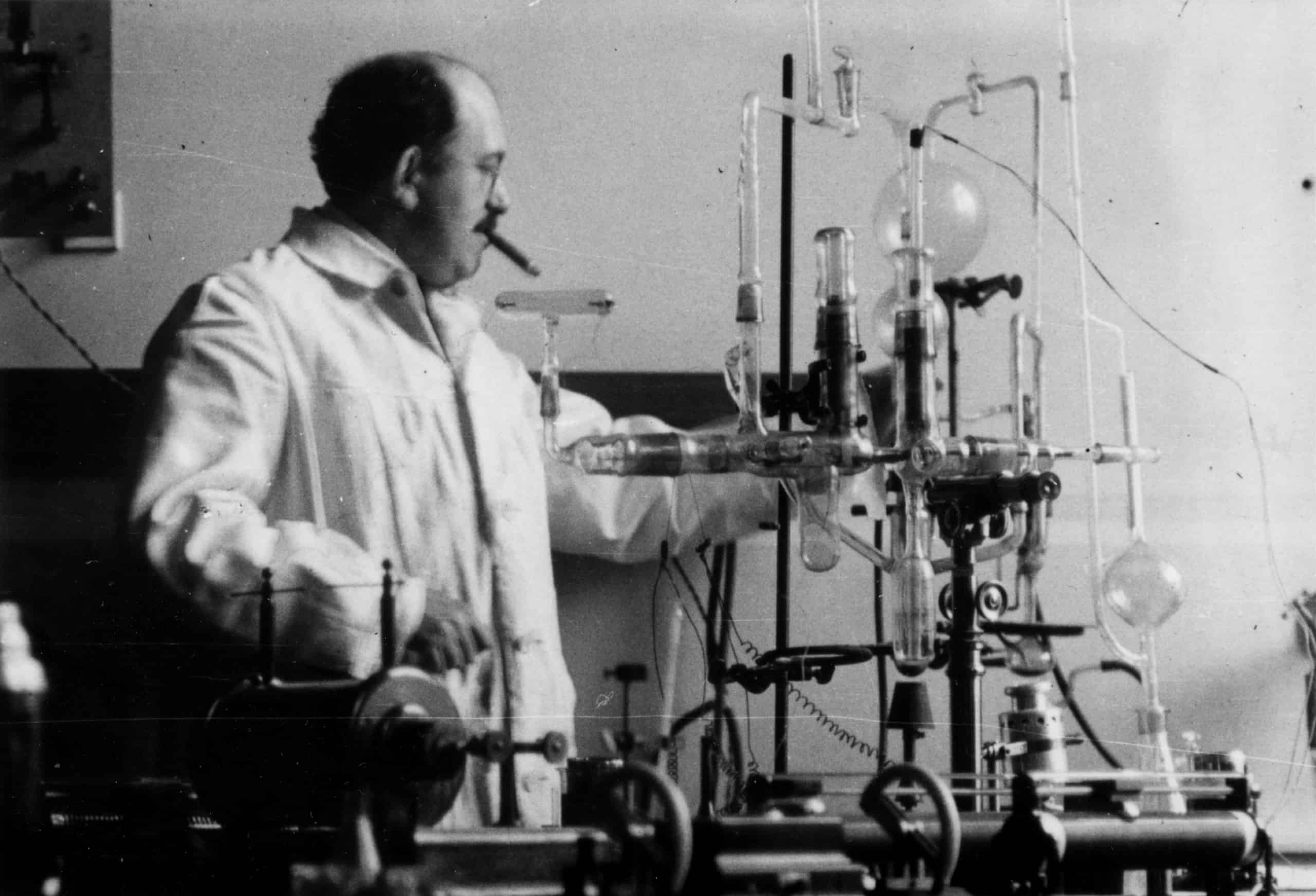
Star player Otto Stern was a physical chemist by training but became interested in physics after being taken under the wing of Albert Einstein at the Charles-Ferdinand University in Prague in 1912. Stern attended Einstein’s lectures and closely followed developments in quantum mechanics. However, he was not convinced that the theory was correct and devised a test, later known as the Stern–Gerlach experiment, to try to disprove it. However, the experiment showed that quantum mechanics was the real deal and Stern was forced to admit that Niels Bohr, one of its founding fathers, was correct. (Courtesy: AIP Emilio Segrè Visual Archives, Segrè Collection)
These ideas included Niels Bohr’s early model of the atom, which was the centrepiece of what is now called the “old quantum theory”. Familiar today as a basic representation of an atom, the Bohr model describes an atom as a dense, positively charged nucleus orbited by negatively charged electrons.
According to classical physics, such electrons should radiate energy and spiral into the nucleus in a matter of picoseconds. As that does not happen in reality, Bohr got around this problem by restricting the electrons to specific atomic orbits, more commonly referred to as orbitals.
Orbital quantization enabled Bohr to explain a phenomenon that had puzzled physicists and chemists for decades – the fact that atoms only absorb and emit light at a discrete set of optical wavelengths. Bohr’s model initially seemed like the right idea, as it allowed him to reproduce a formula for these wavelengths that had been derived in 1888 by the Swedish physicist Johannes Rydberg. The Rydberg formula had given the wavelengths in terms of a series of integers, which we now understand to be the principal quantum numbers of the atomic orbitals.
But as Bohr’s model was studied and honed by leading physicists of the day, something odd became apparent. As a consequence of orbital quantization, it transpired that the component of an electron’s orbital angular momentum along a specific direction must also be quantized. In particular, according to Bohr’s model, an electron in the lowest energy orbital should have only two values of angular momentum along any arbitrary direction. These values would point in opposite directions in space, with no intermediate values permitted.
Known as space quantization, this phenomenon was viewed as even more bizarre than orbital quantization. In fact, Stern was so sceptical of the Bohr model that he vowed to quit physics if it proved to be correct. In 1914, after Stern parted company with Einstein and joined the brand-new University of Frankfurt, a practical opportunity arose for him to put space quantization to the test. Stern realized that if an electron’s orbital angular momentum showed space quantization, then so too would the magnetic moment of an atom.
Testing for space quantization
Stern’s work was initially disrupted by the First World War, when he served in the German army on the Russian front. But on his return to Frankfurt, Stern began experiments on beams of atoms, which had become possible thanks to the invention of the mercury-diffusion vacuum pump by the German physicist Wolfgang Gaede in 1915. This device allowed researchers to create high-vacuum conditions for the first time so that atoms could travel the length of an experimental apparatus without scattering from air molecules.
In 1920 Stern was joined in Frankfurt by Gerlach, who – like Stern – had also served in the First World War. Gerlach had become involved in atomic-beam experiments through his interest in the properties of atoms in magnetic solids. In particular, Gerlach wanted to see if atoms have magnetic moments and had begun thinking about an experiment involving a beam of bismuth atoms travelling through a region with an inhomogeneous magnetic field.
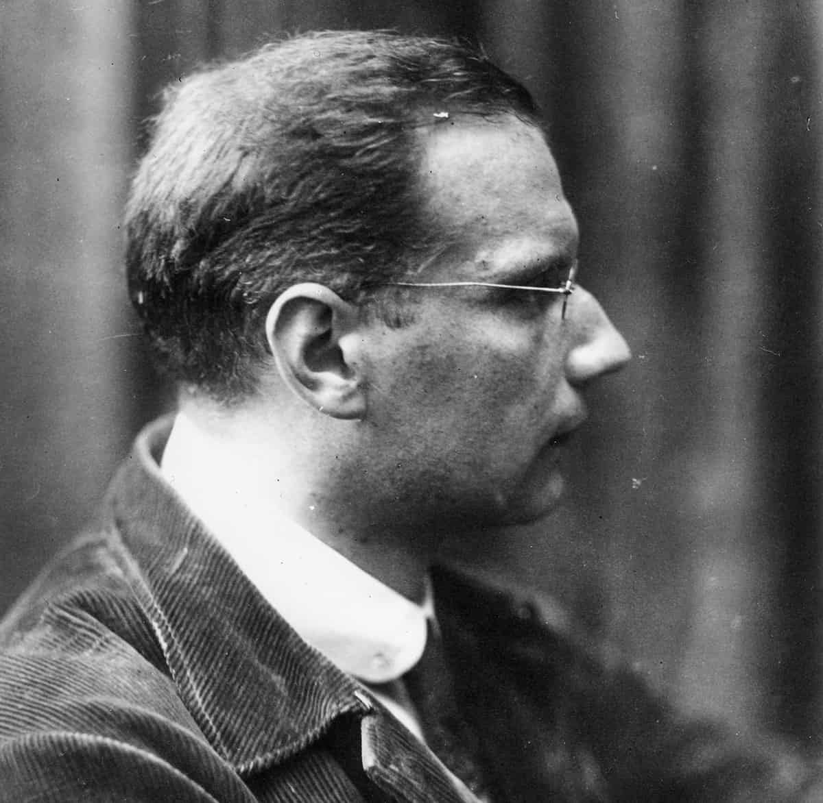
Right direction Born in 1889 in Wiesbaden, Germany, Walther Gerlach gained a PhD in physics from the University of Tübingen in 1912, working on black-body radiation and the photoelectric effect under Friedrich Paschen. At the start of the First World War, Gerlach worked in the university’s medical X-ray lab, creating a device to locate bullets and shrapnel in wounded soldiers. He joined the army in 1915, working with Wilhelm Wien on radio-communications technology and later seeing active service in Belgium. After the war he briefly worked in industry before joining the University of Frankfurt as an experimental physicist in 1920. (Courtesy: AIP Emilio Segrè Visual Archives, Gift of Jost Lemmerich)
If an atom has a dipole magnetic moment, Gerlach reasoned, it will experience a torque due to the magnetic field and will therefore rotate. But if the magnetic field is not uniform, the force at one end of a dipole will be stronger than the torque at the opposite end. That will lead to a net force on the atom, which will deflect as its flies through the inhomogeneous magnetic field – with the size of the deflection revealing the magnitude of the atom’s magnetic moment.
In 1921 Stern realized that such an experiment would be a great way to test for space quantization. If atoms have magnetic moments that can point in any direction (as classical physics would suggest) then the beam of atoms would broaden continuously as it passes through the inhomogeneous magnetic field. However, if the magnetic moments of the atom are space quantized – pointing in opposite directions (up and down) along the inhomogeneous field – then the beam of atoms would be split in two.
As a result, the up and down atoms would be deflected in opposite directions, providing clear evidence for space quantization. So confident was Stern of his idea that he published a paper in the journal Zeitschrift für Physik (7 249) that presented meticulous calculations describing how it could be done using a beam of silver atoms. Gerlach was convinced and started to build his apparatus in 1921. Stern (a theorist) and Gerlach (an experimentalist) proved an effective combination.
Experimental breakthrough
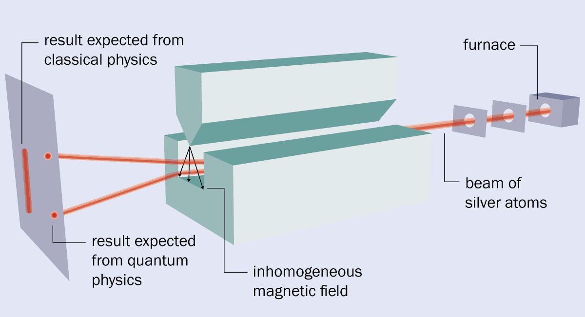
In the original version of the Stern–Gerlach experiment, the two physicists vapourized silver in an oven and allowed some atoms to escape through a hole (see box 1). They then sent the atoms through a pair of collimators, which created a beam that travelled between the two pole pieces of an electromagnet. These pieces provided a magnetic field with the required high level of inhomogeneity because one had a groove cut into it, while the other had a sharp, knife-like edge and was held above the groove.
After passing through the magnet, the beam struck a detector plate where the presence of silver could be revealed by a chemical development process similar to that used in photography. But despite its simplicity, the experiment was fiendishly difficult to undertake.
The apparatus was small – about the size of a fountain pen – and had to be kept under high vacuum using two of Gaede’s diffusion pumps. In fact, the apparatus often broke, making it difficult to achieve the long run time needed to accumulate enough silver on the detector plate to create a visible image.
1 The original set-up
The original version of the Stern–Gerlach experiment was carried out by Otto Stern and Walther Gerlach in February 1922. It involved vapourizing silver in a furnace, with some of the resulting silver atoms sent through two collimators to form a beam. The beam then travelled through a magnet that was built to have a very inhomogeneous field. Classical physics says that when the beam strikes a detector, it should simply broaden out. But Stern and Gerlach found that the beam split into two, which indicated that quantum mechanics is at work. The splitting is caused by the spin of silver’s unpaired electron although at the time it was, incorrectly, seen as proof of so-called “space quantization”.
What’s more, the Stern–Gerlach experiment was expensive, made worse by the hyperinflation that was rampaging through post-war Germany. Money and donated equipment had to be secured from a range of sources including the Physikalischer Verein Frankfurt (Frankfurt Physics Society), ticket sales from popular lectures by Max Born, and donations from Einstein and Henry Goldman – an American banker and son of the co-founder of the financial firm Goldman–Sachs.
At first, Stern and Gerlach only saw their beam broadening when the inhomogeneous magnetic field was switched on. This was not the splitting predicted by quantum mechanics, but was an important achievement in its own right, being the first experimental evidence for atoms having magnetic moments. After Stern left Frankfurt in late 1921 to take up a professorship of theoretical physics at the University of Rostock, Gerlach realized that he could improve the measurement by replacing the round holes in the collimators with slits, which boosted the number of atoms in the beam.
And so, working alone one February night in 1922, Gerlach finally saw the beam splitting that had been predicted. He immediately sent a telegram to Stern saying: “Bohr is right after all”. Gerlach also created a postcard showing the split beam and sent it to Bohr, congratulating him for creating his model of the atom. Their paper presenting the results, published in December 1922 (Zeit. Phys.9 349), provided the first experimental evidence for space quantization in a magnetic field – and thereby crucial evidence for quantum theory.
A new spin on things
The Stern–Gerlach experiment caused an immediate stir in the physics community, with Wolfgang Pauli, for example, quipping that “This should convert even the non-believer Stern”. However, the explanation for the observed beam splitting using Bohr’s model was short-lived.
Other physicists, including Pauli and Paul Dirac, soon realized that the electron has an intrinsic angular momentum or spin – something that was not included Bohr’s model. What’s more, Bohr had been wrong to predict that the ground state of the silver atom has orbital angular momentum; it does not.
Today we know that the splitting that Stern and Gerlach saw in their experiment is actually caused by the spin of silver’s unpaired electron. Indeed, the Stern–Gerlach experiment is now interpreted as evidence of electron spin, rather than as proof of space quantization.
As so often in physics, even supposedly “wrong” results can still lead to progress. What’s more, the work opened the door to a huge variety of other findings, including showing the momentum transfer that occurs if an atom emits or absorbs a photon.
Stern went on to carry out the world’s first matter-wave experiments with atoms when he scattered beams of atoms off the surfaces of crystals, which confirmed the principle of wave-particle duality. Later, in 1933, he measured the magnetic dipole moment of the proton using a set-up similar to the Stern–Gerlach experiment. He found it to be larger than expected, which suggested that the proton is not a point-like particle – as had been assumed at the time – but rather has internal structure. This discovery led Stern to win the Nobel Prize for Physics in 1943.
By that time, Stern – who was Jewish by birth – was based at the Carnegie Institute of Technology in Pittsburgh in the US, having fled Germany in 1933 as Nazi repression of the Jews intensified. While at Carnegie, he extended his work on the magnetic dipole moment of the proton to its heavier cousin, the deuteron. Stern’s lab in Pittsburgh also confirmed Maxwell–Boltzmann velocity distribution of particles in an ideal gas. He died in the US in 1969.
As for Gerlach, he went on to measure the magnetic moment of atomic bismuth and several other metals using the atomic beam technique. He also did experiments on the radiation pressure of light and continued his interest in magnetism and condensed-matter physics. His career later took him to the universities of Tübingen and Munich. Despite being nominated 31 times between 1924 and 1944, Gerlach missed out on a Nobel prize.
[The pair benefitted from] Stern’s ideas on the one hand and Gerlach’s realism and skills in the lab as well as a – sometimes stubborn – determination to make things work
Bretislav Friedrich, Fritz Haber Institute, Berlin
Like Stern, Gerlach was impacted by Nazism – but in a very different way. Although he never joined the Nazi party and rejected the idea of “Jewish science”, Gerlach headed Germany’s nuclear research programme in the final years of the Second World War. He ended up being interned by the Allied Forces at Farm Hall in England along with nine other German physicists suspected of being involved in Germany’s nuclear weapons programme including Max von Laue and Werner Heisenberg.
After the war, Gerlach returned to academic research, spending the bulk of his career back at Munich where he played important roles in rebuilding German science. In 1957, he, von Laue and Heisenberg were part of a group of 18 leading German physicists who signed the Göttingen Manifesto, which rejected a proposal by the then chancellor Konrad Adenauer to arm Germany with nuclear weapons. Gerlach died aged 90 in 1979.
Lasting impact
Apart from shaping the course of modern science, the Stern–Gerlach experiment has also had a huge practical impact. Indeed, Stern is widely viewed as one of the founders of experimental atomic, molecular and nuclear physics, having shown how molecular beams can be used to quantitatively study matter without resorting to spectroscopy. “Sorting states via space quantization is ubiquitous,” says Friedrich, with nuclear magnetic resonance and magnetic-resonance imaging being the most direct descendants of the classic experiment.
Friedrich also credits the Stern–Gerlach experiment for introducing principles that influenced other areas of science, such as the development of the maser and laser. And although Gerlach missed out on a Nobel prize, Friedrich says his recruitment by Stern was a “stroke of luck” for atomic physics, with the pair having exceptional complementary talents. “[They benefitted from] Stern’s ideas on the one hand and Gerlach’s realism and skills in the lab as well as a – sometimes stubborn – determination to make things work.”
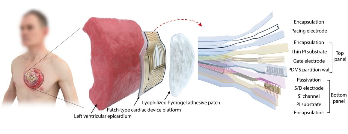
Cardiac patch: Schematic of the device, which comprises an array of pressure-sensitive transistors and biocompatible pacing electrodes with encapsulation layers. The device is attached to the epicardium using a hydrogel adhesive patch. (Courtesy: J C Hwang et al Sci. Adv. 10.1126/sciadv.abq08/CC BY-NC)
Scientists have fabricated and successfully tested an ultrathin patch-type cardiac device that can monitor a heartbeat and apply stimulations as necessary.
Currently, these functions are performed by bulky and intrusive devices such as pacemakers. While these were miracle machines for many decades, the surgery required to implant pacemakers is complicated and can be risky. The new device, on the other hand, has a unique adhesive that allows it to stick directly to a wet organ such as the heart.
The researchers, from Yonsei University in Korea, believe that the platform they have developed may one day replace pacemakers and therefore avoid unnecessary surgical risks. The team has so far only tested the device on a live rabbit model and an artificial heart, but successful results suggest that this is a promising new technology that bears further investigation.
Can this replace a pacemaker?
A key heart problem that the researchers addressed with their research is cardiac arrhythmia, or an irregular heartbeat. The heart can beat too fast, too slow or erratically. Regardless, the cause of cardiac arrhythmia is the improper function of electrical impulses that are supposed to coordinate heartbeats. Sometimes heart arrhythmias are unnoticeable or simply a minor annoyance, but in certain situations they can be life-threatening. If the problem becomes severe enough, the surgical implantation of a pacemaker may be the only remaining option.
The research performed here may give patients a less invasive option, which would give more people the option to monitor and halt cardiac arrhythmias. The new device is a tiny platform consisting of an active-matrix array of pressure-sensitive transistors that can detect heartbeats, along with low-impedance electrodes to stimulate the heart and halt arrythmias. In tests on rabbits, the device was able to detect cardiac arrhythmias and apply suitable electrical treatment to correct the abnormal heart rhythms.
One of the more promising aspects of this new platform is just how thin it is. The sheet of sensors has a total thickness of 38 μm. For reference, a standard pacemaker is about the size of a matchbox and human hairs are generally between 17 to 181 μm thick. Clearly, the platform investigated here is many orders of magnitude smaller than our current best solutions to heart monitoring and stimulation.
In addition to the device’s tiny size, the researchers also developed an unusual method for attaching it to the heart itself. Currently, pacemakers have a generator that attaches to a wire running through a blood vessel in the heart. The new platform works more like a piece of tape stuck on the heart’s epicardium.
The reason this is such an incredible revelation is the same reason that can cause frustration when trying to tape something to a wet surface. Much like putting a piece of labelling tape on a food container with condensation, adhering a patch-type cardiac device to a heart for a long period of time was a challenge. To solve this problem, the researchers looked to nature. Specifically, they found an alginate-based hydrogel adhesive that mimics the adhesive properties of mussels in underwater environments. Not only does this adhesive stick to the wet epicardium, but it also won’t be resorbed by the body, and it is biocompatible and nontoxic for humans.
What’s next?
Of course, there is still work to be done if these kinds of devices are ever to replace pacemakers at a significant scale. To begin, the researchers only performed in vivo testing on rabbits and on an artificial heart. Further work will be required before we can start trusting this technology with human lives.
Additionally, the researchers believe that cardiac arrythmia is only the beginning. They expect this method to expand toward studying and monitoring other heart diseases, such as myocardial infarction, hypertrophic cardiomyopathy, concentric hypertrophy and dilated cardiomyopathy.
The presence of nitrous oxide in the atmospheres of Earth-like exoplanets could be a signature of the presence of extraterrestrial life – according to a study done by researchers in the US led by Edward Schwieterman at the University of California, Riverside.
Using advanced computer models to support their proposal, the team believes that its work could offer important insights for exoplanet studies by current and future observatories – including the James Webb Space Telescope (JWST).
Astronomers know of more than 5000 exoplanets – which are planets that orbit stars other than the Sun – and that number keeps growing. As telescopes improve, astronomers are getting better at determining the compositions of exoplanet atmospheres, and these measurements play an important role in the search for extraterrestrial life. This is done by making spectroscopic measurements on starlight that has passed through exoplanet atmospheres.
In search of life
We have never seen life on another planet, so we do not know exactly how it would affect exoplanet atmospheres. Instead, astrobiologists identify chemicals in Earth’s atmosphere that are associated with the presence of life and search for these “biosignatures”.
This is where nitrous oxide (also known as laughing gas) comes in. While it is not especially common in Earth’s atmosphere today, Schwieterman and colleagues suggest that the gas could have been abundant in previous eras of Earth’s history.
Nitrous oxide is produced by some living organisms on Earth, so it is possible that it could present in the atmospheres of some exoplanets that harbour life. Here on Earth, however, there are natural processes that keep atmospheric nitrous oxide levels very low. However, on other planets an abundance of nitrous oxide could result from low levels of the metal catalysts and biological enzymes that break down the compound. Another possibility is that the stellar radiation received by some exoplanets is not as efficient as sunlight at destroying nitrous oxide. Indeed, nitrous oxide levels in such situations could be high enough to be observed by telescopes like the JWST.
Schwieterman’s team explored this idea by developing a biogeochemical model that quantifies the likely abundance of nitrous oxide in the atmospheres of Earth-like exoplanets orbiting main sequence stars. By coupling their model to photochemical and spectral models, the researchers also calculated that nitrous oxide could build up to detectable levels within a range of atmospheric conditions. This could include the TRAPPIST-1 system, where as many as four planets appear to orbit within the habitable zone of their cold red dwarf host star.
Although nitrous oxide can also be produced by non-biological sources, such as lightning strikes, the team showed that the amounts of gas produced would be orders of magnitude lower than that produced by alien ecosystems. Based on their results, Schwieterman and colleagues hope that the JWST, along with other telescopes actively hunting for signs of life in exoplanetary atmospheres, will add nitrous oxide to the list of viable biosignatures – potentially bringing the discovery of extraterrestrial life a step closer.
Over the past 150 years, our ability to produce and transform engineered materials has been responsible for our current high standards of living, especially in developed economies. However, we must carefully think of the effects that our addiction to creating and using materials at this fast rate will have on the future generations. The way we currently make and use materials detrimentally affects the planet Earth, creating many severe environmental problems.
In this live webinar, we hear presentations from leading researchers within the sustainable materials research community about how the very latest findings are helping address the key challenges faced. This webinar will feature the co-authors of The sustainable materials roadmap, which can be read in full. The roadmap is made up of fantastic contributions from more than 50 notable authors, and highlights sustainability issues within the field of materials and discusses the pathways towards solving them as adopted by the sustainable materials community.
Left to right: Magda Titirici, Agi Brandt-Talbot, Silvia Vignolini, Kong-Yang Lee, Heather Au and Göran Finnveden.
Speakers
Magda Titirici holds the position of chair of sustainable materials energy at Imperial College London. She is the author of more than 250 articles and is included in the Global Highly Cited Researchers (Clarivate Analytics) in each of the past four years. Her current research interests involve sustainable materials with focus on carbon and carbon hybrids produced via hydrothermal processes, waste recycling into advance products, avoidance of critical elements in renewable energy technologies, and the development of truly sustainable clean-energy storage systems.
Agi Brandt-Talbot is a lecturer in the Department of Chemistry at Imperial College London and leads the Sustainable Carbon Solutions research team. She has authored 32 scientific articles with more than 4000 citations and holds three licensed patents. Agi is interested in creating bio-derived materials and chemicals from sustainable biomass and the application of novel tailor-made solvents for more sustainable use of carbon in our economy.
Silvia Vignolini holds the position of professor of chemistry and biomaterials at Cambridge University and is the author of more than 250 research papers. Her research examines photonic structures that occur throughout nature, biomimetic materials, block copolymers for self-assembly, light management for photosynthesis, and much more.
Kong-Yang Lee holds the role of professor in polymer engineering at Imperial College London. His research focuses on the development and manufacturing of novel polymeric materials with a focus on tailoring the interface between two (or more) phases to bridge the gap between chemistry, materials science and engineering.
Heather Au is a Faraday Institution Research Fellow at Imperial College London. Her research is primarily based on exploring sustainable materials for energy storage, and with particular emphasis on the development of engineered carbon hosts for sulphur cathodes in lithium-sulphur batteries.
Göran Finnveden holds the role of professor of environmental strategic analysis at the Division of Sustainability Assessment and Management at the Department of Sustainable Development at KTH Royal Institute of Technology. His current research focusses upon the circular economy, life-cycle assessment methodology, integration of sustainable development in higher-education institutions, and sustainable consumption and sustainable economic development.
About this journal
JPhys Materials is an open access journal highlighting the most significant and exciting advances in materials science.
Editor-in-chief: Prof Stephan Roche, Catalan Institution for Research and Advanced Studies (ICREA) and Catalan Institute of Nanosciences and Nanotechnology (ICN2), Barcelona, Spain.
What can science say about big questions such as how did the universe come into being, or do we have free will? In Existential Physics: a Scientist’s Guide to Life’s Biggest Questions, the theoretical physicist, author and Youtuber Sabine Hossenfelder argues that sometimes science cannot really say much. This is not science’s fault, argues Hossenfelder, but rather it’s because we don’t have enough experimental data to test our hypotheses on some of these weighty issues. What is more, she says that on some of these issues we may never have enough data, putting their resolution beyond science.
Take the origin of the universe. Hossenfelder dissects theories that attempt to describe the Big Bang and what occurred very shortly thereafter. She argues that the problem isn’t with these theories – which she says are mathematically sound – but rather that the theories are based on a dearth of observational data. Astronomers cannot look back far enough in time to glean clues about what happened in the immediate aftermath of the Big Bang, and we can’t currently do experiments that recreate conditions anywhere near those that existed during the birth of the universe.
Hossenfelder dubs these theories as “ascientific” (something that cannot be dealt with using science whatsoever) and goes so far as to say that humans may never have sufficient experimental evidence about the first moments of the universe to test these ideas. As a result, she describes the myriad theories that describe the early universe as “modern creation myths written in the language of mathematics”.
Those familiar with Hossenfelder’s writings and videos will know that she excels at pointing out what she sees as the shortcomings of ascientific thinking in physics. In her latest book she extends these ideas to look at what science can say about a broader range of big questions. For some questions – like do we have free will, and why do we get older and never younger? – Hossenfelder points out that science does indeed have good answers. On other questions – such as does human existence define the laws of physics? – she explains in an entertaining way why science can’t really say much.
So, what is the point of a book with its focus on what we don’t know? I think Hossenfelder wants to make it clear to non-scientists that science is nowhere near to having an answer for every question, and it is perfectly reasonable to conclude that science may never have all the answers. Hossenfelder is careful, however, not to trash the scientific method and leave the reader thinking that science can’t really say much about anything. But she says that scientists should only develop theories that can be tested by observing nature – theories that can be falsified. She also has no problem with theories that can’t be falsified but argues that they are not scientific. Indeed, she believes that such theories can serve a purpose, in the same way that religious beliefs are important.
In these febrile times, some scientists might shudder at this message and how it could affect public trust in areas such as vaccination, where the science is sound. That, of course, is not Hossenfelder’s intention – she rightly believes that a better understanding of the limitations of science will benefit society. This comes across loud and clear in her book, which I found fun to read and really made me think about the scientific method and the big questions in life.
Physicists in Israel have printed a micro-optical element that generates a twisted Bessel beam on the end of an optical fibre. The polymer device consists of a parabolic lens for light collimation and a helical axicon that twists the light. According to the researchers, their work demonstrates how elements that can generate sophisticated beam shapes can be integrated into optical fibres. Such devices could provide tailored light beams for a variety of optical technologies.
A wide range of applications – including communications, sensing and imaging, for example – rely on optical fibres. Light exiting these fibres is usually manipulated and steered using large optical elements. Micro-optics is seen as a way to reduce the size of these elements, expand their function and cut costs. Integrating them directly onto optical fibres could be particularly advantageous.
Shaping light into Bessel beams, a type of twisted light that carries orbital angular momentum, is beneficial due to their resistance to diffraction and large depth of focus. These are promising characteristics for various applications such as optical tweezers and material processing.
“The ability to create a Bessel beam directly from an optical fibre could be used for particle manipulation or fibre-integrated stimulated emission depletion microscopy, a technique that produces super-resolution images,” explains Shlomi Lightman, at the Soreq Nuclear Research Center.
Bessel beams are often created by focusing a Gaussian beam through a cone-shaped lens known as an axicon. Although complex optical elements like axicons have been added to optical fibres before, Lightman and colleagues say that the fabrication processes are challenging. To simplify the process and reduce fabrication time they turned to 3D direct laser writing (3D-DLW).
In 3D-DLW, a photosensitive material is polymerized via a two-photon absorption process using a femtosecond laser. As only the tiny areas where two-photon absorption occurs turn solid, the technique enables the creation of high-resolution 3D elements.
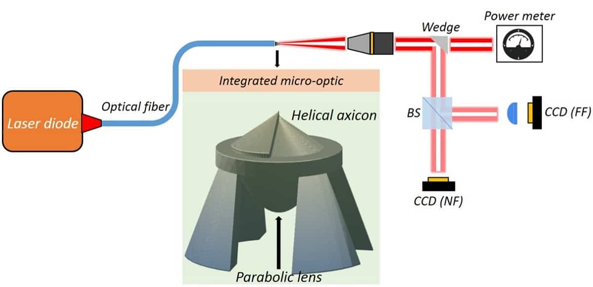
The team printed a 110 µm high, 60 µm diameter optical device on the end of an optical fibre. The device included a parabolic lens with a 27 µm focal length and an axicon with a cone of radius 30 µm and a height of 23 µm. The parabolic lens was designed to align the widely diffracted light from the fibre and focus it into the helical axicon. The axicon had a helical structure designed to add orbital angular momentum to the light.
Once the device was printed, a process that took around four minutes, the researchers spliced the fibre containing the micro-optical device to a fibre laser. They then tested its performance using a purpose-built optical measuring system.
Optical set-up: The measuring system used to analyse the performance of beams shaped by the micro-optical device. (Courtesy: Shlomi Lightman, Soreq Nuclear Research Center)
They found that the device generated a Gaussian–Bessel beam with an initial width of 10 µm. Along a 2 mm distance, this expanded to a width of 30 µm. According to the researchers, a Gaussian beam with identical initial width will reach a width of 270 µm over the same distance, demonstrating that the beam produced by their device is a diffracting-free beam.
The beam of light produced by the micro-optical element was also found to have an orbital angular momentum value of 1 ħ per photon, as expected. The incoming laser beam had no orbital angular momentum.
As the device was printed from organic photosensitive polymers, the researchers were concerned that it may suffer laser-induced damage and limited mechanical stability over time. When they gradually increased the laser power to a maximum optical density of 3.8 MW/cm2 there was no obvious impact on the beam properties. They are now, however, experimenting with this 3D-DLW method on hybrid photosensitive materials that contain a low percentage of polymer. Optical elements printed from such materials could have longer shelf lives and be more resistance to high laser powers, they say.
The team notes that this laser printing technique could also be used for other optical devices. “Our fabrication method could also be used to upgrade an inexpensive lens to a higher quality smart lens by printing a smart small structure on it,” Lightman says.