Stunning images of an Antarctic neutrino detector, a pollinating flower and an aurora-bathed turbine feature in science photography contests

This edition of the Red Folder looks at some images from two science-related photography contests.

Last week the International Union of Pure and Applied Physics (IUPAP) announced the winners of its IUPAP 100 Photo Contest. First prize in the “At a glance” category went to Yuya Makino of Madison, Wisconsin, US who was a “winterover” at the Amundsen-Scott South Pole Station. He was one of two people who monitored the IceCube Neutrino Observatory during the harsh Antarctic winter. In total, 50 people spend the winter at the South Pole, keeping equipment and experiments ticking over.

Makino’s spectacular photo is called “Chasing ghost particles at the South Pole” (above) and it shows a winterover walking towards IceCube, which is bathed in the light of the Southern Lights and the Milky Way.

Surviving by drops

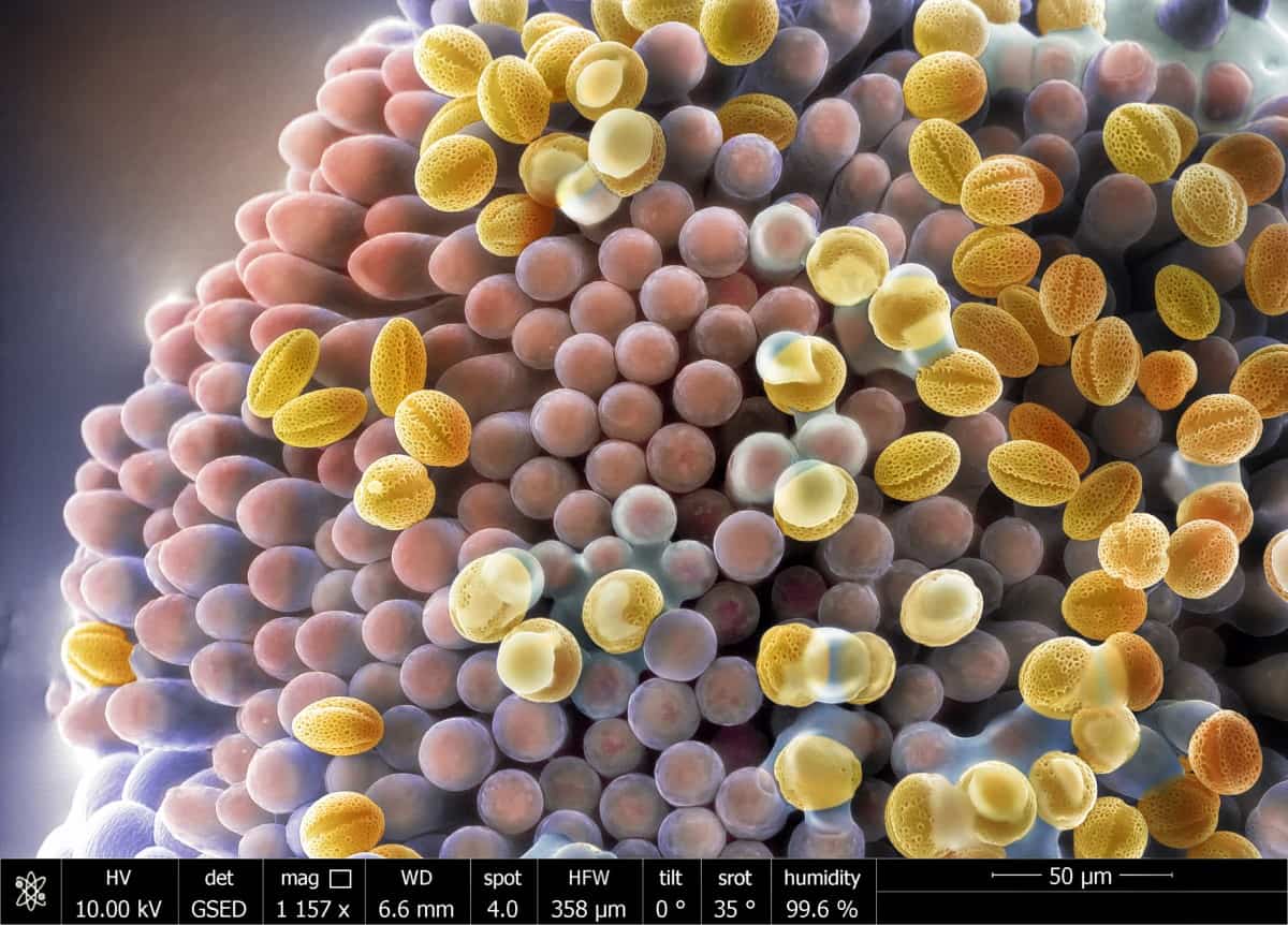
Another of my favourites is “Surviving by drops” by Isabel Sánchez in Granada, Spain (above). This took third prize in the “Beyond our eyes” category and is a colourized environmental scanning electron microscope image. It shows how pollen sitting on the stigma of a flower is activated by condensing water.

Finnish aurora

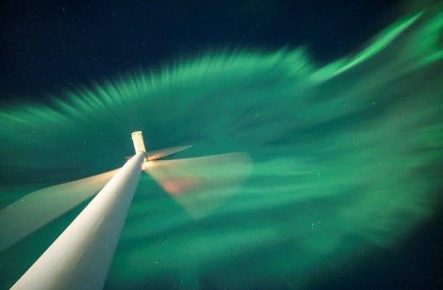
Not to be outdone, this week the UK’s Royal Observatory Greenwich announced its shortlist for its Astronomy Photographer of the Year 2022. Not surprisingly, there were lots of photos of aurorae – indeed the phenomenon had its own category. My favourite entry is the image above taken in Finland by Esa Pekka Isomursu and called “Solar wind power”. It shows a wind turbine illuminated by an aurora, giving the illusion that the two entities are interacting.


Plastic scintillation detectors: real-time dosimetry in the MR-Linac environment

Tumour shape and position relative to healthy tissue will evolve as a cancer patient undergoes a course of radiotherapy – and can even change during an individual treatment session. The ability of MR-guided radiotherapy (MR/RT) systems like the Elekta Unity MR-Linac to detect those changes and adapt therapy accordingly – in effect, helping clinicians to “see what they treat” in real-time – represents a fundamental inflection point for the radiation oncology team.

While it’s still relatively early days for MR/RT adoption, the clinical end-game is already coming into view. Think personalized radiotherapy tailored to the unique indications of each patient – adjusting radiation delivery to address the daily variation in the tumour and surrounding healthy tissue, while enabling the clinician to adapt the plan for tumours that respond rapidly to treatment, as well as those that prove unresponsive to standard doses of radiation. Ultimately, researchers hope that the MR-Linac’s ability to visualize a tumour target with exceptional soft-tissue contrast, both prior to and during treatment, will make it possible to increase the radiation dose to diseased tissue in real-time without damaging adjacent organs-at-risk (OARs) and other critical structures.

Real-time adaptation

In the vanguard of this adaptive MR/RT research effort are Martin Fast and colleagues at UMC Utrecht in the Netherlands. For context, UMC Utrecht was the first research hospital to “go clinical” with high-field MR/RT – back in 2017 – and the centre’s radiation oncology department now has three Elekta Unity systems in routine daily operation. “We’re currently doing adaptation just before treatment starts and sometimes midway through a course of radiotherapy,” explains Fast, associate professor of medical physics at UMC Utrecht. “Where we’re heading, though, is continuous online adaptation of radiation delivery – a breakthrough that will allow us to take account of changes in tumour shape and position as a result of respiratory and cardiac motion.”

Fundamental to success here is a convergence of core enabling technologies. For starters, there’s the ability to capture the tumour and its environment “on the fly” using the MR-Linac’s real-time imaging capability. Put simply, novel MR imaging sequences are able to map a wide range of tumour motion – when the patient breathes, for example, or as a result of peristaltic activity along the digestive tract – which opens the way to dynamic tracking of the linac’s multileaf collimator (MLC), continuously reshaping the treatment beam so as to confine dose to the tumour target while avoiding OARs and surrounding healthy tissue.

Reliable, end-to-end QA is equally fundamental for the successful translation of breakthrough technologies into the radiotherapy clinic. A key building block in the MR/RT QA workflow at UMC Utrecht is the QUASAR MRI4D, an MR-safe, programmable motion phantom from Modus QA, a Canadian supplier of QA solutions for radiation oncology. As well as being a preferred QA vendor, Modus has a long-standing R&D partnership with the UMC Utrecht team – a dialogue that’s proved instrumental in addressing another piece of the MR/RT QA puzzle. “We were brainstorming new ways of measuring dose dynamically,” adds Fast, “and Modus introduced us to Medscint and their real-time, small-field dosimetry solution based on plastic scintillation detectors.”

Making light work of MR/RT QA

Based in Québec City, Medscint is a young and growing technology company combining expertise in photonics, scintillation dosimetry and medical physics. In terms of specifics, the start-up’s plastic scintillators combine near-water-equivalence and real-time response with high spatial resolution and MR-Linac compatibility. The scintillation detectors – known commercially as the HYPERSCINT Research Platform – also offer multipoint capability with a compact footprint (0.5 mm long, 0.5 mm diameter), which makes them ideal for small-field dosimetry and novel phantom developments.

After exploratory conversations at the end of last year, the UMC Utrecht/Medscint collaboration has proceeded at pace. The partners jointly presented on the characterization of the scintillation detectors in an MR-Linac environment at the Canadian Organization of Medical Physics (COMP) Annual Scientific Meeting last month. Meanwhile, new results demonstrating multipoint time-resolved plastic scintillation dosimetry on an MR-linac will feature at the AAPM Annual Meeting in Washington DC next week. This latest work is part of a collective effort – also involving scientists and engineers from Elekta and Modus – to road-test an experimental set-up in which a prototype 3D-printed hybrid cassette (containing four scintillation detectors and one EBT3 film) is incorporated in the QUASAR phantom to quantify MLC tracking for lung stereotactic body radiotherapy.

“We want to use a dosimeter that will integrate easily with the QUASAR motion platform,” explains Prescilla Uijtewaal, a PhD student at UMC Utrecht who co-led the design and validation work on the hybrid cassette. What’s more, the plastic scintillation dosimeter allows the end-user to see how dose is developing in real-time without being influenced by MR image acquisition, the scanner’s magnetic field or the motion of the phantom. Unlike other detectors, the HYPERSCINT platform can also be used regardless of its orientation in a magnetic field environment. “In that sense,” adds Uijtewaal, “it’s a pretty straightforward dosimetry device, because we don’t have to worry about all these dependencies.”

Fast, Uijtewaal and colleagues are already in talks to extend the strategic partnership with Medscint, with plans to evaluate more real-time dosimetry use-cases in the MR-Linac environment. One work-in-progress initiative involves the use of deformable inserts for the QUASAR phantom, mirroring the complexity and 4D deformation that tumours are subjected to as a result of motion within the body – and notably so for radiotherapy treatments in the cardiac region.

“The heart’s motion is not just rigidly translating – it’s deforming with each heartbeat,” explains Fast. “As such, we plan to integrate the HYPERSCINT detectors with a realistic model that supports time-resolved dosimetry while taking account of this type of complex motion and deformation.”

Big opportunities in small-field dosimetry

François Therriault-Proulx

François Therriault-Proulx, president and CEO of Medscint, was one of the company’s co-founders in 2016, along with colleagues Simon Lambert-Girard (chief science and technology officer) and Jonathan Turcotte (chief product, sales and marketing officer). Prior to establishing Medscint, Therriault-Proulx spent eight years as an academic scientist working on the fundamentals of scintillation dosimetry, spanning PhD and postdoctoral research positions at Université Laval, Quebec City, and the University of Texas MD Anderson Cancer Center, Houston. He talked to Physics World after his presentation at last month’s AAPM Summer School on Small-Field Dosimetry, SRS and SBRT.

How did delegates respond to your talk at the AAPM summer school?

There were around 180 attendees at the summer school – mostly medical physicists. For many delegates, my presentation offered the first opportunity to learn about plastic scintillation detectors in terms of the fundamental science and enabling technologies under development. As such, the event was an ideal forum for Medscint to get the word out to the clinical physics community, raising awareness about the HYPERSCINT Research Platform, the capabilities of our in-house product development team, also our R&D partnerships on cutting-edge applications like adaptive MR/RT and FLASH radiotherapy.

Why are scintillation detectors a good fit for small-field dosimetry?

Compact-footprint, water-equivalent plastic scintillator technology has a lot to offer as treatment fields get smaller and geometrically more complex – for example, in SRS treatments of metastatic tumours in the brain. With no need for small-field correction factors to characterize device behaviour, our detectors provide a real-time measurement tool that combines high linearity with respect to dose and dose rate. That wide linear dynamic range is relevant at both ends of the treatment spectrum, whether for novel low-dose-rate irradiation schemes or ultrahigh-dose-rate FLASH applications.

What are the priorities for Medscint at the AAPM Annual Meeting this month?

First up, we’re looking to establish new collaborations with radiotherapy research groups, though our direction of travel is shifting towards cross-disciplinary teams with a focus on clinical translation. Second, we want to gather granular feedback from clinical end-users on their evolving QA requirements for next-generation radiotherapy modalities. We’ll also be exploring potential synergy with other technologies and partnership opportunities with relevant manufacturers.

Characterization of Li-ion battery thermal runaway in ESSs and EVs

Want to learn more on this subject?

Thermal runaway is an undesired occurrence with lithium-ion cells and batteries when improperly designed, manufactured or used. With the size of these batteries increasing exponentially and their use in confined spaces becoming more common, it is imperative to fully characterize the nature and products of thermal runaway. In this webinar, Judy Jeevarajan of the Electrochemical Safety Research Institute (ESRI) at UL Research Institutes provides details on the need to carry out such characterizations; methods to take the test articles into thermal runaway; the worst case events observed; and products of fire and smoke for test articles studied by ESRI. Finally, Jeevarajan provides some recommendations on addressing the prevention and propagation of thermal runaway in Li-ion batteries.

Want to learn more on this subject?

Judy Jeevarajan is vice-president and executive director of the Electrochemical Safety Research Institute (ESRI) at UL Research Institutes (formerly Underwriters Laboratories, Inc.). She has worked in the field of batteries for more than 25 years, with a primary focus on lithium-ion chemistry.

Jeevarajan serves in the technical working groups and committees for standards organizations such as UL, Society of Automotive Engineers, International Civil Aviation Organization/Society of Aerospace Engineers, Radio Technical Commission for Aeronautics, International Electrotechnical Commission, and American National Standards Institute. She currently leads an effort under the American Institute of Aeronautics and Astronautics to develop a space safety standard for battery systems. She also serves as a member of the Informal Working Group and Dangerous Goods Panel under the United Nations. Jeevarajan is a member of the Great Lakes Energy Institute Advisory Board at Case Western Reserve University.

Before joining Underwriters Laboratories Inc., she worked for NASA at the Johnson Space Center (JSC) in Houston for 12 years, serving as group lead for Battery Safety and Advanced Technology. Prior to becoming a civil servant at NASA, Jeevarajan worked onsite for five and a half years at NASA-JSC for Lockheed Martin Space Operations.

Jeevarajan earned an MS in chemistry from the University of Notre Dame and PhD in chemistry (electrochemistry) from the University of Alabama. She has won numerous NASA awards, including the NASA Exceptional Service Medal and the NASA-NESC Engineering Excellence Award. She also received the 2019 American Institute of Aeronautics and Astronautics Aerospace Power Systems Award, and India Energy Storage Alliance Woman Leader of the Year 2020–Energy Storage Systems Award.


Graphene tattoo provides cuffless
blood-pressure monitoring

Wrapping a cuff around a patient’s arm and inflating it to measure blood pressure is one of the most routinely performed medical tests. It provides a quick and reliable assessment of cardiovascular health, as blood pressure is an independent predictor of all-cause mortality. But such arm cuffs are bulky and uncomfortable, making them impractical for continuous monitoring outside of clinics.

For this reason, researchers are developing cuffless alternatives with the goal of unlocking new possibilities for patient diagnostics and management, as well as providing new understanding of physiology. However, none of these tools has become a mainstay yet.

One option, acoustic sensors, slide during movements and are too large to be easily incorporated into untethered ambulatory sensors. Meanwhile, optical modalities such as smart watches are limited by the low penetration of light into tissues, which hinders their ability to capture haemodynamic parameters in the arteries. Studies also show that optical sensors are sometimes inaccurate when used with darker skin tones or larger wrists.

Graphene tattoos and machine learning

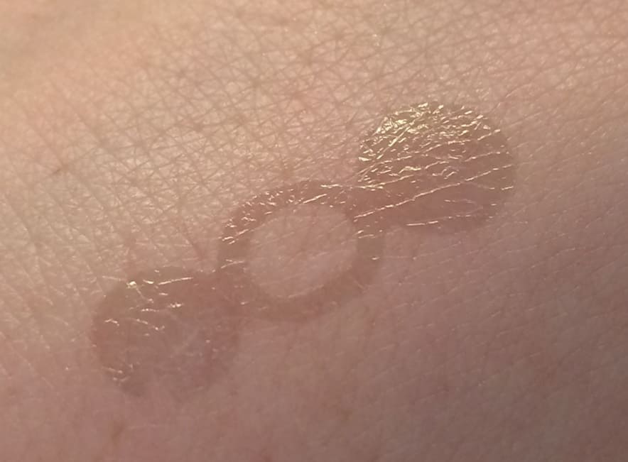
A group of researchers from the University of Texas and Texas A&M University, led by Roozbeh Jafari and Deji Akinwande, circumvented these impediments by developing a sticky and stretchable graphene electronic tattoo that is comfortable to wear for long periods and does not slide around. They describe the new blood-pressure monitor in Nature Nanotechnology.

Graphene electronic tattoo

Graphene, one of the strongest and thinnest materials in existence, is similar to the graphite found in pencils but with the carbon atoms precisely arranged into layers just one atom thick.

“The sensor for the tattoo is weightless and unobtrusive. You place it there. You don’t even see it, and it doesn’t move,” says Jafari.

The device performs measurements by injecting a low-intensity electrical current into the skin and then analysing the body’s response, known as the bioimpedance. The electrical signal penetrates deep into the skin and propagates through the path of least resistance: the blood vessel, as blood is ion-rich and thus a better conductor than the surrounding fat and muscle cells. The signal that is collected reveals variations in bioimpedance, which are correlated with blood-pressure variations.

The researchers also used the device to measure pulse wave velocity, the speed at which blood travels in the arteries. They then used the bioimpedance and pulse wave velocity data as inputs for a common machine learning algorithm (AdaBoost) to predict diastolic (minimum) and systolic (maximal) blood pressure points.

Grade-A classification performance

To assess the accuracy of the tattoo, the researchers enrolled seven volunteers, attached sensors above their radial arteries and asked them to perform a series of activities known to change blood pressure (hand grip, cycling on a stationary bike and the Valsalva breathing manoeuvre). They measured reference blood pressures using a medical-grade blood pressure cuff.

In total, the researchers recorded 18,667 data points and split the data into 89% for training and 11% for testing, a process known as cross validation. The measurement accuracy – 0.2±4.5 mm Hg for diastolic pressures and 0.2±5.8 mm Hg for systolic pressures – was equivalent to grade-A classification, according to the IEEE standard for blood-pressure monitoring devices.

Further, some activities were combined with sweat-inducing walks outside at 38 °C or push-ups; none of the sensors degraded electrically after exposure to light and heat or contact with water or sweat. The sensor was able to monitor arterial blood pressure for more than 300 min, 10-fold longer than reported in previous studies. The sensors could also be used to record other vital signals, such as breathing respiratory rates, and could be placed on other locations, such as the tibial and carotid arteries and the jugular notch.

Looking forward, there is still some work needed to estimate central aortic blood pressure, which differs from peripheral blood pressure and is thought to be a superior indicator of cardiovascular events. Likewise, assessing the whole pressure wave over a cardiac cycle as opposed to single points could provide additional information on blood vessel functions and cardiac performances.

Aliens could use quantum signals to communicate with Earth

The Search for Extraterrestrial Intelligence (SETI) might want to add quantum communication to its list of ways for aliens to get in touch. According to calculations by researchers at the University of Edinburgh in the UK, quantum signals would be a viable means of establishing contact across interstellar distances – a result that also suggests we might need to update our technology to recognize any such signals coming in our direction.

This finding might seem surprising, given that setting up quantum links here on Earth has proven no easy task. Such links are based on creating entanglement between individual nodes and teleporting quantum states between them, but these states are fragile, and their tendency to decohere – that is, to lose their quantum nature – limits the stability of the links. Interstellar links, therefore, represent a bold step forward. Could quantum information survive the hostile space environment during a journey towards an interstellar receiver?

Effects of interstellar disturbances

To answer this question, the Edinburgh researchers calculated the likely impact of various disturbances a quantum signal could encounter. One such disturbance is gravity, which could cause quantum states to decohere and signals to lose fidelity. However, the researchers computed that a photon could travel 127 light-years before such decoherence comes into play, meaning that a considerable number of stars with known exoplanets are within reach.

The impact of space travel on the fidelity, or quality, of a quantum signal is slightly different, because decoherence is not the only contributor. “High fidelity” means being able to fully process a quantum signal once it is received. This parameter can be quantified by considering a relativistic effect known as Wigner rotation that can change the signal’s phase, resulting in a loss of fidelity while coherence remains intact. However, the researchers note that if the receiver knows the signal’s origin, they would in principle be able to estimate the magnitude of this effect and calculate the signal’s original phase.

Besides gravity, several other factors could disrupt the quantum state of a photon. Interstellar space contains a distribution of electrons, photons, hydrogen atoms and some heavier elements. Locally, such particles can also come from our own Sun. But when the researchers calculated the probability of a signal photon interacting with any of these, they found that the mean free path distance was larger than the observable universe, meaning no considerable interaction can be expected. Photons at X-ray wavelengths, in particular, have longer mean free paths through scattering and absorbing media such as gas and dust, and are less susceptible to interference from large magnetic fields, making them favourable for quantum communication.

ET teleport home?

Finally, the researchers considered the question of why an extraterrestrial civilization might choose quantum communication over classical signals. According to Arjun Berera, a physicist at Edinburgh and lead author of a paper on the research in Physical Review D, there are some benefits. One is that the quantum nature of the signal would be a sign that it comes from an intelligent source rather than a natural process. Another is that quantum communication makes it possible to pack a lot of information into the signals, especially when utilizing higher-dimensional entangled states.

Michael Hippke, an expert on interstellar communication who is affiliated with Germany’s Sonneberg Observatory, calls the new research “an excellent contribution to the field” because it shows that quantum photons can travel over interstellar distances without losing coherence. As for whether other civilizations (if they exist) could communicate with quantum light, Hippke, who was not involved in the latest research, describes the idea as plausible. “We should look for that,” he says. He adds that identifying the X-ray region of the electromagnetic spectrum as a potential carrier is important, though he notes that any attempt to detect such a signal would have to be carried out in space because the Earth’s atmosphere absorbs most X-rays.

Berera says that the team’s next step is to establish whether any natural astrophysical sources could also produce coherent quantum photon states. “It would be an important question to answer before we start focusing our attention on the quantum route for finding ETs,” he says.

Wallpaper made of moth wings is an excellent absorber of sound

When moth wings are used to coat hard, artificial surfaces, they can significantly reduce the reflection of incoming ultrasound, researchers in the UK have shown. Without making any modifications to the wings’ scale structures, Marc Holderied and colleagues at the University of Bristol showed how the natural metamaterial performs remarkably well as natural soundproofing.

Conventional soundproofing materials tend to be porous, and to be effective they must be thicker than about 10% of the wavelength of the sound they are blocking. Metamaterials made of specially designed structures can be thinner than 1% of the wavelength they absorb, but these tend to operate over a very narrow band of frequencies. While broadband metamaterials have been created, they tend to be much thicker.

To create thinner broadband sound absorbers, some researchers are looking to the wings of moths for inspiration. Bats hunt moths using echolocation, so some moth species have developed a remarkable ability to absorb the high-frequency sound waves bats produce. The insects do this using microscopic scales that decorate both sides of their wings.

Range of sizes

These scales come in a broad range of sizes – each with a characteristic resonant frequency. This allows the wings to absorb sound across a wide range of frequencies, making them far more effective than conventional sound-absorbing materials. Previous studies have shown how moth wings absorb sound waves as the insects travel through the air. In their study, Holderied’s team looked at how the wings absorb sound when attached to an aluminium disk.

Typically, such a hard, manmade surface will reflect most incoming sound. In contrast, the researchers observed that the moth-wing coating reduced this reflection by up to 87% at the lowest frequencies they tested. The ultrasound used by the team had wavelengths some 50 times longer than the thickness of the wings.

The team then had a closer look at how the sound was absorbed. They removed the scales from the upper side of the wing and discovered that this caused the absorption to vary with the orientation of the incoming sound. While its performance remained high when the bald side faced the incoming sound, it broke down almost completely in the orientation. By recreating this scenario in simulations, Holderied’s team showed that the metamaterial’s performance strongly depends on the presence of scales in the air gap between the wing membrane and the hard surface beneath.

The sound waves absorbed by moth wings may be beyond the range of human hearing, but by adapting the design to absorb lower frequencies, Holderied and colleagues hope that new artificial metamaterials inspired by their structure could soon be developed. These structures could lead to new breakthroughs in high-performance soundproofing: potentially leading to coatings for walls, vehicles, and noisy machinery that takes up a fraction of the space required by existing materials.

The research is described in Proceedings of the Royal Society A

How we know climate change causes extreme weather, celebrating the 10th anniversary of the Higgs-boson discovery

In this episode of the Physics World Weekly podcast, the climatologist Fredi Otto explains why scientists can say with confidence that certain extreme weather events such as floods and heatwaves are more likely to have happened because of climate change. Otto is at the Grantham Institute for Climate Change and the Environment, Imperial College London. She has recently published a review paper in the journal Environmental Research: Climate called “Extreme weather impacts of climate change: an attribution” and speaks to Physics World’s James Dacey about her research.

Also in this episode, we chat about the discovery of the Higgs boson, which was announced 10 years ago by physicists working on the Large Hadron Collider at CERN. Physics World is celebrating the anniversary by publishing a month-long series of articles about particle physics, so stay tuned to website.

Researchers produce first in vivo images of brain inflammation using MRI

Imaging brain inflammation

Chronic inflammation of the brain is linked to a range of increasingly common degenerative brain diseases, such as Alzheimer’s and Parkinson’s. Evidence suggests that neuroinflammation contributes to the progression and worsening of such diseases.

However, the current diagnostic tool used for monitoring brain inflammation, positron emission tomography (PET), involves ionizing radiation, which may increase the patient’s risk of developing cancer. The delivered radiation dose also makes it impractical to perform longitudinal research studies or repeat testing during treatments. As such, there’s a need to develop an efficient imaging modality that will not worsen the condition of patients with neuroinflammation.

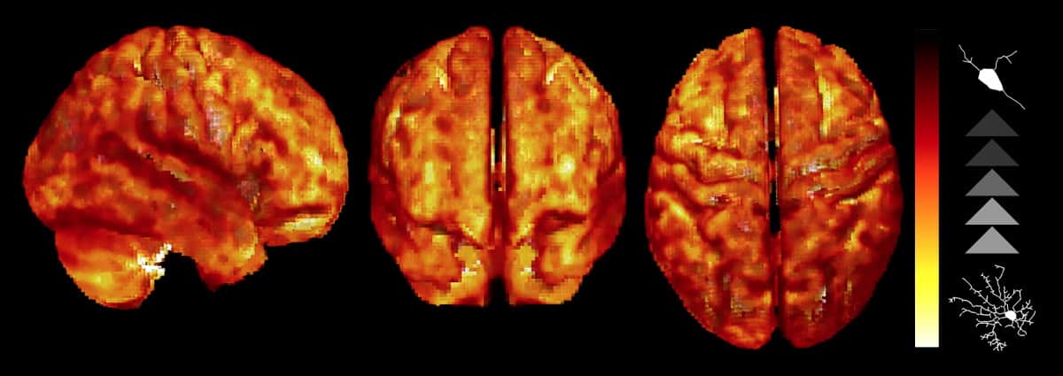
Researchers from Alicante, Spain, have developed a non-invasive method for visualizing brain inflammation using diffusion-weighted magnetic resonance imaging (DW-MRI). The team, led by Silvia De Santis and Santiago Canals from the Institute of Neurosciences, a joint centre of the Spanish Superior Research Council (CSIC) and the Miguel Hernández University (UMH), devised a series of MR data acquisition sequences and mathematical models to detect changes in the activation of two brain cell types associated with neural inflammation: astrocytes and microglia.

Raquel Garcia-Hernandez and Silvia De Santis

DW-MRI enables the collection of images from microstructures within the brain with a high resolution by utilizing the random motion of water molecules within brain tissue. Most previous research use of DW-MRI has focussed on the brain’s white matter and axons, but to investigate chronic inflammation, the researchers were interested in imaging the brain’s grey matter.

To focus on the all-important astrocytes and microglia, they therefore had to adapt and design advanced DW-MRI sequences, for use in combination with mathematical models based on the biological knowledge of the functional tissues within the brain.

The scientists tested their model on rats, using an established technique for inducing inflammation (administering lipopolysaccharide), which first activates the microglia, followed by a delayed response from astrocyte cells, allowing independent investigation of the two cell types. The MRI scans showed specificity to both the microglial and astrocyte activation in grey matter in vivo.

Secondly, the researchers used the method in human participants in a proof-of-concept experiment, scanning six healthy volunteers across five occasions. They found that the pattern of microglial cell density significantly correlated with the MRI parameter of stick fraction. The results highlight the model’s ability to detect glial biomarkers and confirmed reproducibility between scanning sessions.

The researchers hope that their work will enable better characterization of brain tissue microstructure during inflammation, with higher resolution and the possibility of longitudinal study without a dose of ionizing radiation. They believe that this could transform the diagnosis and treatment monitoring of the many diseases associated with an inflammatory glial response.

The research is published in Science Advances.

3D quantum spin liquid discovery gains theoretical support

For decades, quantum spin liquids were seen as largely hypothetical, with real-world examples thought to exist only in unusual systems such as quasi-one-dimensional chain-like magnets and a handful of two-dimensional materials. Then, in 2019, researchers at Rice University in the US and McMaster University in Canada found experimental evidence that a pyrochlore magnet, Ce2Zr2O7, could be a long-sought-after example of a three-dimensional quantum spin liquid (3D QSL). This “experimental conjecture” has now received further support from physicists at Rice and Florida State University in the US. Working with colleagues at the Max Planck Institute for Physics of Complex Systems in Dresden, Germany, the researchers used a combination of theoretical techniques to conclude that Ce2Zr2O7 (CZO for short) is indeed a 3D QSL.

QSLs form when atoms in solid magnetic materials cannot arrange their magnetic moments (or spins) into a regular and stable pattern. This “frustrated” behaviour is very different from that of ordinary ferromagnets or antiferromagnets, which have spins that point in the same or alternating directions, respectively.

Quantum mechanics explains this frustration by suggesting that the orientation of the spins is not rigid. Instead, the spins constantly change direction in a fluid-like way, producing an entangled ensemble of spin-ups and spin-downs. Thanks to this behaviour, a QSL will remain in a liquid state even at temperatures near absolute zero, where most materials freeze solid.

“Sleuthing out” the parameters of a theoretical model

In 2019, two experimental groups – one led by Pengcheng Dai at Rice and the other by Bruce Gaulin at McMaster – concluded, based on data from thermodynamic and neutron scattering experiments, that CZO behaved in a way consistent with a 3D QSL. In the latest work, a team led by Florida State’s Hitesh Changlani set out to prove its status beyond reasonable doubt.

To do this, Changlani and colleagues analysed the raw data from both experiments, seeking to determine the optimal parameters of a so-called dipole-octupole model of cerium magnetic moments with strong spin-orbit coupling. Finding these parameters would establish what kind of QSL exists in CZO – if indeed one exists at all.

“This was a daunting task and the simple ‘mean-field’ treatment we initially attempted did not work,” Changlani says. In its place, the team turned to an alternative known as the finite temperature Lanczos method, together with a classical–quantum correspondence previously developed by some of the team’s members, to perform quantum calculations of the material’s specific heat and magnetization.

This approach allowed the researchers to model the material’s molecular spin dynamics (based on the Landau-Lifshitz equation) and compare their results with those from the earlier inelastic neutron scattering experiments. According to the team, the modelled parameters “capture[d] the salient features of the experimental data both qualitatively and quantitatively”.

“Upon taking into account all the experimental data on CZO and previous theoretical proposals for QSLs on the pyrochlores, we were able to ‘sleuth out’ the parameters of the theoretical model that could indeed describe all the observations,” Changlani explains, adding that the model was first proposed in a paper in Physical Review Letters in 2014. “From everything we know, it would appear that the 2019 experimental conjecture was correct.”

“Worth looking at more closely”

Until now, many quantum materials researchers thought that for a material to have exhibit strong quantum fluctuations, and hence a predisposition towards QSL behaviour, it needed to have reduced dimensionality – that is, its structure needed to consist of weakly coupled planes that could be thought of as 2D-like. “Our work challenges this notion,” Changlani tells Physics World. “We have provided evidence that 3D pyrochlores are an example of frustrated magnets worth looking at more closely.”

In terms of practical applications, Changlani says it is too early to say what they might be. Although some versions of quantum computing are based on the use of fractionalized excitations realized in QSLs, he cautions that “we do not know yet whether CZO could fit the bill here”.

According to team member Andriy Nevidomskyy, the results are more exciting for fundamental physics – including a possible connection between QSLs and magnetic monopoles, which are hypothetical particles that carry an isolated north or south magnetic pole.

“Of course, in classical physics one can never isolate just one end of a bar magnet,” he says. “The north and south monopoles always come in pairs. But in quantum physics, magnetic monopoles can hypothetically exist, and quantum theorists constructed these almost 100 years ago to explore fundamental questions about quantum mechanics.

“As far as we know, magnetic monopoles don’t exist in a raw form in our universe,” Nevidomskyy continues. “But it turns out that a fancy version of monopoles does exist in these cerium pyrochlore quantum spin liquids. A single spin flip creates two fractionalized quasiparticles called spinons (particles that would carry an electron’s spin) that behave like monopoles and wander around the crystal lattice.”

The researchers say they also found evidence that monopole-like spinons were “created in an unusual way” in CZO. Due to the material’s tetrahedral arrangement of magnetic atoms, it might develop octupolar magnetic moments – that is, spin-like magnetic quasiparticles with eight poles — at low temperatures. “Our research showed spinons in the material were produced from both these octupolar sources and more conventional, dipolar spin moments,” Nevidomskyy concludes.

Full details of the research are reported in npj Quantum Materials.

Keep on keeping on: why we must persevere in our study of the Higgs boson

Cartoon of 'How it started' and 'How it's going' showing excited celebrations in the first frame, dejection in the second

2012: we found the Higgs boson

The discovery of the Higgs boson was a massive triumph. Not just for the physicists who spent decades designing, building, tuning and operating CERN’s Large Hadron Collider (LHC) and its huge detectors. It was also a triumph for scientific imagination.

Physics had done something bold. We looked at the patterns of the particles and said: “This would make more sense if there was another piece, right here.” As if the universe were a jigsaw puzzle and we could imagine the shape of the next piece.

And we found it! The discovery of the Higgs boson showed us that our imagination, with a healthy dose of deduction, can reveal the shape of reality.

And we had many more ideas too. Important pieces of the puzzle were still missing from our understanding of the fundamental particles, and physicists had interesting new ideas for what those pieces could be. Could the Higgs boson’s mysteriously small mass, for example, be explained by a swarm of new particles with names like gluinos, sbottoms, photinos and staus?

We were intoxicated by the power that our imagination and our scientific tools had allowed us to have, and we looked forward to finding many new particles in the next decade at the LHC.

2022: We’re still hoping to see something (anything!)

Ten years later, we haven’t found any more puzzle pieces. Despite 10 trillion (1013) trips for particles around the LHC rings, and 100 quadrillion (1017) proton collisions, no new particles have been discovered. Had we been too clever with our ideas? Not clever enough? Was it a mistake to think that imagination could guide us to new discoveries? Was it a mistake to keep running the LHC? Or to plan for more powerful, future accelerators?

A decade of experiments has confirmed what we already knew: research is exploration, where discoveries are never guaranteed. Nothing that we’ve found at the LHC in the last 10 years has generated the kind of fanfare and excitement that came from discovering the Higgs boson. But that’s not why we do it.

We explore because we are not content with just thinking about beautiful ideas of what might be. We want to know what is real. It’s the same reason we land rovers on Mars or send spacecraft to scan the moons of Jupiter. Because to explore is to venture into the unknown. Imagination motivates exploration, it doesn’t replace it.

Sometimes you have to step into the unknown to confirm what is, and isn’t, there. Remember that not finding new particles can often be as significant as finding them; we should never underestimate the power of null results – and who said physics was ever straightforward or easy? The LHC’s work will surely guide us through the next phase of particle-physics exploration as we continue to look for new pieces of the grand puzzle of nature.

Copyright © 2026 by IOP Publishing Ltd and individual contributors