Migrating birds use information extracted from the Earth’s magnetic field to target the same breeding grounds year after year, with the field’s inclination angle, in particular, acting as a “stop sign” telling them they have reached their destination. This conclusion, based on a new analysis of data obtained by attaching metal rings to the legs of birds and then tracking their movements, adds to a growing body of knowledge about field-based navigation in migratory animals.
Although there is considerable evidence that some species of birds use the Earth’s magnetic field to navigate, the precise mechanism is still not fully understood. Some theories invoke an inherited “clock and compass” vector system to explain how birds can return to their breeding grounds with extraordinary precision, but the question of how they know when and where to stop was unresolved.
In an attempt to answer it, Joseph Wynn and Tim Guilford of the University of Oxford in the UK and colleagues at the University of Oldenburg, Germany turned to a long-term experiment on Eurasian reed warblers (Acrocephalus scirpaceus). These migratory songbirds fly across the Sahara each year to spend the summer in Europe, and between the years 1940 and 2018 more than 17,000 of them were “ringed” to allow researchers to monitor their movements over many years and large geographical areas.
A single magnetic inclination angle
The team’s analyses suggest that the warblers register, or learn, a single magnetic inclination angle – effectively the degree of “dip” between the Earth’s magnetic field and its surface – before setting off on their journeys. Later, they use this angle as a unique coordinate telling them they have reached their breeding site. While different locations across the globe have the same inclination angle, the team say that the warblers solve this problem by stopping at the first place where they encounter the right inclination, according to their inherited vector system.
This explanation may not be all there is to it, however. Because the Earth’s magnetic field in a given location changes slightly from one year to the next, the magnetic parameter values that are characteristic of an individual bird’s breeding site will exist in a somewhat different location on its return trip. In a paper in Science describing the work, the researchers acknowledge that these magnetic variations need to be taken into account. “Nonetheless, we believe that our findings provide evidence for an unconventional mechanism of long-distance navigation, both within birds and migratory animals more generally,” they conclude.
Space junk – debris left by humans in space – is a growing danger for satellites and space missions orbiting the Earth. It turns out that the Moon also has space junk and in this episode of the Physics World Weekly podcast, Roberto Furfaro and Vishnu Reddy of the University of Arizona talk about the challenges of tracking lunar space junk and identifying its origins.
Also this week, the teacher David Marshall talks about the sometimes byzantine process of correcting errors in physics textbooks, curricula and exams. He also shares some of the more bizarre mistakes he has found over the years – including one about bouncy light, which has yet to be corrected.
I woke up in Germany at 5 a.m. CET on Thursday 24 February 2022, two hours after the first bombs had landed in my hometown Kyiv. The day before in a group meeting at the University of Bayreuth, my PhD supervisor asked my wife (also a PhD student) and me if we were scared about the situation in Ukraine. My wife said that she was worried about her parents and possible war. I was calm; I believed no-one could be so insane as to start a war in Europe in the 21st century.
Early Thursday morning my wife was sleeping and I decided not to wake her up, she deserved to have a few more hours of peace. I immediately messaged both our parents, but no-one answered.
We spent the entire morning writing invitation letters so our parents could come to Germany. I also wrote to my PhD supervisor to get some documents confirming our status to help our parents cross borders. He responded quickly and said that we should stay at home and do everything possible to help our families.
We scrolled through Telegram channels all day, monitoring the situation
The first day after the conflict began was spent in despair. We were scared of war and knew we could not do anything being so far from Ukraine right now. We scrolled through Telegram channels all day, monitoring the situation. In Ukraine as in the other post-Soviet countries, Telegram is the most popular messaging service and almost everyone uses it. No wonder it became the main source of information for us.
The Ukrainian government created official Telegram channels, where our president Volodymyr Zelensky reports every day about the current situation. In addition, every region of Ukraine has an official channel, where they try to report about the status of each region. People began to organize themselves through Telegram to attack Russian propaganda websites and send photos of Russian military columns moving through Ukraine to military officials to guide attacks.
Sleeping in a corridor
The next day I wrote to my former undergraduate adviser. He was in Ukraine with his family in the centre of Kyiv. I was happy to discover that he is now supervising a friend, who is doing a PhD with him. However, life is not normal: my adviser’s family is sleeping in the corridor of their building so that they as far from the windows as possible. Nevertheless, he was optimistic. I told him that the deadline for nominations for the Philipp Schwartz Initiative of the Alexander von Humboldt Foundation has been extended until 18 March for Ukrainian scientists. This is a programme for scientists who face considerable and ongoing personal danger in their home countries, inviting them to work in Germany. I also told him that I could help him with finding a host at my German university. He told me that he would do it only if Russia occupies Ukraine, otherwise he is going to stay.
On the third day after war began, our despair had been replaced by anger
On Sunday 27 February, the third day after war began, our despair had been replaced by anger. We were angry about war; angry about the fact that we cannot do anything except donate to the Ukrainian cause; angry about the bomb attacks; and angry about Russia’s president Putin.
Our university began to stir. On the Bayreuth campus, students started collecting warm clothes and medicine for people in Ukraine. My wife received an e-mail from a professor she met only once who said he would be happy to accept students from Ukraine for short research visits and asked for help to spread the news. In social networks, I started seeing more and more research groups worldwide encouraging Ukrainian students and scientists to apply for internships and visits.
Ukrainian science has been declining steadily following the collapse of the Soviet Union, funding is scarce and corruption has been eating into what little money is left. The equipment is old and researchers have to wait a long time to make measurements and do experiments. I remember how proud the scientists from a Ukrainian research institute were when they showed us an electron microscope that they received as a gift from Japan in the early 1980s.
I believe that Ukrainian science will rise in the future, as a part of the European scientific family
What is happening in Ukraine is a tragedy, but people all over the world are helping and the scientific community is no exception. Science should not have borders and it is wonderful that more and more science is being done in collaborations. I believe that Ukrainian science will rise in the future, as a part of the European scientific family and the bonds between scientists will grow stronger.
A new study aimed at quantifying how plants take up plastic nanoparticles from the soil has revealed that the plastics accumulate mainly in the roots rather than the shoots. The technique used to trace the nanoparticles involves materials known as lanthanide chelates, and the researchers who developed it say it could be a versatile way to analyse the interactions between nanoplastics and plants.
Tiny fragments of plastic are everywhere – in the ocean, in our food, even on the summits of mountains. The smallest of these fragments, known as nanoplastics, are thought to be more hazardous to life because their small size enables them to penetrate cell membranes. Since the continued large-scale production of plastics means that concentrations of nanoplastics are unfortunately likely to increase, it is important to understand their impact on the environment and the potential risk to human health.
Because nanoplastics can interact with plants in many ways, scientists need to be able to follow how these particles accumulate and move through the plant’s structure. While many studies have investigated the way that plant protoplasts – that is, cells with their walls removed – take up nanoplastics, the mechanisms for uptake and translocation of nanoplastics through large-scale plant structures remain poorly understood. Quantitative information on the rate at which plants uptake nanoparticles and then transport them is particularly lacking.
Studies on lettuce and wheat
In the present work, researchers led by Yongming Luo of the Chinese Academy of Sciences studied how two crops, lettuce and wheat, took up 200-nm-diameter polystyrene particles doped with a europium chelate, Eu-β-diketonate. Polystyrene is the one of the most commonly produced polymers in the world and is widely employed both in food packaging and as a “soil conditioner” to stabilize soil surfaces and help them retain moisture. It has been detected in organic fertilizers, sewage sludge and wastewater.
To mimic different environmental conditions, Luo and colleagues grew their lettuce and wheat in hydroponic cultures and in sandy soil. They quantified the doped polystyrene particles in the plants using a technique known as inductively coupled plasma mass spectrometry. As europium is a very rare element and does not naturally occur in plants, every signal they detected represents a particle that the plant took up. They also visualized the particles using background-free time-resolved fluorescence imaging, and confirmed their presence using scanning electron microscopy.
The team’s analyses revealed that polystyrene-europium particles accumulated mainly in the roots of the plants, while transport to the shoots was less than 3% for 5000 μg of polystyrene particles per litre of exposure. Willie Peijnenburg, a researcher at Leiden University in the Netherlands who was also involved in the study, explains that finding more plastic in the root than the shoot means that only a small number of particles end up in the edible parts of the plants.
The researchers, who report their work in Nature Nanotechnology, say they now plan to apply this technique in microcosm or mesocosm experiments to enhance the sensitivity of their nanoplastic tracing and detection methods.“We need to carefully monitor potential lanthanide leaching from the particles in the systems we studied due to the complex environmental conditions, as well as due to the presence of a wide number of (micro)organisms,” team member Lianzhen Li tells Physics World.
Science fiction has always explored scientific possibilities, both current technology and the furthest reaches of what could still be described as science. The most interesting SF uses science as a means to explore society, psychology and other aspects of being human, which often means any physics involved isn’t explored in depth. Even “hard science fiction” – depicting science that is possible and central to its plot – rarely goes into great scientific detail.
In that regard, The EXODUS Incident by Peter Schattschneider is an exception to the rule, not only including lengthy discussions of physics (and indeed other sciences) within its pages, but also featuring a bulky appendix with abundant background detail to the physics explored. It’s also a lively crime thriller set in the future.
Schattschneider is a physicist based at TU Wien who has spent much of his career both writing science fiction and using classic SF in his lectures. So he is well placed to blend complex physics into storytelling.
The novel opens with an academic-paper-style abstract followed by social-media messages (Schattschneider wisely doesn’t specify what platform they’re being posted on), which tell us that the Earth is suffering from catastrophic climate change, a series of pandemics and of course war. In what might be the last chance to save humanity, Europe is sending a spaceship called Exodus to establish a colony in the Proxima Centauri system, on a planet identified as habitable by the Breakthrough Starshot project initiated by Yuri Milner and Stephen Hawking (the novel doesn’t mention the real-life project’s third partner Mark Zuckerberg).
After that set-up, I was a little confused to find the narrative is not initially set on a spaceship, but instead follows two police detectives in Austria investigating a serial murderer. They are living in a future where Vienna is unbearably hot, meat consumption has been outlawed by the EU and the population is declining fast, but otherwise their police work is familiar from any police procedural you might read or watch on TV. Until, that is, lead investigator Oliver Storm is given access to AI virtual-reality tools to help him explore crime scenes.
Schattschneider’s depiction of Europe in the future is scarily believable. As well as climate catastrophe, there are frequent military check points, border wars (including between the UK and Ireland – an interesting, albeit unnerving, detail) and resource scarcity. Everyone who can is moving further from the equator to temperate climes. One of the fundamentalist groups we encounter call themselves “Thunberg adepts”, who are still struggling to be heard in their call for real action to help the planet.
Schattschneider’s depiction of Europe in the future is scarily believable. As well as climate catastrophe, there are border wars and resource scarcity
The hero, Storm, is a smug, misogynistic character yearning for a past when he could own a car, travel freely and eat as much meat as he wanted to. In a nod to classic fictional detectives, he’s a loner with a shady past and manages to have sex with every woman he takes a shine to, despite being generally rude to them. He is also, importantly, curious about everything he encounters so that over the course of the novel various specialists can explain complex science and technology to him, as a cypher for us readers.
Sadly, Storm does reflect a certain old-fashioned patriarchal tone to the novel as a whole. The small number of women characters have no identifying characteristics beyond their appearance; there are no gay, trans or non-binary characters; men are known by their surname, women by forename; and men appear to be in charge of everything (though perhaps this is a deliberate part of the dystopia that Schattschneider has created).
I decided after a few chapters that it’s best not to worry too much about the minimal character building, as this is not Schattschneider’s strong suit. When he tries to add character detail it’s clumsy and stands out from what is otherwise a strange but enjoyable murder mystery with a futuristic SF backdrop, which develops into fully embraced SF with the crime investigation as the backdrop. And while individual characters are lacking in depth, Schattschneider’s explorations of wider psychological themes are handled well, particularly isolation and conspiracy theories.
The plot goes to some (for me) unexpected places that explore a range of ideas social, political and psychological. Schattschneider’s influences are clear, from The Matrix to Arthur C Clarke, and many of these are acknowledged in fictional conversations between characters.
This novel is part of Springer’s Science And Fiction series, a collection of both hard science fiction and analysis of SF by scientists. It’s an interesting idea for an academic publisher to pursue, but I couldn’t help noticing that the series’ large roster of editors are all men, and of the 46 books published in the series since 2014, only three are written by women. SF has historically been a tough market to break into for authors who aren’t cis white men, but in recent years that has changed significantly and it’s a shame for a major publisher not to follow that trend.
Despite its flaws The EXODUS Incident is gripping and thought-provoking. The technical details of the Exodus spaceship are particularly thorough. It is propelled by a Bussard ramjet engine and last year Schattschneider co-authored a paper on the feasibility of such a system (Acta Astronautica 191 227) – concluding that the engine could be made to work but would achieve much lower speeds than previous studies suggested.
For physics fans, the appendix is the real treasure trove, as here Schattschneider produces a fictional mission report with all the technical details, from the ramjet engine to the type of vegetation that might survive on the exoplanet and its weather systems. But don’t skip ahead to the back of the book as it is full of spoilers.
Glioblastomas are the most common and deadliest tumours of the central nervous system. Standard-of-care for these tumours typically involves some combination of surgery, radiation therapy and chemotherapy, but patients still often survive only a few months following treatment. Some newer immunotherapy drugs that show promise in other cancers have shown little to no benefit for patients with glioblastomas.
In part, this is because immunotherapies, which boost a person’s immune system so that it can fight cancer, do not cross the blood–brain barrier well. Another challenge is that the tumour microenvironments of glioblastomas suppress the immune system, making it challenging for the immune system to recognize and attack cancerous cells.
A recent study published in ACS Nano presents a novel combination therapy, investigated in mice, that may address both challenges. The therapy, when combined with a burst of radiation, halted glioblastoma growth and prolonged mouse survival.
“We overcame these hurdles by using extracellular vesicles,” says Bakhos Tannous from Harvard Medical School. Tannous, who is senior author on the ACS Nano paper, says that extracellular vesicles (EVs) are “known to facilitate intercellular communications governing diverse processes, such as immune response”.
Therapeutic EVs
EVs are naturally released from many cell types and carry different types of cargo, such as proteins, nucleic acids, lipids and metabolites, from a parent cell. At tens of nanometres to almost 10 µm in size, the smallest EVs can cross the blood–brain barrier and aren’t recognized as invaders.
“They [EVs] are naturally secreted by every cell in the body and therefore are not foreign molecules that induce immune rejection such as solid lipid nanoparticles, for instance,” says Tannous, who is also director of the Experimental Therapeutics Unit and the Viral Vector Core Facility at Massachusetts General Hospital.
Therapeutic uses for EVs arrived on the scene when researchers realized that EVs taken up in a target cell can alter its behaviour. Since this discovery, researchers have demonstrated that EVs can be used as a vehicle to deliver drugs throughout the body.
But EV-based therapies alone are not enough to treat glioblastoma, Tannous’ team notes.
An unwanted brake
Radiation therapy is perhaps the most important nonsurgical treatment for glioblastoma. While radiation sensitizes tumours that fail to generate T cell responses (which help kill cancerous cells), not all responses to radiation are beneficial. Sometimes, infiltrating immune cells are recruited into a tumour in response to radiation. These cells increase the amount of a critical protein called PD-L1.
PD-L1 is often described as a brake that keeps the body’s immune responses under control. Elevated expression of PD-L1 can trick the body’s immune system into thinking that a cancerous tumour isn’t a harmful, foreign substance. As a result, therapy can be less successful than it might be in the absence of elevated levels of PD-L1.
Tannous’ team has introduced a combination immunotherapy that inhibits PD-L1 and induces an immune response in the body to kill cancer cells. Tannous and his collaborators, also based at Nanjing Medical University, the University of Balamand and Brigham and Women’s Hospital, introduced small interfering ribonucleic acids (siRNAs) into EVs to reverse the anti-immunogenic effect that occurs when tumours are primed with radiation.
“By loading EVs with siRNAs against PD-L1 and injecting them in tumour-bearing mice primed with a burst of radiation, we can reverse this effect and induce T cell activation and anti-tumour immunity,” Tannous explains. “The burst of radiation was essential not only in recruiting immune cells, but also in increasing the uptake of these EVs by the tumour and its microenvironment.”
New combination therapy
The team produced the EVs using a human neural progenitor cell line and modified them with a peptide (cyclic RGDyK) that targets brain tumours and helps the EVs penetrate the blood–tumour barrier. The researchers then introduced siRNAs into the EVs’ membranes to help ensure that the body’s immune system responds to the tumour.
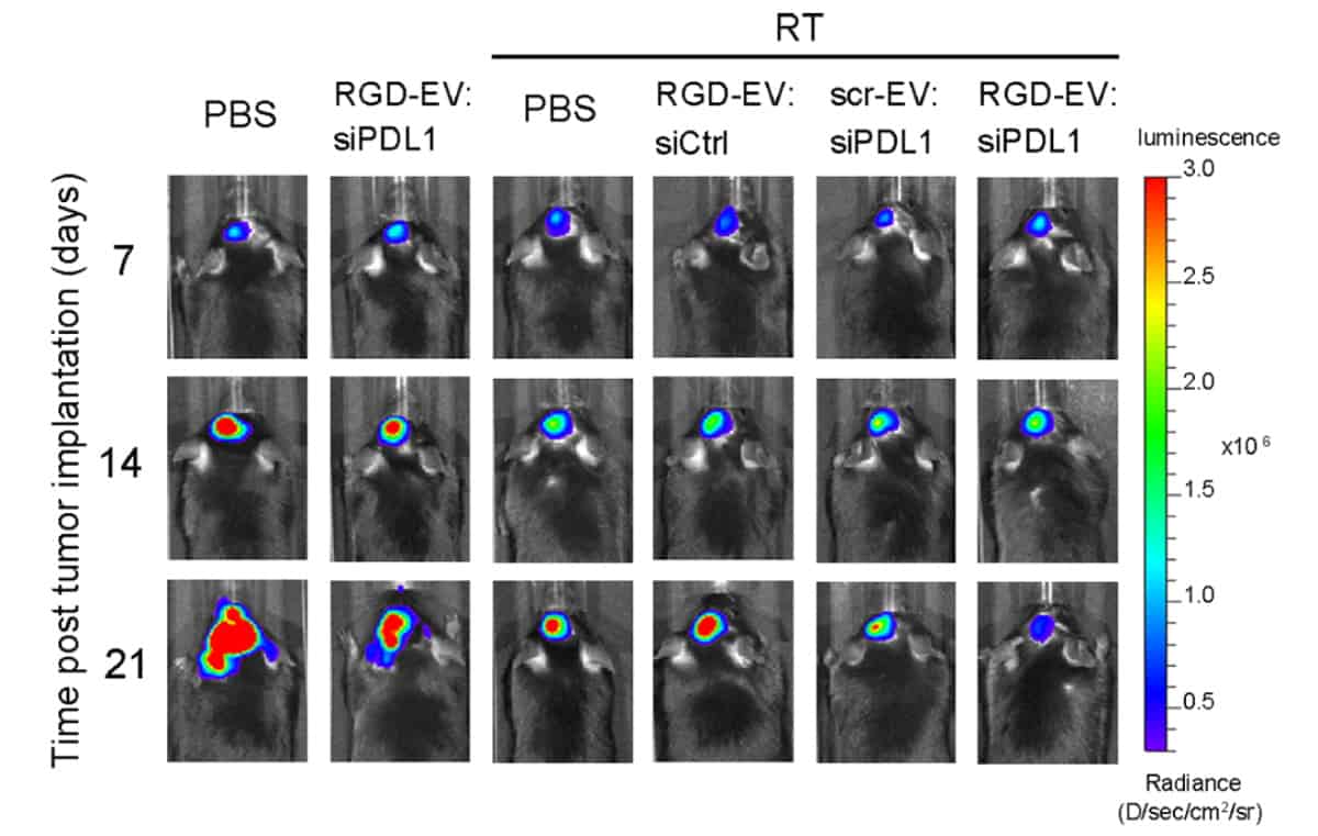
The researchers then put their combination therapy to the test. They injected mice with murine glioblastoma cell lines. Murine tumours were primed with a 5 Gy burst of radiation, analogous to stereotactic radiosurgery in a clinical setting, seven and 14 days following tumour cell injection. The mice received an injection of the combination therapy, unmodified EVs or saline (as a control) on days 10, 12, 17 and 19 following tumour cell injection.
The team found that priming the murine glioblastoma tumours with a burst of radiation enhanced the delivery of EVs to the tumours. The combination therapy also halted tumour growth and prolonged mouse survival. Results further suggest that using EVs allowed the immunotherapy to cross the blood–brain/tumour barrier, recruiting immune cells to the tumour site and inhibiting the expression of PD-L1. Fluorescence imaging studies showed that stronger fluorescence signals, and therefore more EVs, were observed in mice brains that had been irradiated relative to those that were not.
Large-scale production
The researchers say that their decision to isolate EVs from a human neural progenitor cell line, rather than stem cells or dendritic cells, was an important one. Using a human neural progenitor cell line allows them to produce EVs in larger quantities for larger studies and clinical trials.
Another important decision was to use copper-free click chemistry to modify EVs to include the brain-tumour-targeting peptide on the EV surface. Copper-free click chemistry, unlike some cell engineering methods, is suitable for in vivo applications, is fast and can be used for large-scale production of modified EVs. Previous work from the group found no obvious toxicity or tissue damage using these methods.
Now, Tannous’ team is scaling up EV production and labelling. The researchers are also working to further improve the EV delivery system and are testing how EVs can deliver nucleic acid therapies to brain tumours.
A close analysis of data from NASA’s Parker Solar Probe has revealed that electromagnetic “whistler waves” – so named because early radio operators mistook these eerie, descending sounds for a person whistling – are unexpectedly absent from certain regions of the Sun’s upper atmosphere. The discovery could lead to a better understanding of the physics of the solar wind, and thus to more accurate predictions of space weather and how it might affect us here on Earth.
The solar wind is a stream of energetic, charged particles – mostly electrons and protons – ejected from the Sun’s upper atmosphere, or corona, in all directions. Learning more about the dynamics of the solar wind is important because these charged particles perturb the Earth’s magnetic field when they collide with it. Such perturbations are known as space weather, and they can damage satellites, impact communications technologies and GPS signals, and even cause power outages on the ground at high latitudes.
Corona heating
The Parker Solar Probe was launched in 2018 with a mission to learn more about the solar corona and how heat moves through it. In the latest study, Cynthia Cattell and colleagues at the University of Minnesota Twin Cities in the US performed a statistical analysis of data from all of the probe’s solar “encounters” (as its orbits of the Sun are known) so far, focusing on observations made when the probe was especially close to the Sun.
Previous analyses had revealed that between about 35 solar radii (one solar radius is a little less than 696,000 km) and the Earth’s orbit at 215 solar radii, the solar wind contains “whistler waves”. These electromagnetic waves help regulate the way heat flows from the corona, and they also play an important role in the Van Allen radiation belts that surround the Earth.
Signatures of an ambipolar electric field
In regions closer to the Sun, however, the Minnesota researchers found no evidence of whistler waves. At less than about 30 solar radii, they instead spotted signatures of a different, electrostatic wave. The electrons in the solar wind in this region also showed evidence of being affected by an ambipolar electric field created partly by the Sun’s gravity. This effect is somewhat like the one that occurs near the Earth’s poles, where the solar wind is accelerated, Cattell explains.
The absence of whistlers implies that they cannot be responsible for controlling the heat flux in this region of the solar corona, she adds. “This heat flux is carried by what is called ‘strahl’ (the German word for beam or ray) electrons and the limiting of the heat flux is due to scattering of strahl by the whistlers so it is not a beam anymore,” she tells Physics World. “The whistler electric field rotates in a right-hand sense about the solar wind magnetic field at the same rate as the electrons do. So, electrons moving in a range of speeds see a constant electric field and are accelerated.”
Cattrell thinks the work should help scientists make better predictions of space weather. “If you don’t understand the details of energy flow close to the Sun, then you can’t predict how fast the solar wind will be moving or what its density will be when it reaches Earth,” she notes. “These are some of the properties that determine how solar activity affects us.”
Understanding the flow of heat is important also in many other astrophysical settings, including accretion discs, other stellar winds, and the interstellar medium, she adds.
The researchers, who report their work in Astrophysical Journal Letters, say they now hope to better understand why whistler waves are absent so close the Sun, how the electrons accelerated by the gravity-associated electric field might excite other waves and how that, in turn, impacts the solar wind. Meanwhile, Parker’s encounters are getting closer and closer to the Sun. In 2024 it will fly to within 6.08 million kilometres from our star – the closest any spacecraft has ever ventured.
Wouldn’t it be amazing to have a tool like Google Earth but for the human body, where you could zoom in from a full organ down to its cellular structures? That’s now becoming a reality thanks to the Human Organ Atlas project, and physics is key to this innovation. This short videos explains how the Human Organ Atlas project emerged from the COVID-19 pandemic and how it could help medical scientists in a range of clinical contexts.
Discover more about the Human Organ Atlas in this feature, originally published in the March issue of Physics World.
In this age of information, we expect to have knowledge at our fingertips. If we’re looking to obtain a first impression of someone, many of us head straight to their social-media pages. If we want to understand a new topic, we don’t buy a textbook – most of the basics are waiting for us on Wikipedia. And if we want to explore a new city, we can do much of it by moving around in Google Earth. Information that was once costly or exclusive is now free to all.
But what about medical images? Suppose you want to explore what a real human heart looks like, from the entire organ down to the smallest blood vessels. Currently, for most of us, that’s impossible. True, a heart surgeon could obtain radiological images of a patient’s heart, and order biopsies of specific volumes. But even then, the doctor will be easily frustrated by the limitations of individual imaging methods.
Clinical computed tomography (CT), which uses X-rays to build up 3D images slice by slice, is restricted to millimetre resolution. So too is magnetic resonance imaging (MRI), which peers inside the body using magnetic fields and radio waves. Microscopy of biopsies, meanwhile, is usually limited to millimetre-sized volumes. The dream of seeing an organ – or the entire human body – with micron or near-micron resolution has simply been out of the question, whether you are a specialist or not.
Not any more. For the last two years, dozens of scientists in Europe have been busy compiling the most detailed 3D views of real organs ever seen. Like a Google Earth of the human body, the Human Organ Atlas, as the team’s project is known, is both simple and astonishing. Its goal is to create a freely accessible, online image bank of highly “zoomable” human organs, revealing everything from their biggest features (on the scale of centimetres and metres) all the way down to micro-scale structures.
The project has already led to the creation of 3D images of lungs, a brain, a heart, a kidney, a spleen and a liver. By 2025 the Human Organ Atlas team wants to have imaged an entire human torso and, not too far beyond that, an entire human body. The work is impressive for scientists and non-scientists alike – so much so that the project is being bankrolled by some high-profile funding agencies in the UK, EU and US. Even Google has taken an interest.
One scientist who has been collaborating on the project is Danny Jonigk, a lung pathologist at Hannover Medical School in Germany. He feels as if he has spent his entire career doing research under candlelight, only for someone “to suddenly switch the lights on”. Then there’s Daniyal Jafree, a medical student at University College London (UCL) in the UK, who’s doing a PhD in kidney imaging. When he heard what was being developed elsewhere at UCL, Jafree couldn’t quite believe it. “I thought that sounds ambitious,” he says. “Then I saw the images.”
X-rays at your service
The Human Organ Atlas project wouldn’t be possible without physics. It began at the European Synchrotron Radiation Facility (ESRF) in Grenoble, France, which has been one of the world’s foremost X-ray light sources since it opened more than 30 years ago. Unlike the X-rays delivered by a clinical CT scanner, synchrotron X-rays have high energy and a high spatial coherence. That means their waveforms remain very much in phase with one another as they propagate, allowing researchers to exploit minute changes in X-ray phase to produce tomographic (section-by-section) 3D images of very high detail and contrast (see box below).
For many years, this phase-contrast X-ray technique has delivered incredible reconstructions of biological specimens. In 2011, for example, ESRF beamline scientist Paul Tafforeau helped produce what is still the most detailed scan ever of the inside of a skull of an early human ancestor, Australopithecus sediba. More recently, he has produced scans of small dinosaur fossils, ancient human teeth and even mummified crocodiles.
Seeing more deeply Claire Walsh from University College London (left) and Paul Tafforeau from the European Synchrotron Radiation Facility in Grenoble, France, are among the scientists to have developed the new imaging technique of Hierarchical Phase-Contrast Tomography (HiP-CT). Originally used to scan donated human organs, including lungs from a patient who died from COVID-19, the technique is central to plans for a zoomable Human Organ Atlas. It will provide 3D reconstructions of entire intact organs that can then be explored anywhere down to the cellular level. (Courtesy: ESRF/Stef Candé)
Then, in 2020, two things happened. The first was that the ESRF finished commissioning a new, “fourth-generation” source, making it the world’s brightest synchrotron lab. More than a decade in planning and construction, the Extremely Brilliant Source (EBS) delivers X-rays that are 100 times brighter than before, and 100 times more coherent in the transverse (horizontal) plane, making them almost laser-like at low energies. The EBS has done wonders for tomographic imaging, enabling users to scan bigger objects, in more detail and at a greater range of scales.
The second big event of 2020 was, of course, the COVID-19 pandemic. For many scientists, the pandemic brought research to a full stop. Not for Tafforeau. Unexpectedly, he received a call from Peter Lee, a regular ESRF tomography user at UCL, who in turn had been approached by Jonigk. Could the ESRF be of help, Lee wondered, in reconstructing lung tissue samples from people who had died after catching COVID-19? It was a great question and almost overnight Tafforeau switched from study-ing ancient fossils to human organs.
“The COVID-19 pandemic changed a lot of things for many people,” Tafforeau recalls. “I realized that several imaging techniques that we originally developed for palaeontology could open access to a new level of imaging precision on complete human organs. Then, while developing the techniques further, we realized that it may be a game-changer for biological imaging in general.”
Swiftly, Lee composed an international, multidisciplinary team to see what could be done: synchrotron imaging scientists at UCL and the ESRF; mathematicians and computer scientists at UCL; medical scientists at Hannover Biobank, as well as the universities of Mainz and Heidelberg in Germany. As the apparent potential of the new tomographic imaging grew, so did the breadth of the collaboration: it now includes more than 50 people.
The scientists called the technique hierarchical phase-contrast tomography (HiP-CT), thanks to its ability to provide 3D reconstructions of entire intact organs that can then be explored anywhere down to the cellular level. As a result, the technique bridges the gap in scales between clinical CT and MRI, and the microscopy of biopsies. In November 2021 the project was formalized as the Human Organ Atlas, with a goal to provide a reference database of organ imagery that is accessible to all.
Hierarchical Phase-Contrast Tomography (HiP-CT) in a nutshell
Most simple imaging methods – including conventional computed tomography (CT) – involve measuring the loss of intensity (the attenuation) of an electromagnetic wave as it passes through a sample. In 1953, however, the Dutch physicist Frits Zernike won the Nobel Prize for Physics for developing an alternative – and potentially more illuminating – imaging method that involves measuring shifts in the phase of electromagnetic rays.
Zernike’s “phase-contrast” microscopy was initially fit only for visible light. But in 1965 it started being extended to X-rays too thanks to the work of Ulrich Bonse and Michael Hart – two physicists at Cornell University in the US – who used a crystal interferometer to convert phase changes into interference patterns.
Limitations with interferometers meant that phase-contrast X-ray imaging of biological samples had to wait until the 1990s through the efforts of Atsushi Momose at Hitachi and Tohoru Takeda at the University of Tsukuba, Japan, and others. At roughly the same time, Anatoliy Snigirev and others at the European Synchrotron Radiation Facility in Grenoble, France, realized they could deduce phase changes without an interferometer, simply from the interference of highly coherent synchrotron X-rays in free space. By combining many propagation phase-contrast 2D images in CT mode, they were able to produce 3D reconstructions of small biological samples with far more detail than that available from clinical CT scanners.
With the upgrade of the ESRF to a “fourth-generation” X-ray source in 2020, “hierarchical” phase-contrast CT (HiP-CT) became possible. The lab’s ultra-coherent X-rays provide information on phase changes over very long propagation distances up to 40 m, allowing samples of up to 2.5 m × 1.5 m in size – including human organs, torsos, even entire bodies – to be imaged in 3D at micron resolution.
The atlas in action
A video of a human brain, as imaged by HiP-CT, gives an impression of the technique’s capabilities (figure 1). It starts off conventionally enough, moving through cross sections of the entire organ. Here the brain looks like it does with a clinical CT scan, albeit at some 50 times the resolution. The various lobes are clearly visible, as are some of the external blood vessels. Then the “camera” zooms in to the back of the brain, the cerebellum, perfectly transitioning from big to small.
At 5 μm resolution, the smallest features of white and grey matter come into view; at 2.5 μm resolution, the tiniest blood vessels can be discerned. Even pyramid-shaped cells can be seen, known as Purkinje neurons, which are largely responsible for human motor function. Finally, the view retreats and the reconstruction morphs to depict blood vessels only. Now the incredible density and complexity of the brain’s “vasculature” become apparent. As the system that delivers and receives the oxygen, glucose and metabolic waste, it keeps every one of us alive and thinking.
The HiP-CT video looks like the cutting-edge CGI you see in sci-fi blockbusters – yet it is perfectly real. What’s more, as all the raw data have been collected and post-processed, it’s possible for scientists to explore different parts of the brain at will. In fact, the sheer wealth of information in the imagery is so great that interpreting it is a major problem in itself. The team divides the work, with Tafforeau in charge of reconstructing the images and the UCL team trying to make sense of them.
1 The Human Organ Atlas in action Stills from a video of a human brain imaged using Hierarchical Phase-Contrast Tomography (HiP-CT) shows what the technique can do as you zoom in and back out. a A cross-section of the entire organ reveals lobes and some external blood vessels. b A closer image of the back of the brain, the cerebellum. c At 5 mm resolution, white and grey matter come into view. d At 2.5 mm resolution, you can see the tiniest blood vessels. e As the view retreats, the reconstruction depicts the blood vessels only, revealing the full complexity of the brain’s ‘vasculature’. f Zooming back out again, the blood vessels in situ. See videos on YouTube. (Courtesy: ESRF/HiP-CT: C L Walsh, P Tafforeau, W L Wagner et al.)
“It’s a little bit overwhelming, like being a kid in a sweetie shop,” admits Claire Walsh, a biophysicist in UCL’s computational analysis team. “Medics and histologists can tell us when something looks weird, but we have to quantify that: exactly how weird?” One example is the size of alveoli, which has in the past been used to indicate the seriousness of lung disease. Previously, says Walsh, the alveoli were assumed to be roughly spherical, like grapes on a vine. But the new technique reveals them to be more irregular.
As a result, the researchers have had to define new parameters, with input from their medical collaborators, to capture the potential of the new information. Joseph Jacob, a chest radiologist who joined the UCL team early in the pandemic, stresses the scale of the interpretation challenge. “When I first saw the images, I felt amazement and apprehension – probably apprehension more than amazement,” he says. “It was definitely what I wanted to work on, but the complexity of labelling it – obviously it would only be possible with computer science.”
Fortunately, Jacob knows how vital the image-processing of X-ray data is, having developed algorithms to stitch together hundreds of CT images to see the lung in detail. He believes the reward now is well worth the labour. “[This new technique] is going to show us things we never knew existed,” he says, which could be vital given how medicine is a very “organ-centric” discipline. “As a chest specialist, I just look at the lungs – I don’t look at the heart, for instance. But disease doesn’t necessarily work that way. If you could image a whole torso, you could understand how disease is affecting other organs; it would be a much more rounded approach.”
The way ahead
As things stand, almost all the organs in the atlas have been imaged at the ESRF’s long-serving BM05 beamline. In December 2021, however, the team acquired its first HiP-CT images at BM18 – a new ESRF beamline that has been designed to maximize the benefits of the EBS for microtomographic images of large objects. Although the beamline won’t be fully operational until the end of 2022, it will eventually be able to image a torso – and even an entire human body.
Imagine one day being able to explore, in virtual reality, human bodies of all ages, backgrounds, states of health and disease. As Lee points out, the damage wrought by new diseases could then be easily compared with that of existing conditions, to indicate possible known methods of treatment. People could see what sort of processes might be going on inside themselves. Medics could entertain pure curiosity, without having to resort to the knife.
We are not there yet, but preliminary images have already given some indication of the benefits of the large-scale, detailed view of HiP-CT. Reconstructions of several lungs from COVID-19 victims have revealed heterogeneous damage that appeared on previous clinical CT scans merely as a fuzzy, ground-glass texture (Nature Methods18 1532). The result is helping to determine whether it is the connectedness of lung damage, or the sheer amount of it, that is the cause of death by the virus.
Meanwhile, Jafree is keen to find out if HiP-CT can help us to give us a better understanding of the kidneys, the organs he specializes in. We know that the number of blood-vessel networks, or glomeruli, is a proxy for general kidney function. But no-one knows how losing some of these networks affects those that remain, or whether their volume or shape affects kidney health. “HiP-CT allows us to look at things in a different way,” says Jafree. “It also encourages [students] like me to learn some of the image-analysis techniques. We need that expertise to generate something meaningful for biology and medicine – and we have an incentive now.”
Vital signs These HiP-CT images of human kidneys could reveal if the volume or shape of blood-vessel networks affects the onset of kidney disease. (Courtesy: ESRF/HiP-CT: C L Walsh, P Tafforeau, W L Wagner et al.)
Sarah Teichmann, a cellular geneticist at the Wellcome Sanger Institute in Hinxton, UK, says she was “blown away” by the first HiP-CT images she saw, letting her view the cellular structures inside organs in exquisite detail, before zooming out to see the whole tissue. “Not only do these images and videos give a new appreciation of the beautiful complexity of the human body,” she says, “they are also stocked full of information about how our bodies work.”
Teichmann believes that the whole-organ or whole-body approach could benefit our understanding of diseases such as cancer. She also reckons that the Human Organ Atlas project ties in well with the Human Cell Atlas – an international consortium that she co-founded to create a comprehensive reference map of all human cells. “[It] could help us to see where these cell types – which we characterize at the molecular level – fit into the bigger picture of the organ. This will help to bridge the gap between cells and systems, painting a more holistic picture of the human body.”
Beautiful times
Alongside the huge scientific impact of this imaging technique, there is also an inherent beauty to the images taken by the Human Organ Atlas. In December 2021 National Geographic magazine picked a HiP-CT image of a lung as one of its favourite science images of the year. Francesco Sette, the physicist who has been director-general of the ESRF since 2009, has even compared the advancement of the technique with Leonardo da Vinci’s anatomical drawings of the early 16th century.
Those drawings gave unprecedented insights into the workings of the human body, especially its biomechanics. It is not yet clear what the ramifications of the Human Organ Atlas will be, although the concept is proving popular. The collaboration’s Nature Methods paper has been downloaded more than 50,000 times, and is in the top 1% of Nature articles in terms of its Altmetric score, or reach.
The project is also gaining some serious backers, not least a $2.75m donation from the Chan-Zuckerberg Initiative (CZI), which was set up by Facebook founder Mark Zuckerberg and his wife Priscilla Chan. The CZI is independent of Facebook – which may be a good thing, as the Atlas team is just beginning a collaboration with Google to make its database available to the public. According to Lee, the plan is to create something like an anatomical version of Google Earth, with 3D “satellite” resolution of 40 μm resolution for a whole organ, and a 3D “street view” resolution down to 1 μm to expose individual cells.
After Google Earth, Google Maps and Google Sky, perhaps it is fitting that one day we will have a Google Body search tool too.
Placing large-scale solar farms on the Arabian Red Sea coastal plain could dramatically increase rainfall in this arid part of the world, a new modelling study claims. According to the researchers, simulations show that such installations could change the reflectiveness of the land enough to unsettle coastal air circulation. The resulting changes in local weather patterns and rainfall could potentially produce enough water to meet the annual needs of five million people. Although idealized, the team says its research points to the feasibility of freshwater recovery from sea breezes by land surface geoengineering.
As the world warms, water security is becoming a major concern. This is a particular issue in hot, arid parts of the world like the Middle East. Many countries in this area, such as Saudi Arabia, are in the middle of a water crisis. They have little rainfall and are exhausting underground aquifers. Water desalination is already widely used to increase freshwater supplies, but current methods are unsustainable due their high energy use. Desalination also unlikely to be able to meet future water demands.
There is increasing interest in the idea of artificially increasing rainfall over the Arabian Peninsula, using techniques such as cloud seeding. There is a lot of water in the air in the region because the Red Sea loses 0.7 teratonne of water every year through evaporation. This is equivalent to nearly 8% of the mass of all water vapour in Earth’s atmosphere. On the Arabian Red Sea coast, sea breezes blow this water across the land, but little of it falls as rain. Instead, it is transported south towards the equator and the middle of the Indian ocean.
Changing albedo
Georgiy Stenchikov, an expert in climate and atmospheric modelling at King Abdullah University of Science and Technology in Saudi Arabia, and his colleagues wondered how changing surface albedo – the reflectiveness of the land – over the region would impact water transport and change rainfall patterns. The idea, Stenchikov told Physics World, is to try to utilize this vast natural freshwater resource by increasing precipitation.
In their latest work, published in the Journal of Hydrometeorology, Stenchikov and colleagues ran a series of numerical simulations in a regional weather research and forecasting model. They focused on three scenarios: extensive forest planting, increased albedo and decreased albedo.
The simulations show that afforestation and increased surface albedo along the Arabian Red Sea coastal plain would reduce rainfall. Sea breezes in the region are driven by the horizontal thermal contrast between land and sea. The warmer land and cooler sea create a pressure gradient that pushes moist sea air towards the land. The models show that afforestation and a more reflective surface cools the land, which dampens sea breezes and reduces the movement of water vapour from sea to land.
Strengthening sea breezes
Conversely, decreasing surface albedo would increase rainfall. It would warm the coastal region, strengthening sea breezes and increasing water vapour transport to the shore, the researchers found. The increase in convection currents over the land increases vertical mixing in the lower atmosphere, particularly of water vapour, the models show. This increases humidity, cloud formation, and instability and turbulence. “The basis for all this is increased water surface flux,” Stenchikov says.
Solar panels are known to alter the surface energy balance by absorbing solar energy and heating up. The team found that a reduction in albedo that corresponds with the large-scale installation of solar panel plants would increase rainfall in the region by around 1.5 gigatonne in the dry summer season. This is linked to an almost doubling of wet days from July to September, compared with current conditions. Smaller reductions in albedo would also have significant impacts on the number of wet days and rainfall volumes.
Stenchikov says that this proof-of-concept study shows that the idea could work, but more analysis is needed. “We have to work further to try to optimize this scheme, looking at the sizes [of solar installations needed] and optimal distribution for this specific area,” he explains.
Stenchikov says that such schemes could in theory work in any coastal region with sea breezes. They will work best, he explains, in areas with strong sea breezes, high levels of evaporation from the sea and large water vapour fluxes.