
I spy: a virtual reality system in the MRI

Virtual reality in an MRI scanner

The bore of a magnetic resonance imaging (MRI) machine can be intimidating. The claustrophobic atmosphere, paired with knocking sounds that are sometimes louder than an aircraft flyover, can frighten even those comfortable with medical procedures. Children are especially susceptible to these fears, and as a result, MR scanning failure rates can be as high as 50% in young children.

MR technicians employ many simple methods to distract patients during imaging, including audiovisual systems, which they can also use to stimulate brain activity for functional MRI scanning. However, these techniques fail to remove the sensation of entering the MRI bore or assuage the jarring sensations that arise during its function. A research team centred at King’s College London envisioned a technology that would immerse patients in a game environment, distracting them and even replacing the sensations of being inside an MRI machine. The team, led by MRI specialists Kun Qian, Tomoki Arichi and Jo Hajnal, published the study findings in Scientific Reports.

The group investigated a virtual reality (VR) system that replaces the visual scene seen by a patient with immersive and tailored content, in an attempt to alleviate claustrophobia and allow more effective scanning of vulnerable groups like children. The system consists of a 3D-printed headset, a projector and multiple MRI-compatible cameras used to record the patient’s eye movements. The cameras allow the subject to control a VR environment using gaze tracking.

VR system utilizing gaze tracking

The gaze tracking system acts as the crux of the VR system, as it allows immersive control of the environment without bodily movement. First, the cameras obtain images of the patient’s eyes and surrounding areas. A computer then identifies the coordinates of the eye corners and pupils on these images and feeds this information into a progressive interaction interface. The patient then calibrates the system by gazing at dots on the screen. Their gaze coordinates at these times are fed into a regression model, which is constantly updated, improving robustness as they interact with the scene.

Participants reported more attention diversion when playing the video game than they did while watching a film, and no subjects required selection support from carers while exploring the system.

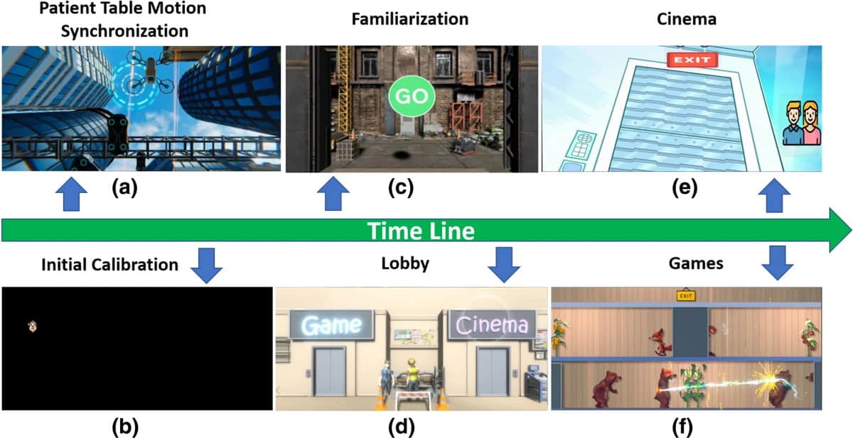
The researchers note that maintaining congruence with external physical stimuli was especially important to the subject’s immersion experience. Therefore, in addition to standard audio-visual stimuli from the VR system, the system integrated external stimuli into its storyline and function. For example, the system integrates the loud knocking and vibration of the MRI scanner by introducing construction characters into the scene, whose tools would be likely to cause said disturbances. If the subject gazes at these characters, they apologize for causing the disruption.

Further, to fully immerse the user in the virtual environment from the start, the system synchronizes the environment with the movement of the patient table sliding into the bore. In this way, subjects of the study noted that they were left without the impression of even having entered the scanner bore.

This technology demonstrates the potential to act as an immersive, engaging escape for patients requiring MRI scans. The researchers call for more in-depth testing of the VR system, especially for integration of full MRI exams and systematic studies with children.

Blood test detects brain tumours at an early stage

Detecting a brain tumour at the earliest possible stage enables faster treatment and safer surgery, which are essential to improve the patient’s chance of a good clinical outcome. But brain tumour diagnosis is a difficult task, as common symptoms such as headaches or memory change are not specific to cancer. As such, many tumours remain undetected until they are larger or of a higher grade. A research team in the UK has now demonstrated that spectroscopic liquid biopsy can detect both small and low-grade gliomas – and could increase the likelihood of early diagnosis.

Liquid biopsies are a minimally invasive diagnostic tool in which small samples of blood are analysed. For cancer diagnostics, most liquid biopsies detect genetic material such as circulating DNA. But early-stage tumours can have extremely low levels of cancerous genetic material in the blood, and for brain tumours, the blood–brain barrier creates further limitations.

The researchers are developing an alternative approach. Instead of detecting specific genetic material, they analyse the molecular composition of a blood sample using attenuated total reflection Fourier transform infrared (ATR-FTIR) spectroscopy combined with machine learning algorithms.

“The spectroscopy is used as a broad assessment of the entire composition of the blood sample, which contains over 20,000 molecules,” explains first author Ashton Theakstone from the University of Strathclyde.

Serum sample studies

In their latest study, described in Cancers, Theakstone and colleagues examined 177 blood serum samples from patients with varying sizes of brain tumours, including 90 patients with high- and low-grade gliomas, as well as 87 asymptomatic controls. They measured the tumour volumes using MR imaging and divided the patients were into two groups depending on their MRI parameters (T1-weighted with contrast enhancement, or T2-weighted/FLAIR).

The researchers performed the liquid biopsies by depositing 3 µl of each patient’s serum onto an optical sample slide and collecting nine spectra per patient, which typically took 15 minutes. They collected spectra in the wavenumber range 4000–450 cm-1, with spectral analysis focused on the fingerprint region (1800–1000 cm−1).

The team first used principal component analysis (PCA) to examine spectra from the T1 group, all of whom had grade IV glioblastoma tumours, and from control patients. PCA provides clear visualization of any variation between these datasets and identifies important wavenumber regions within the spectral data. The analysis revealed a distinction between tumours and controls, which the researchers used to determine the wavenumber bands responsible for this separation.

“Any variances within particular wavenumber regions correspond to certain functional groups that are known within the literature,” Theakstone explains. “For example, the region between roughly 1700 to 1500 cm-1 corresponds to the amide I and amide II of proteins, therefore variances within this region relate to fluctuations in protein content within the blood.”

Cancer classification

Next, the team examined the ability of three classification models (random forest, partial least squares-discriminant analysis (PLS-DA) and support vector machine) to differentiate these high-grade tumours from controls. For each model, the data were split into a training set, used to identify biosignatures, and a test set.

The PLS-DA model had the greatest predictive ability, with a sensitivity of 98.5%, specificity of 95.1% and balanced accuracy of 96.8%. The team repeated this process with the T2/FLAIR group, which included mostly low-grade (grade II) tumours and some grade III tumours. Again, PLS-DA performed the best in differentiating tumours from controls, with a sensitivity of 88.7%, specificity of 94.7% and balanced accuracy of 91.7%.

To minimize sensitivity and specificity errors whilst also minimizing analysis time, the researchers repeated each classification 51 times. They note that the first iteration for each classification model gave correct predictions for all patients within the T1 cohort and the majority of the T2/FLAIR group.

Significantly, the model correctly identified a patient with a tumour size of just 0.2 cm3 as a cancer patient for each iteration and for all nine of their recorded spectra. “Therefore, we can confidently state that this model will identify tumours as small as 0.2 cm3 with 100% success rate in this particular example,” says Theakstone.

The researchers conclude that their spectroscopic liquid biopsy shows great promise as a potential diagnostic tool for early diagnosis of brain tumours. Importantly, and unlike other liquid biopsies, this approach appears insensitive to tumour volume. They note that the test could be used to fast track patients who need medical imaging, and are continuing this research to deliver an early-stage triage tool for brain cancer detection.

Furthermore, Glasgow-based Dxcover, a spin-out from the University of Strathclyde, is commercializing an early detection platform based on the spectroscopic liquid biopsy technique.

“This breakthrough is a watershed moment in the development of early cancer detection,” says Matthew Baker, Dxcover’s chief technical officer and co-founder, in a press statement. “The study demonstrates the effectiveness of our Dxcover Brain Cancer Liquid Biopsy at detecting even the smallest brain tumours, which is great news for the care of future brain cancer patients, increasing treatment options and potentially extending life expectancy.”

An inventory of cosmological mysteries

The Eridanus supervoid

Over thousands of years, humans have never tired of pondering the universe and our place in it. But as our knowledge has advanced, the specific questions we ask have changed. What’s Eating the Universe and Other Cosmic Questions by physicist and writer Paul Davies is a whistle-stop tour of the biggest mysteries that cosmologists are investigating today.

Each of the 30 chapters is devoted to a different question. The chapters are only about two to five pages long each, but they are not there to provide answers, nor even to deeply explore any theories. The author briefly explains where each question arose from, and some of the suggested solutions, occasionally opining on whether he thinks any are convincing.

This structure makes for a nice overview of the state of cosmology. After all, science is driven by questions, so summarising the questions that scientists are currently asking is a good way of describing the state of the field. Some chapters look at problems I had already heard of, such as the mysterious fine-tuning of fundamental constants that allows life to exist. Others were new to me – the eponymous chapter details an unexpected void-like cold spot that astronomers have found in the constellation of Eridanus. Speculations follow that our universe might be spontaneously engulfed by a collision with another, or by the quantum vacuum decaying to a lower energy level. If you can bear to contemplate such scary prospects, this book is a fun way of making sure you’re all caught up on where cosmology is at today.

  • 2021 Allen Lane £16.99hb 192pp

Polycrystalline thermoelectric breaks record for heat conversion efficiency

Researchers in Korea and the US have created the most efficient thermoelectric material to date. The material, polycrystalline tin selenide, boasts a heat-to-electricity conversion efficiency of nearly 20%, and could be used in devices that capture waste heat from industrial power plants as well as heat generated by combustion engines in cars, ships and tankers.

Over 65% of the energy produced worldwide is lost as waste heat. Thermoelectric power generators, which are semiconductor-based electronic devices, can turn this heat into electricity via the Seebeck effect. However, to do this well, they need to be made from materials that have an extremely low thermal conductivity while also being good electrical conductors. This is a tricky combination to achieve, especially since the devices may also be exposed to heat sources as hot as 400-500 °C, and thus need to be robust to high temperatures.

Poor mechanical properties

In 2014, a team led by chemist and materials designer Mercouri Kanatzidis at Northwestern University discovered that single-crystal tin selenide (SnSe) was better at converting waste heat to useful electricity than any other known material, with a record-high thermoelectric figure of merit, ZT, of 2.6 at 924 K. Subsequently, another research group found that the ZT of bromine-doped n-type single SnSe crystals was even higher, reaching 2.8 at 773 K. However, the single-crystal version of SnSe is tricky to synthesize and has poor mechanical properties, so it cannot be mass produced.

The polycrystalline form of SnSe, in contrast, is a simple, binary, inexpensive and Earth-abundant material with good mechanical properties. Its conduction properties should also be in its favour: polycrystalline samples are generally understood to have lower lattice thermal conductivities than single crystals thanks to the additional scattering of phonons (vibrations of the crystal lattice) at crystal grain boundaries. Surprisingly, though, other researchers found that the lattice thermal conductivity for polycrystalline SnSe was higher than for the corresponding single-crystal version.

Removing the “skin”

The Northwestern team’s initial measurements of polycrystalline SnSe proved similarly disappointing. However, when the researchers took a closer look at their sample, they found that a thin layer of oxidized tin had formed on its surface. This heat-conducting “skin” is 150 times more thermally conductive than tin selenide itself, so Kanatzidis and In Chung of Seoul National University developed a new synthesis technique to minimize its presence. Their approach involved reducing the tin starting material as well as the tin selenide compound using hydrogen and argon and then annealing the ensemble to high temperatures.

The resulting material has a heat-to-electricity conversion efficiency of nearly 20%, with a ZT of 3.1 at a temperature of 783 K. This is far higher than any other bulk thermoelectric system studied to date, which is good news since the commercialization of thermoelectric technology has been seriously limited by low ZTs and the presence of toxic elements like lead (Pb) or rare ones like telluride (Te). Indeed, the ZT of most thermoelectric materials is less than 2, while that of PbTe is around 2.5.

The material in this work, which the team describes in Nature Materials, is a p-type material. Since thermoelectric devices require a pair of p- and n-type materials with similar thermoelectric properties, members of the team say they now hope to develop an n-type counterpart.

Flexible X-ray detector constructed without harmful heavy metals

A wearable, flexible X-ray detector that is constructed without using harmful heavy metals has been developed by researchers from China and the US. The prototype, which is made from metal–organic frameworks layered with gold electrodes and plastic, could provide a safer and more environmentally friendly route to the next generation of radiology devices.

Conventional radiation detectors for X-ray imaging – whether integrated into large, immobile instruments like CT scanners or the small bitewing detectors used by dentists – are typically fashioned into rigid panels. For some applications, however, flexible sensors would be more appropriate, for example in allowing detectors to conform to rounded body parts or to be moulded into the inside of confined spaces. To this end, researchers have turned to so-called metal–organic frameworks – flexible, semiconducting materials that respond to electromagnetic radiation by producing a measurable electric current.

Unfortunately, however, many of these metal–organic frameworks have a drawback that they share with conventional, rigid X-ray detectors: they are partly made from lead, the mining and use of which brings both environmental and health risks.

“Although the encapsulated detectors present a low risk of exposure to patients, there is a significant health risk to the manufacturing and maintenance personnel involved,” explains mechanical engineer Shenqiang Ren of the State University of New York at Buffalo.

In their study, Ren and colleagues produced a lead-free metal–organic framework by mixing a nickel chloride salt with 2,5-diaminobenzene-1,4-dithiol (DABDT) for several hours. The nickel linked the DABDT molecules, resulting in a compound that was more sensitive to 20 keV X-rays (suitable for use in diagnostic imaging applications) than recently reported flexible detectors made from amorphous selenium, Cs2TeI6 or gallium(III) trioxide. To make a basic sensor array, the team sandwiched individual pixels of their metal–organic framework between gold electrodes before attaching each to a plastic surface and linking them up with copper wiring. Finally, they coated the whole device in a flexible, silicone-based polymer.

Proof-of-concept

Tests imaging an aluminium letter “H” placed on the detector array confirmed that a significantly lower current output was recorded by the pixels covered by the metal shape than those that were unimpeded. The detector, Ren says, “manifests a high detection sensitivity and low detection limit, demonstrating a proof-of-concept wearable X-ray detector for radiation monitoring and imaging”.

While the researchers’ prototype featured only a 12 x 12 grid – and measured 6 x 6 cm – the design is inherently scalable to both higher resolutions and pixel densities. “Traditional X-ray imagers are rigid and uncomfortable to wear on the body or to place near the examined organ,” Ren explains. “The flexible detector shown in this work could be mounted onto objects with curved geometries and complex surfaces, such as wearable electronics.” Alongside applications for medical imaging, the flexible sensor could also be used for dosimetry measurements and for non-destructive object scanning.

“The need to implement large area, low-power operated and conformable radiation detectors has recently triggered a quest for novel material platforms able to reliably detect ionizing radiation,” comments Beatrice Fraboni, a physicist from the University of Bologna, Italy, who was not involved in the present study. The metal–organic framework developed by Ren and colleagues, she adds, is a “novel and promising candidate that well compares with the other material platforms so far proposed for this task – organic semiconductors and perovskites – and grants a lead-free, and thus sustainable, option”.

Imalka Jayawardena – an engineer from the University of Surrey who was also not involved in the study – agrees, noting that metal–organic frameworks have also shown promise in other areas, including photodetection and energy storage. “This work certainly shows the potential of such material systems for future X-ray detector technologies that should not only have high sensitivity but can also curved to conform to complex geometries,” he says.

With their initial study complete, the researchers are now moving to explore the structural design and controlled sensing performance of a variety of different metal–organic frameworks, alongside improving the resolution of their detector design and exploring the potential for large-scale manufacturing.

The study is described in Nano Letters.

Planning for space travel

For much of the last year and a half, planning international travel, sometimes even national travel, has been for most of us wishful thinking. But Bergita and Urs Ganse, the authors of The Spacefarer’s Handbook, have something even more ambitious on their minds. They are scientist siblings, Urs a theoretical space physicist, and Bergita a space physician, and their book is a thorough guide to space travel for biological beings.

From building resilient spacecraft, to the physics of orbital navigation and atmospheric re-entry, it leaves no stone unturned. The chapters about life in space demonstrate just how much more complex it is to send a person into space than to send a machine, detailing how everything from sleep cycles to our sense of taste are affected by a microgravity environment.

The chapter on space medicine might be off-putting for some would-be space farers, with sections on “puffy face-bird legs syndrome”, “shrinking hearts” and “G-measles”. However, the exploration of the psychological impacts of space travel may be surprisingly relevant for readers right now – astronauts are not the only ones who have found themselves enclosed in a small space with very few other people during the last year.

The handbook was originally written in German but has been translated and published in English this year. The occasional phrasing betrays this, but it does not detract from the book as a fun and informative read. There are many pictures and diagrams, which help to explain both the challenges of these far-reaching voyages, and the ingenuity that might help us to achieve them.

  • 2020 Springer £19.99pb 307pp

The great upheaval in physics – through the eyes of a scientist who was there

If you have studied physics beyond school, you will likely have heard of the Rayleigh–Jeans law – an equation that accurately approximates the spectral radiance of blackbody radiation at long wavelengths. The equation is named after the English physicists John Rayleigh and James Jeans, the latter being the subject of a new biography: Sir James Jeans: Scientist, Philosopher and Musician, written by his son Christopher Jeans (co-authored with Alexandr Kozenko).

This book details Jeans’ extensive research in physics and astronomy, but, as the title suggests, it also explores his family life and his love of music. With its large dimensions and its numerous images, it feels like something of a family photo album – one in which the photos often feature household names like Albert Einstein and Marie Curie. Some pages even feel like a virtual museum, with photographs of intriguing artefacts, such as the chair in which Jeans did all his writing, and a letter from his correspondence with Ernest Rutherford.

In this comprehensive biography, Jeans’ personality comes through, as does the author’s fascination with his eminent father’s life. For those of us without such an immediate connection to the book’s subject, it offers a tangible record of the great upheaval in physics that happened in the last century, through the life of a scientist who was there at the heart of it.

  • 2021 Cosmogonic Press £25pb 294pp

Wooden satellite to launch in 2023, glowing thread is made from wood

Wood is an amazing material. It can be strong, flexible or lightweight – and sometimes all of the above depending upon the tree. Wood can be used on its own, or it can be processed to create materials with an even wider range of desirable properties.

So, it should come as no surprise that scientists in Japan are looking at how wood could be used in space. Previous studies have shown that wood can withstand the extremes of temperature experienced in space – it holds up well from in the -150–150 °C range – and it is not affected significantly by near-vacuum conditions.

Now, Koji Murata at Kyoto University’s Biomaterials Design Lab and colleagues are sending several samples of wood to the International Space Station (ISS). The samples are destined for Japan’s Kibō research module on the ISS, where they will be placed outside in space.

The team is particularly interested in how wood is eroded by collisions with atomic oxygen, which is present in low Earth orbit. The ultimate goal of Murata’s team is to build a wooden satellite called LignoSat, which they hope to launch in 2023.

Enhanced glow

One amazing property of wood that the Japanese team will not be studying is the material’s ability to glow in the dark – at least faintly. Researchers in China and the UK have devised a way of enhancing this phosphorescence by crosslinking lignin molecules from wood with an acrylic polymer. The result is a thread that will glow for about 1 s when exposed by UV light – which is nearly 1000 times longer than the natural glow of wood.

The team is interested in glowing wood because most phosphorescent materials are either toxic or difficult to work with. They used their thread to create luminescent textiles that they say could be used to prevent the counterfeiting of luxury goods.

The team hope that biodegradable phosphorescent materials can be made from lignin, which is an abundant waste product of the paper industry. The team includes Tony James at the UK’s University of Bath and Zhijun Chen at the Engineering Research Center of Advanced Wooden Materials at China’s Northeast Forestry University. They describe their research in Cell Reports: Physical Science.

The physics of rollercoasters

Rollercoasters are so thrilling because our bodies frequently experience changing forces. To a physicist, they also offer a perfect example of classical physics in action, which can be used as a teaching tool. This video offers a brief introduction to rollercoaster mechanics and how technology developments are bringing new opportunities for thrill-seekers. Find out more by reading the article “Twists, turns, thrills and spills: the physics of rollercoasters”, originally published in the August issue of Physics World.

Solar polarization paradox resolved at last

A paradox that puzzled a generation of solar and atomic physicists – and occasionally pitted theories from one field against the other – has been resolved. The paradox concerns the polarization of light at a specific point in the solar spectrum, and previous attempts to explain it required either an extreme reduction in the Sun’s magnetic field (which solar physicists deemed unlikely) or changes to the physics of atom-photon interactions (which atomic physicists did not observe). The latest research, however, indicates that no such adjustments are needed. Instead, careful numerical modelling showed that the discrepancy between solar observations and atomic theory vanishes after complex, previously neglected interactions are taken into account.

Most sunlight is at least partly linearly polarized, and the reasons are generally well-understood. Because atoms in the solar atmosphere absorb more photons from “behind” them (that is, from the direction of the Sun), conservation of angular momentum means that some of their spin sublevels become more populated than others. When the atoms then decay back to their ground state, they emit more photons from these populated sublevels, leading to polarized emissions.

The paradox of the sodium D1 line

The current polarization puzzle originated in 1996, when astrophysicists Jan Stenflo of ETH Zurich in Switzerland and Christoph Keller (then at the US National Solar Observatory in Arizona) used an instrument called ZIMPOL (Zurich Imaging Polarimeter) to study solar radiation at a wavelength of around 589nm. This wavelength corresponds to a perturbation in the Sun’s spectrum caused by sodium atoms in the solar chromosphere absorbing photons and then emitting them from multiple sublevels of an excited state.

While theory predicted that emissions from this so-called sodium D1 line should not be polarized, Stenflo and Keller’s observations suggested otherwise. “If the line in question were not polarizable, you would see a decrease in polarization with respect to the baseline, because the transition would simply be absorbing [partially polarized] radiation and re-emitting non-polarized radiation,” explains solar physicist Ernest Alsina Ballester of Switzerland’s Università della Svizzera Italiana (USI), now working at the Instituto de Astrofísica de Canarias (IAC) in Tenerife, Spain. “What you get is polarization added.”

Two years later, Egidio Landi Degl’Innocenti of the University of Florence, Italy tried to explain this anomaly by noting that emissions from the D1 line could be polarized if the ground state involved in this transition was itself polarized. However, Landi Degl’Innocenti readily admitted to a problem with this proposal: it could only be true if the magnetic field in the solar chromosphere was less than 0.01 G. Any higher, and the ground state would be depolarized by the so-called Hanle effect. Unfortunately, such a tiny magnetic field conflicted with observations of other spectral lines (largely by Stenflo and Keller) as well as arguments from plasma physics suggesting that the field must be orders of magnitude stronger. “If the chromosphere is magnetized there was no available explanation for this spectral line to be produced,” summarizes Alsina Ballester. “This became known as the paradox of the sodium D1 line.”

A search for explanations

Over the next quarter-century, various researchers proposed creative solutions to this paradox. Stenflo and others suspected that the quantum theory of scattering needed modification. “This seemed to be more compelling than the idea that the chromosphere was truly unmagnetized, which would have led to many problems in our current understanding of the solar atmosphere,” Alsina Ballester says. Atomic physicists duly designed laboratory experiments to test this idea using broadband radiation, lasers and other light sources, but nothing conclusive was found.

The first real hint appeared in 2013, when Luca Belluzzi and Javier Trujillo Bueno of the IAC showed that if the radiation anisotropy the sodium atoms perceive varies with wavelength across the tiny absorption bandwidth of the D1 transition, this could produce polarized emission without a polarized ground state. Previously, Alsina Ballester explains, “it had typically been assumed that it was a good approximation to neglect these variations”. The only problem was that at the time, there was no theoretical framework that would have allowed Belluzzi and Trujillo Bueno to incorporate a magnetic field into this model and thereby extract predictions to compare with astronomical observations.

Linear polarization of sunlight

In 2017, Véronique Bommier of the Paris Observatory developed such a framework. According to Alsina Ballester, a key aspect of Bommier’s work is that it accounts for frequency correlations between incoming and scattered radiation. Armed with this information, Belluzzi (now at the USI), Trujillo Bueno and Alsina Ballester constructed a detailed prediction of the polarization of the light from the sodium D1 line. They then made their own observations using ZIMPOL-3 – a third-generation version of Stenflo and Keller’s instrument – and found almost perfect agreement with their predictions.

Scattering theory enters the mainstream

Bommier is impressed with the new research, which appears in Physical Review Letters. “They obtain excellent agreement between theory and observation, and I think that this is, in itself, a kind of proof that this is a polarization profile of the sodium Dline,” she says. In her view, however, the study’s most important result is that the researchers observed no net polarization when they integrated their results across the entire absorption spectrum of the D1 line (opposite polarization is seen at different wavelengths, producing a sum consistent with zero). This differs from Stenflo and Keller’s original observations, and Bommier argues that the net polarization they saw may have been an observational artefact. “This was the paradox,” she says. “There was a disagreement between quantum mechanics and Stenflo’s original observations.”

The study’s authors declined to comment on this suggestion, saying only that “a careful analysis is still needed”. Stenflo’s response is to point out that the new observations show that the polarized D1 profile shape varies with spatial location on the Sun, and at some places there is more net polarization than at other places. “The authors selected one particular observed D1 profile and showed that it could be explained in terms of their theory when accounting for the spectral structure of the pumping radiation in the scattering process,” Stenflo says. “While this is a significant result, they have not yet demonstrated that the theory can provide a consistent explanation of all types of observed D1 profiles.”

Nevertheless, Stenflo hails the present paper as a step forward for the field. In the past, he notes, the D1 paradox was seen as a fringe problem. Now that new generations of solar telescopes are making the polarization of solar emissions increasingly observable, Stenflo thinks astronomers are beginning to appreciate that the D1 line provides diagnostic access to complex magnetic fields that govern the dynamics and heating of the solar corona. “The resolution of the two-decade old solar paradox that is provided by the present paper is a major step in moving polarized scattering physics into the mainstream of solar physics,” he concludes.

Copyright © 2026 by IOP Publishing Ltd and individual contributors