Lithium (Li) metal has been considered an ideal anode for high-energy rechargeable Li batteries, while Li nucleation and growth at the nanoscale remain mysterious as to achieving reversible stripping and deposition. A few decades of research have been dedicated to this topic.
Breakthroughs in novel electrolytes have been seen in the last few years, where the efficiency of lithium deposition exceeds 99.6%. In this illustration, cryogenic-transmission electron microscopy (Cryo-TEM/Cryo-FIB) was used to reveal the evolving nanostructure of Li deposits at various transient states in the nucleation and growth process, in which a disorder-order phase transition was observed as a function of current density and deposition time. More importantly, complementary techniques such as titration gas chromatography (TGC) reveal significant insights about the phase fraction of solid electrolyte interphases (SEI) and electrochemical deposited Li (EDLi).
While cryo-EM has made significant contributions to enabling lithium metal anodes for batteries, its applications in electrochemical interphases such as those in solid state batteries, beyond lithium batteries, are still in their infancy. Therefore, a few new perspectives will be discussed about how future advanced imaging and spectroscopic techniques can help to accelerate the innovation of novel energy storage materials and architectures.
DrY Shirley Meng received her PhD in Advance Materials for Micro & Nano Systems from the Singapore-MIT Alliance in 2005, after which she worked as a Postdoctoral Research Fellow and Research Scientist at the Massachusetts Institute of Technology. She currently holds the Zable Chair Professor in Energy Technologies and Professor in Materials Science & Nanoengineering at the University of California San Diego. She is the Principal Investigator of the Laboratory for Energy Storage and Conversion research group. Starting in 2005, Dr Meng served as the Founding Director of the Sustainable Power and Energy Center, until she was named Inaugural Director of the Institute for Materials Discovery and Design in 2020. She has received prestigious awards including the 2020 Michael Faraday Medal of the Royal Chemical Society; 2019 International Battery Association Battery IBA Research Award; 2018 and 2019 Blavatnik Awards for Young Scientists Finalist; 2018 American Chemical Society ACS Applied Materials & Interfaces Young Investigator Award; 2017 IUMRS-Singapore Young Scientist Research Award; 2016 ECS C W Tobias Young Investigator Award; and 2011 NSF CAREER Award. Dr Meng is a Fellow of The Electrochemical Society; Editor in Chief of MRS Energy & Sustainability; and co-founder of Unigrid LLC. She is also the author and co-author of more than 210 peer-reviewed journal articles and two book chapters, and holds five issued patents.
Train of thinking Thought experiments involving trains are a helpful way of exploring Einstein’s theory of relativity. (Courtesy: iStock/Leonid Andronov)
Einstein’s special theory of relativity may be the best example there is of fact being stranger than fiction. I don’t know what human imagination could have conjured such a bizarre construct, unprompted by hard experimental data.
In their new book, Reimagining Time: a Light-Speed Tour of Einstein’s Theory of Relativity, artist Tanya Bub and her physicist father Jeffrey Bub guide you as you follow in Einstein’s footsteps towards his ground-breaking revelations. Rather than simply reeling off facts, the book is structured around a series of drawings that represent Einstein’s thought experiments, with a second-person narration that places you, the reader, at the heart of the experience.
It starts with the axiom that “light goes at one speed” (as you find written on a laser gun when you wake up on a train in empty space), and takes you from there through to the main implications of special relativity, including the universal speed limit, momentum conservation and the one equation in the book: E = mc2.
Reimagining Time takes nothing for granted, so anyone would be able to follow it. But those who have already learnt what’s in the book might also enjoy it as a fun exploration of their knowledge, one that gives the reader space and encouragement to really be awed by the strangeness of nature.
This book also has possibly the best dedication I have ever read: “For Einstein, who liked trains”.
Boron neutron capture therapy (BNCT) is a biologically guided radiotherapy modality that has shown significant promise in clinical trials for the treatment of malignant brain tumours and locally recurrent head-and-neck cancers – complex indications that are difficult to address using conventional radiotherapy techniques. Now, clinicians and physicists at Helsinki University Hospital in Finland – supported by a network of industry partners including Neutron Therapeutics, Siemens Healthineers and BEC – are aiming to take BNCT into the clinical mainstream by exploiting a compact, accelerator-based neutron source that forms part of a purpose-built treatment unit within Helsinki’s Comprehensive Cancer Center.
Commissioning of the accelerator-based BNCT facility is already well advanced, with an initial clinical trial scheduled for mid-2022 on a small cohort of patients (around 30 or so) with inoperable head-and-neck cancers. “The Finnish Radiation and Nuclear Safety Authority has approved the new facility for neutron-beam commissioning,” explains Liisa Porra, project manager of the work-in-progress facility and a medical physicist at Helsinki University Hospital. “Over the coming months,” she adds, “the project team’s focus will shift to verification and validation of the neutron source and the end-to-end treatment workflow, including the patient-positioning robot and in-room CT scanner.”
Biological targeting
In terms of fundamentals, BNCT uses a non-invasive two-step process to target cancer at the cellular level while minimizing collateral damage to surrounding healthy tissue. The first step of the treatment sees patients infused with a tumour-seeking drug (most commonly the boronated amino acid boronophenylalanine) containing a non-radioactive, enriched isotope of boron (10B). Subsequently, the tumour target volume is exposed to a beam of low-energy neutrons, which split the 10B atoms into alpha particles and 7Li nuclei – highly ionizing particles that cause significant damage to the DNA of cancer cells.
Owing to the enhanced accumulation of the 10B carrier in tumour cells, BNCT is characterized by steep dose gradients between cancerous tissue and normal tissue (with significantly more radiation dose deposited in the tumour). “The radiobiology is such that BNCT can be used to treat areas that have been previously irradiated with conventional radiotherapy as well as tumours located adjacent to sensitive organs like the brain stem or the spinal cord,” notes Heikki Joensuu, professor of radiotherapy and oncology at Helsinki University Hospital and senior clinical consultant to the Helsinki BNCT facility.
Joensuu, for his part, is one of the main-movers within Finland’s pioneering BNCT development programme, having worked on clinical trials at the FiR 1 nuclear research reactor in Espoo from 1999 up to its closure in 2012. “We treated more than 200 patients at FiR 1 with malignant brain or head-and-neck cancers,” he explains. “The results demonstrated the clinical efficacy and safety of the technique, while yielding best-practice approaches for BNCT dosimetry and patient/machine QA.”
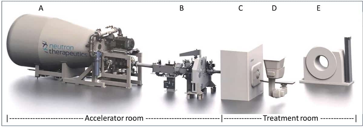
Figure 1: The key enabling technologies in Helsinki University Hospital’s BNCT facility. (A) compact proton accelerator; (B) proton beam optics; (C) beam-shaping assembly; (D) robotic couch; (E) rail-mounted CT scanner. (Courtesy: Neutron Therapeutics)
Yet it was the reliance on access to a modified nuclear reactor – a common feature of all BNCT clinical studies over recent decades – that hitherto limited the clinical potential of BNCT R&D efforts. “Our new accelerator-based BNCT facility is located on the hospital campus – a strategic breakthrough that will transform the logistics and economics of patient treatment with neutrons,” Joensuu adds. “Other key innovations – the use of robotic patient positioning and in-room CT for image guidance, for example – will deliver greatly enhanced treatment accuracy, streamlined workflows and increased patient throughput. Having a CT capability in the treatment room will also support future research studies on functional imaging, including the use of functional CT perfusion to evaluate treatment response to BNCT.”
Taking BNCT into the clinic
At the heart of Helsinki’s hospital-based BNCT facility is nuBeam, a compact accelerator-based neutron source from Neutron Therapeutics, a specialist BNCT equipment maker headquartered in Danvers, Massachusetts. The neutron source is housed in a dedicated accelerator and beamline room (65 m2) and comprises a number of discrete building blocks: a single-ended 2.6 MeV electrostatic proton accelerator designed to operate at 30 mA; a beam transport system; an online proton-beam monitoring system; a rotating solid lithium target for neutron generation; a beam shaping assembly (with circular beam delimiter sizes from 8 cm to 20 cm); and an online neutron-beam monitoring system (see figures 1 and 2).
Treatment planning, meanwhile, is based on full Monte Carlo (MC) simulation using CT, MR and PET images, with the planning software defining the patient geometric model including target volumes, organs at risk and their tissue compositions. Worth noting as well that the treatment dose is controlled via direct measurement of the neutron fluence by the online beam-monitoring detectors (rather than relying on calibrated proton current data).
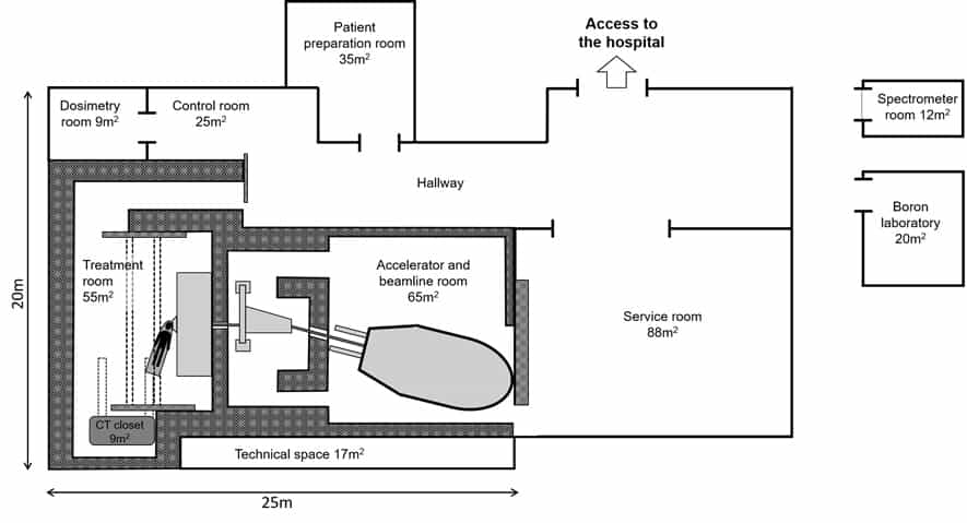
Figure 2: Layout of the Helsinki BNCT facility. The accelerator and beamline, treatment room, control room and patient preparation room are all located on the ground floor. Laboratories and offices are on the upper floor, with restricted access for approved clinical personnel. (Courtesy: Helsinki University Hospital)
Downstream from the nuBeam source, the patient treatment room (55 m2) is fitted out with a robotic patient positioning system, exacure, from German medical technology specialist BEC. The custom-modified industrial robot can not only be positioned in all three dimensions, and with six degrees of freedom, but also be moved along the ceiling towards and away from the neutron-beam nozzle. An integrated optical tracking system monitors the treatment couch position 500x per second to apply corrections (accurate to ±0.5 mm) for patient positioning and CT imaging. The latter is performed with a rail-mounted Siemens Healthineers SOMATOM CT Sliding Gantry, which takes in-room CT images for comparison with the planning images to ensure precise patient set-up ahead of treatment delivery.
“Patient positioning for BNCT is even more challenging than for conventional radiotherapy modalities,” explains Mikko Tenhunen, chief medical physicist at Helsinki University Hospital. “The horizontal nature of the neutron beam means we will often require two or even three unique couch set-ups to deliver a range of treatment fields.”
More broadly, implementing a new BNCT workflow from scratch brings its own unique set of challenges. Any equipment placed near the neutron beam – the positioning robot, for example – is covered with neutron-absorbing material to inhibit activation, while all patient-support devices – such as pillows, cushions and restraints – are tested for susceptibility to neutron activation before being cleared for use in the treatment room. The treatment facility is also equipped with a high-purity Ge gamma spectrometer for neutron activation analysis; paired ionization chambers with water and PMMA phantoms for estimating neutron and gamma-ray absorbed dose to reference tissue; and an inductively coupled plasma optical emission spectrometry device for analysis of blood-boron concentration prior to treatment.
Due diligence
Right now, Tenhunen and colleagues are laser-focused on equipment commissioning in advance of the BNCT clinical trial next summer. “All of the main subsystems of the BNCT facility have been tested separately,” he says, “while end-to-end validation of the treatment workflow will follow later this year. Ultimately, clinical success is all about how the various subsystems integrate together to deliver optimum outcomes across treatment planning, treatment delivery and treatment management.”
The outlook, it seems, is bright for the clinical application of BNCT technology. “The new Helsinki facility will be the first in-hospital BNCT treatment system in Europe,” concludes Joensuu. “It’s a significant landmark – one that we hope will promote clinical validation and wide-scale adoption of BNCT by cancer treatment centres around the world.”
Liisa Porra will present a progress report on the Helsinki BNCT programme at the Biology-Guided Adaptive Radiotherapy (BiGART 2021) virtual symposium on 6 October.
The human brain can do tasks such as image recognition much more efficiently than a computer. This is why Kwabena Boahen is developing an electronic architecture called Neurogrid, which mimics how our brains process information. In this episode of the Physics World Weekly podcast, the Stanford University researcher explains how he and his colleagues are blending analogue and digital technologies to create neuromorphic computers of the future.
The speed at which global solar-energy capacity has increased over the past two decades is astonishing and a testament to the hard work and innovation of scientists such as Giulia Grancini at the University of Pavia. The chemical physicist explains why her current research is focussed on hybrid perovskite materials, which show great promise for the next generation of solar panels.
This podcast also looks at some of the special holiday-themed content on Physics World, including a feature article about the physics of rollercoasters.
Modern consumer electronics can produce magnetic fields strong enough to interfere with implanted medical devices, but the effect only poses a low risk to patients. That is the conclusion of Seth Seidman and colleagues at the US Food and Drug Administration’s Centre for Devices and Radiological Health , who measured variations in magnetic field strength at varying distances from the latest models of a smartphone and a smart watch. Based on their findings, the researchers suggest that these electronics should be kept at least 15 cm away from implanted devices.
For patients suffering from heart rhythm disorders, implantable pacemakers and cardioverter-defibrillators can deliver life-saving electrical pulses that cause the chambers of the heart to contract and pump blood at an appropriate rate. When an implant wearer undergoes a procedure where strong electromagnetic interference is possible, or a medical treatment where implanted devices must be temporarily suspended, devices are put into “magnet mode” to prevent inaccurate heart rate tracking and the delivery of inappropriate electrical pulses in response.
As a standard, the magnet mode is activated by a simple doughnut magnet, with a strength of roughly 9 mT (around 200 times the strength of Earth’s magnetic field). However, it can also be inadvertently triggered by any static magnetic field encountered by the patient, with a strength exceeding roughly 1 mT.
Rare-earth magnets
Such strengths were once rare from household items, and only found near devices like stereo speakers and cordless tools. Today, however, they can also be generated by powerful rare-earth magnets such as neodymium – which are small enough to fit inside headphones, door locks, and smartphone speakers.
In their study, Seidman’s team assessed the influence of these fields by placing an iPhone 12 and Apple Watch 6 at varying distances from a magnetic field detector. They found that at proximities of less than 1 cm, the detector encountered fields significantly stronger than 9 mT – which could easily trigger magnet modes inadvertently. This strength attenuated as the distance increased, and dropped below 1 mT beyond 2 cm.
From their results, the researchers conclude that modern consumer electronics currently pose a low risk to people with implanted medical devices. They add that they are not aware of any cases where the effect has placed a patient in danger. Yet as technology continues to advance, they expect that the risk will grow as stronger fields become increasingly common.
As a result, Seidman and colleagues say that steps should be taken to ensure patients and healthcare providers are aware of the potential risks, and suggest simple preventative measures to ensure patient safety. These include keeping consumer electronics such as smartphones and watches at least 15 cm away from any implanted devices, and not carrying them in pockets covering the devices.
The organizers of ESTRO 2021 will once again welcome delegates to join its live conference in Madrid, Spain, while also offering full digital access to anyone unable to attend in person. Running from 27 to 31 August, the event will enable both onsite and online delegates to share knowledge, interact with speakers and other attendees, and learn about the latest technical innovations.
All registered attendees will be able to access the digital platform, which has been updated this year to enable greater interactivity and enhanced online participation. To be able to host the live conference, the venue capacity has been reduced by half to enable social distancing, with 2500 spaces available on a first come, first serve basis.
According to conference chair Ben Slotman, head of radiation oncology at AmsterdamUMC in the Netherlands, this year’s event aims to raise the visibility of submitted papers. Highlighted contributions will take place in a plenary setting, greater emphasis has been placed on the discussion of poster presentations, and the interdisciplinary track has been expanded compared with previous editions. “We want to provide the best possible congress experience under all circumstances, and to foster broad and continuous participation,” he comments in his welcome letter.
Both live and online delegates will have the opportunity to discuss the latest technical developments and methodologies with leading equipment vendors. The conference will host Europe’s largest industry exhibition in radiation oncology, while online attendees will have full access to three days of industry symposia. Some of the latest innovations are highlighted below.
Motion phantom boosts testing capabilities in MR-guided radiotherapy
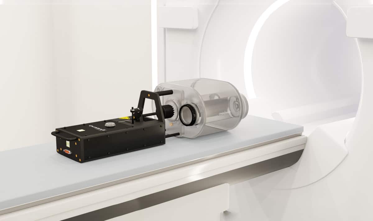
The QUASAR MRI4D from Modus QA is an MR-safe motion phantom that has been designed to meet the advanced physics testing needs of adaptive MR-guided radiation therapy. The MRI4D makes it possible to develop, test and validate advanced 4D treatment delivery protocols on MR-guided machines.
The QUASAR MRI4D from Modus QA has been used with Elekta’s Unity MR-linac to demonstrate the feasibility of real-time adaptive radiotherapy treatments. (Courtesy: Modus QA)
Interchangeable inserts are available for imaging, planning, targeting, dosimetry and delivery quality assurance (QA). Software included with the phantom provides all the tools needed to create, import, edit and stream patient-specific respiratory waveforms.
Modus QA has worked with early adopters of both the Elekta Unity and ViewRay MRIdian to provide a range of testing capabilities. In a poster originally presented at MR in RT 2021, a collaboration between Modus QA, Elekta and researchers at UMC Utrecht in the Netherlands and Uppsala University Hospital in Sweden describe how the MRI4D was used in a prototype software platform designed to demonstrate the feasibility of real-time adaptive (RTA) treatments using both intensity-modulated and stereotactic radiation therapy. The prototype platform achieves full 3D motion modelling, deformable multileaf-collimator tracking and dose accumulation with low latency, enabling a full RTA treatment with instant QA.
Sun Nuclear highlights integrated and independent QA solutions
During ESTRO 2021 Sun Nuclear will present its solutions for integrated and independent quality management at its exhibitor booth and in a lunch symposium on 29 August (13:00 CEST, Room N107+108) that will feature three clinical presentations.
The first two speakers will describe the use of Sun Nuclear’s SunCHECK platform to streamline QA, detect errors, and standardize quality management among clinical users, sites and machines:
Improving QA Effectiveness with an Integrated, Automated Platform Presenter: Nuria Jornet, Hospital de la Santa Creu i Sant Pau, Barcelona, Spain
Clinical Experience of a Comprehensive and Automated Machine QA Platform Presenter: Greg Martin, Clatterbridge Cancer Centre, Liverpool, UK
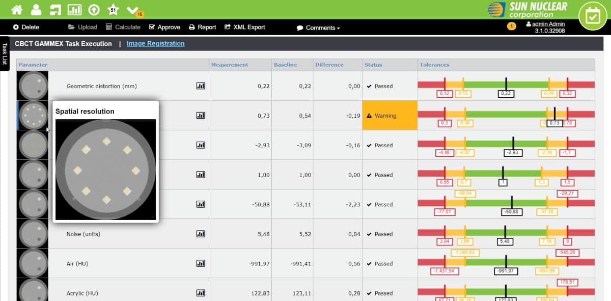
With the latest release of the SunCHECK platform, users have even more options for customizing the tasks and templates for machine QA. The latest version also provides access to advances in patient QA for secondary dose calculations in photon, electron and brachytherapy plans.
The SunCHECK platform enables tasks and templates for machine QA to be standardized across users, sites and machines. (Courtesy: Sun Nuclear)
The third presentation in the symposium will offer an insight into building a strong end-to-end stereotactic programme using SRS MapCHECK, Sun Nuclear’s film-less array for patient QA.
End-to-End Testing for SRS/SBRT with a High Density Diode Array Presenter: Nestor Chinillach, Hospital IMED, Valencia, Spain
SRS MapCHECK now includes CyberKnife machine QA, making it the only device that offers both machine QA and patient QA on a CyberKnife.
Online solution offers fast and accurate measurement of MR image distortion
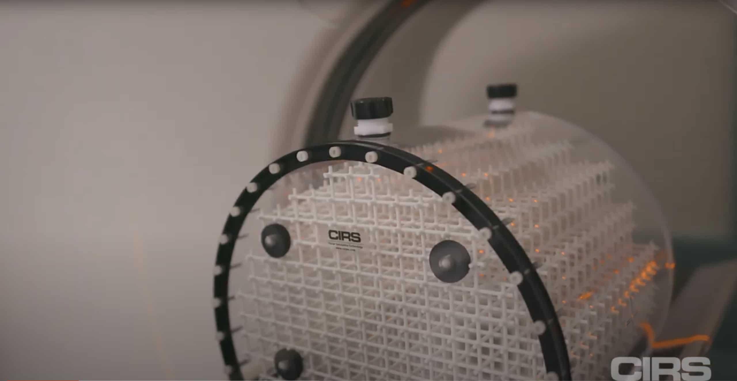
New from CIRS is Distortion Check, a cloud-based solution that has been designed to quantify distortion in MR images quickly and automatically. Used in conjunction with CIRS MRI grid phantoms, the software offers a rapid solution for accurately measuring distortion throughout the entire image volume.
When used with MRI grid phantoms from CIRS, Distortion Check offers fast and accurate measurement of MR image distortion. (Courtesy: CIRS)
The software first detects all grid intersections, and then registers either a CAD or CT scan ground truth to each of these MR-detected control points. An interpolation is then used to generate the 3D distortion vector fields, with the density of control points optimized to bring interpolation close to linearity.
Results can be reported in a variety of output formats, including scatter plots, contour plots, and box and whisker plots for trending analysis, while DICOM overlays can also be imported to TPS or any other third-party software. The cloud-based solution can be accessed from any machine and with any operating system, enabling easy collaboration and review of results.
The software algorithms have been designed to work with any grid configuration. CIRS also exploits proprietary 3D printing techniques that enable grid phantoms to be easily modified to meet customer requirements.
Giving fish-like robots adjustable tails makes them much more efficient swimmers, scientists in the US have discovered. As well as providing insight into how real fish swim, the researchers say their findings could enable the development of swimming robots that can carry out more complex missions than is possible with current technologies.
Real fish swim efficiently over a wide range of speeds. Biologists have long suspected that this is partly because they actively tune the stiffness of their tails to accommodate different conditions. However, while various evidence hints at such speed-dependent tail stiffening, it is hard to measure it directly, says Qiang Zhong, an engineer at the University of Virginia.
“Fish use muscle to flap their body and also use that muscle to control stiffness,” Zhong explains. “Basically, they use one organ to control stiffness and flap their body, so it is really hard to tell how the muscle plays a role in the body stiffness, because we can’t really isolate that.”
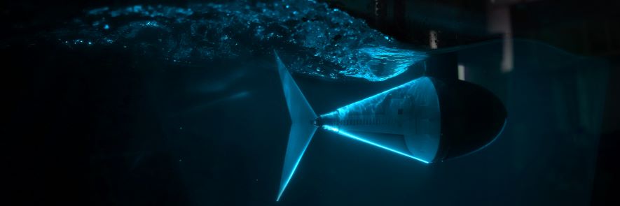
A robot with a spring in its tail
To test the tail-stiffening theory, Zhong and his colleagues decided to build a swimming robot with independently controlled tail stiffness. Their tuna-like robot, which they describe in Science Robotics, has a fixed head and a tail that moves from side to side to provide thrust. At the end of the tail is a joint attached to a passive fin. The stiffness of this joint can be adjusted by tensioning a tendon-like spring; pulling harder on the spring increases torsional stiffness.
When the researchers played around with the stiffness of the robot’s tail joint and combined their findings with mathematical modelling, they found that both swimming speed and efficiency are closely tied to tail stiffness. When the frequency of tailbeats is low, the swimming speed increases in tandem with the frequency. Above a certain frequency, however, the distance travelled with each tailbeat (and thus the robot’s efficiency) starts to drop if tail stiffness remains constant.
Internal spring A photo of the tuna-inspired robot in the water channel. (Courtesy: Qiang Zhong & Daniel Quinn, University of Virginia)
To maintain the relationship between frequency and speed (and thus keep the robot’s efficiency high), the researchers found that the tail needs to stiffen. Indeed, the relationship between speed and optimal tail stiffness proved remarkably simple: to maximize efficiency, tail stiffness needs to scale with swimming speed – or tailbeat frequency – squared. “If you want to flap your tail twice as fast – twice the frequency – you should pull on that tendon with four times the strength,” says team leader Dan Quinn.
Variable missions
While robots with stiff tails are good at swimming fast, Zhong notes that once their tailbeat frequency drops, so does their efficiency. “The benefit of using tunable tail stiffness is to help the robot always stay at a peak efficiency, as much as possible,” he says. This is important because while real fish are good at multi-speed missions (such as swimming rapidly to a reef and then slowing down to feed), swimming robots are generally “terrible” at them, Quinn adds.
To test the effectiveness of a tuneable tail for this type of mission, the researchers put their robot in a tank with flow speeds that varied from 0.1 to 0.65 metres per second over a period of 15 minutes and directed it to maintain its position by adjusting its tailbeat frequency. When they compared the tunable tail with tails that were moderately stiff, completely rigid, and fully de-tensioned, they found that the tunable tail used 16, 41 and 55 per cent less energy respectively. The loose and rigid tails also proved unable to reach the high speeds needed later in the mission.
Quinn says that the team’s findings could make it possible to deploy large swarms of small, cheap fish-like robots. Thanks to the multi-speed efficiency of their adjustable tails, such robots could be deployed remotely, travel to their mission quickly and then slow down to complete tasks such as climate and environmental measurements or large-scale mapping.
The team are keen to explore how their findings translate to different-sized swimming robots, particularly as their modelling suggests that tunable tail stiffness becomes more important in bigger fish. Qiang tells Physics World that he would also like to explore advanced ways of controlling stiffness, such as artificial muscle networks. This would enable him to investigate the impact of tunable stiffness all over the robot’s body, which is likely to be closer to how a real fish behaves.
A quantum bit based on a vibrating carbon nanotube and a pair of quantum dots could be unusually resistant to noise. Although the new nanomechanical qubit is currently at the proposal stage, calculations by Fabio Pistolesi of the French National Centre for Scientific Research (CNRS) at the University of Bordeaux and colleagues in the US and Spain indicate that its so-called “decoherence time” – a measure of how long fragile quantum information can survive in a noisy environment – would be remarkably long, making it an attractive platform for quantum computing.
Quantum computers can, in principle, solve certain problems much faster than classical computers because they exploit a quantum system’s ability to be in a superposition of two or more states (as opposed to classical bits that have only 0 and 1 states). Promising candidates for such quantum bits, or qubits, include superconducting circuits, trapped ions, defects in solid materials and “artificial atoms” known as quantum dots.
New qubit platform
In the proposed nanomechanical qubit, a suspended carbon nanotube acts as a resonator, and its vibrations couple to a double quantum dot that forms within the nanotube itself. This double quantum dot has discrete electronic states, and the coupling between them allows the resonator to become strongly anharmonic – that is, the frequency of its oscillations depend strongly on their amplitude. In such an oscillator, even the tiniest change in the resonator’s amplitude is easily detected. This amplitude, Pistolesi explains, can then be used to store quantum information.
“Essentially the minimum possible oscillating amplitude (the quantum ground state) corresponds to the 0 of the qubit, while the next smallest amplitude (the first excited state) corresponds to 1,” he says. “These two states could easily be read out by a microwave signal. The fact that the frequency of the oscillator changes when its amplitude changes allows us to detect and manipulate the qubit.”
The qubits in such a platform would stay coherent for a long time, Pistolesi tells Physics World, because the information is stored in their mechanical oscillation amplitude and the oscillator can perform millions of oscillations before it starts to become damped.
Generating anharmonicity
While researchers knew that qubits based on such a mechanical oscillator would have a long coherence time, they were not sure how to introduce enough anharmonicity into them to make them controllable. Pistolesi and colleagues had previously found strong anharmonicity in a similar system (a carbon nanotube coupled to a single electron transistor). They undertook their present study to find out if they could also generate such anharmonicity in an oscillating nanotube coupled to a double quantum dot.
As well being a promising qubit platform, the oscillators could also be used as highly accurate quantum sensors thanks to their sensitivity to classical forces. Such devices might be employed to detect faint changes in acceleration, gravity, magnetic moments and electric fields.
The researchers, who report their work in Physical Review X, now plan to fabricate the qubit they have proposed and test its performance experimentally.
Academics with children become less productive and do work of lower impact as their childcare responsibilities increase. That’s the finding of a survey of 11,226 researchers around the world, which revealed the same trend for both male and female “lead” parents. But because women academics are much more likely to be the primary care providers – even when reporting equal sharing of parenting – it means that they suffer more scientifically when having children
Based on researchers who have written papers indexed in Clarivate Analytics’ Web of Science, the survey reveals that women play a much bigger role in raising children, with almost a third (30.6%) of female respondents being the primary care providers for their children, compared with just 3.9% of men fulfilling that role. More than half (52.0%) of women and 57.1% of men say they share childcare responsibilities in a “dual” parenting model. However, only 17.4% of women play a minor or “satellite” role in parenting, compared to 38.9% of men.
For women, it doesn’t matter if you are a lead parent, a dual parent or a satellite parent – the effect [of having children] is severe.
Gemma Derrick, Lancaster University
In terms of the number of papers they produce, both men and women report a similar drop in productivity when they are single or lead parents, compared with dual parents. Dual fathers, however, are more productive than dual mothers and have a smaller “parenting penalty”. The same goes for men who take a secondary role in parenting, who write more papers than secondary mothers. These patterns are also mirrored in terms of the scientific impact of their papers as measured by citation data.
“For women, it doesn’t matter if you are a lead parent, a dual parent or a satellite parent – the effect [of having children] is severe,” says Gemma Derrick, an education-policy expert at Lancaster University in the UK, who carried out the survey with colleagues in Canada, the Netherlands and the US . “But for men it is graded along whether you take a lead, dual or satellite parenting role. Men can choose to be a parent part-time or full-time with various effects on their productivity, but women don’t seem to have that choice.”
Questions on household duties in the survey, such as who feeds, bathes or plays with their children, shed some light on these differences. In nearly every parenting-related task, female dual parents are, in reality, more likely than male dual parents to report being the primary care provider. “We saw a lot of women,” Derrick says, “who, after completing the survey, went hang on ‘I realise I take on far more responsibilities than my partner, so maybe we are not in a dual parenting partnership after all’.”
A lot of women went hang on ‘I realise I take on far more responsibilities than my partner, so maybe we are not in a dual parenting partnership after all’.
Gemma Derrick, Lancaster University
Sharing of childcare is more equal, however, for women who divvy up their parenting duties with another academic. Mothers who share the workload with non-academic partners, in contrast, do far more childcare. That trend is reflected in scientific productivity, with women sharing parenting with academics writing more papers than those with non-academic partners. Quite why this should be is not clear, but open-ended responses to the survey suggest the reason lies in academic work being seen as more flexible.
Given that the level of childcare responsibilities is the main cause of gender differences in academic productivity, the authors suggest that policies should be introduced to let parents shift more easily between professional and parenting responsibilities. Academic institutions could, for example, provide on-site childcare and lactation rooms and also restructure conferences for the benefit of parents.
An ultra-precise quantum sensor based on trapped beryllium ions is up to 20 times better at detecting weak electric fields than previous atomic devices. By introducing entanglement between the collective motion of the ions and their electronic spin, a collaboration led by the US National Institute of Standards and Technology (NIST) demonstrated that the ion displacement sensitivity in the presence of an electric field was an order of magnitude greater than for classical protocols with trapped ions. With further improvements, the technology could even be used in the search for dark matter.
Quantum sensors can detect and measure signals that are undetectable with their classical counterparts. They are thus a promising tool in many areas of fundamental science, including biological imaging as well as physics. Of the many different systems being pursued as quantum sensors, trapped ions could be particularly favourable due to experimenters’ precise control over their parameters and their ability to introduce entanglement into the system.
The Ion Storage Group at NIST, led by John Bollinger, decided to exploit these properties for measuring very weak electric fields. “We realized our ion crystal can be incredibly sensitive to electric fields,” explains Kevin Gilmore, a former graduate research assistant at NIST and the lead author of a paper describing the research. “We found a protocol that exploits our ability to produce quantum entangled states and is very sensitive to small displacements of the ions driven by weak electric fields. It’s a neat demonstration of how quantum effects can be used to gain an advantage over classical systems.”
Entanglement enhances sensitivity
The experiment traps approximately 150 beryllium ions in a two-dimensional plane with a combination of magnetic and electric fields known as a Penning trap (see image above). The motion of the ion crystal can be thought of like a drumhead, where all ions collectively move together. In the NIST group’s system, this collective motion, known as the centre-of-mass vibrational mode, is entangled with the collective electronic spin of the ions before the experiment begins.
After entangling the centre-of-mass vibrational mode with the collective electronic spin, the centre-of-mass mode is excited with an oscillating electric field. The system is un-entangled, and the motional information of the ions – in other words, their displacement – is then stored in their individual spins, which are straightforward to measure. Hence, the motional behaviour of the ion crystal can be directly inferred from the spin measurements. With this capability, the experiment can then measure electric fields very precisely.
“Our measurements surpass classical limits of sensitivity to electric fields (and displacements of the crystal),” Gilmore says. “This means that we demonstrate a sensing advantage by using quantum effects (i.e., entanglement), which is non-trivial.” He adds that the device might even be sensitive enough to detect dark matter candidates known as axions, which are thought to produce tiny oscillating electric fields.
Building bigger crystals
Now that the proof of principle has been established, the group are working towards creating sensors with larger ion crystals. While they currently trap approximately 150 beryllium ions in a two-dimensional plane, their goal is to create a three-dimensional crystal with around one million ions.
“This would greatly enhance our electric field sensitivity,” says Gilmore. “Simultaneously, we hope to reduce the dominant noise source for this experiment, which is frequency fluctuations of the centre-of-mass mode. It’s possible that using a 3D crystal may result in better overall cooling of the ions, which in turn could reduce the centre-of-mass frequency fluctuations.”

 Dr Y Shirley Meng received her PhD in Advance Materials for Micro & Nano Systems from the Singapore-MIT Alliance in 2005, after which she worked as a Postdoctoral Research Fellow and Research Scientist at the Massachusetts Institute of Technology. She currently holds the Zable Chair Professor in Energy Technologies and Professor in Materials Science & Nanoengineering at the University of California San Diego. She is the Principal Investigator of the Laboratory for Energy Storage and Conversion research group. Starting in 2005, Dr Meng served as the Founding Director of the Sustainable Power and Energy Center, until she was named Inaugural Director of the Institute for Materials Discovery and Design in 2020. She has received prestigious awards including the 2020 Michael Faraday Medal of the Royal Chemical Society; 2019 International Battery Association Battery IBA Research Award; 2018 and 2019 Blavatnik Awards for Young Scientists Finalist; 2018 American Chemical Society ACS Applied Materials & Interfaces Young Investigator Award; 2017 IUMRS-Singapore Young Scientist Research Award; 2016 ECS C W Tobias Young Investigator Award; and 2011 NSF CAREER Award. Dr Meng is a Fellow of The Electrochemical Society; Editor in Chief of MRS Energy & Sustainability; and co-founder of Unigrid LLC. She is also the author and co-author of more than 210 peer-reviewed journal articles and two book chapters, and holds five issued patents.