A team of researchers from Peking University in China has fabricated an optical analogue of “magic-angle” graphene bilayers in a photonic nanocrystal. They have used the structure to create a completely new type of highly-efficient nanolaser.
Graphene is a flat crystal of carbon just one atom thick. When two such sheets are placed on top of each other with a small angle misalignment, they form a Moiré superlattice. At a twist angle of 1.08°, the material becomes highly correlated and begins to show properties such as superconductivity at low temperatures.
At this so-called magic angle, the way in which electrons move in the two coupled sheets changes because they are now forced to organize themselves at the same energy. This leads to “flat” electronic bands, in which electron states have exactly the same energy despite having different velocities.
This flat band state makes an electron dispersionless – that is, its kinetic energy is completely suppressed and it cannot move in the Moiré lattice. The result is that electrons slow down almost to a halt and become localized at specific positions along the coupled sheets, where they can strongly interact with one another. This is the effect that gives rise to the abovementioned superconductivity, as well as producing many exotic and unexpected phenomena such as correlated insulator states and orbital magnetism.
Stopped-light nanolasing
Nanolasers are key to developing integrated photonics. They work by confining light in a nanocavity and emitting coherent light within a very narrow spectral range through optical amplification, after passing through the cavity multiple times to increase its gain.
Researchers have designed many nanolaser schemes over the years, with very different optical cavity designs, including nanodisc lasers, nanowire lasers, plasmonic nanolasers and photonic crystal nanolasers. However, the cavities of these nanolasers require materials with highly differing properties or disorder/defects to localize a light field.
The laser developed by Ren-Min Ma and colleagues works in a very different way. The new device makes use of dispersionless light stopping in an optical magic-angle graphene-like lattice of nanoholes in a semiconductor membrane. The membrane consists of InGaAsP multi-quantum wells, which act as the active gain medium.
No need for a “cavity”
The researchers introduced two graphene-like photonic crystals into the same semiconductor membrane. When they twisted one with respect to the other at an angle of 2.65°, they found that the coupling between the two photonic crystals created a completely flat-band dispersion, which stops and localizes light – just like magic-angle twisted graphene. This effect does away with the need for a conventional laser cavity.
Ma and colleagues optically pumped their laser to induce gain in the structure. They say they unequivocally observed lasing at around 1.5 µm, a wavelength that is important for telecommunications applications.
A completely new design for nanolasers
As well as representing a completely new design of nanolaser, the device also has many advantageous properties. For one, it has a threshold of only 0.037 mW in pump power. This is much lower than nanodisc lasers, nanowire lasers or plasmonic nanolasers and is on a par with state-of-the-art photonic crystal defect nanolasers made from the same gain materials. It also boosts a higher “quality factor over mode volume” (a figure of merit usually used to characterize laser cavity quality) compared with the types of lasers mentioned above. Indeed, at more than 400 000, its quality factor is among the highest of all kinds of nanolaser cavities, says Ma.
“Our scheme provides a novel flexible and robust platform to construct high-quality nanocavities for lasers, nanoLEDs, nonlinear optics and cavity quantum electrodynamics,” he adds. “It could be used in applications in integrated photonics, near-field spectroscopy and sensing.”
The researchers say that they will now be studying the exotic light–matter interaction in their new platform. “We will also be pushing forward applications for the device,” Ma tells Physics World.
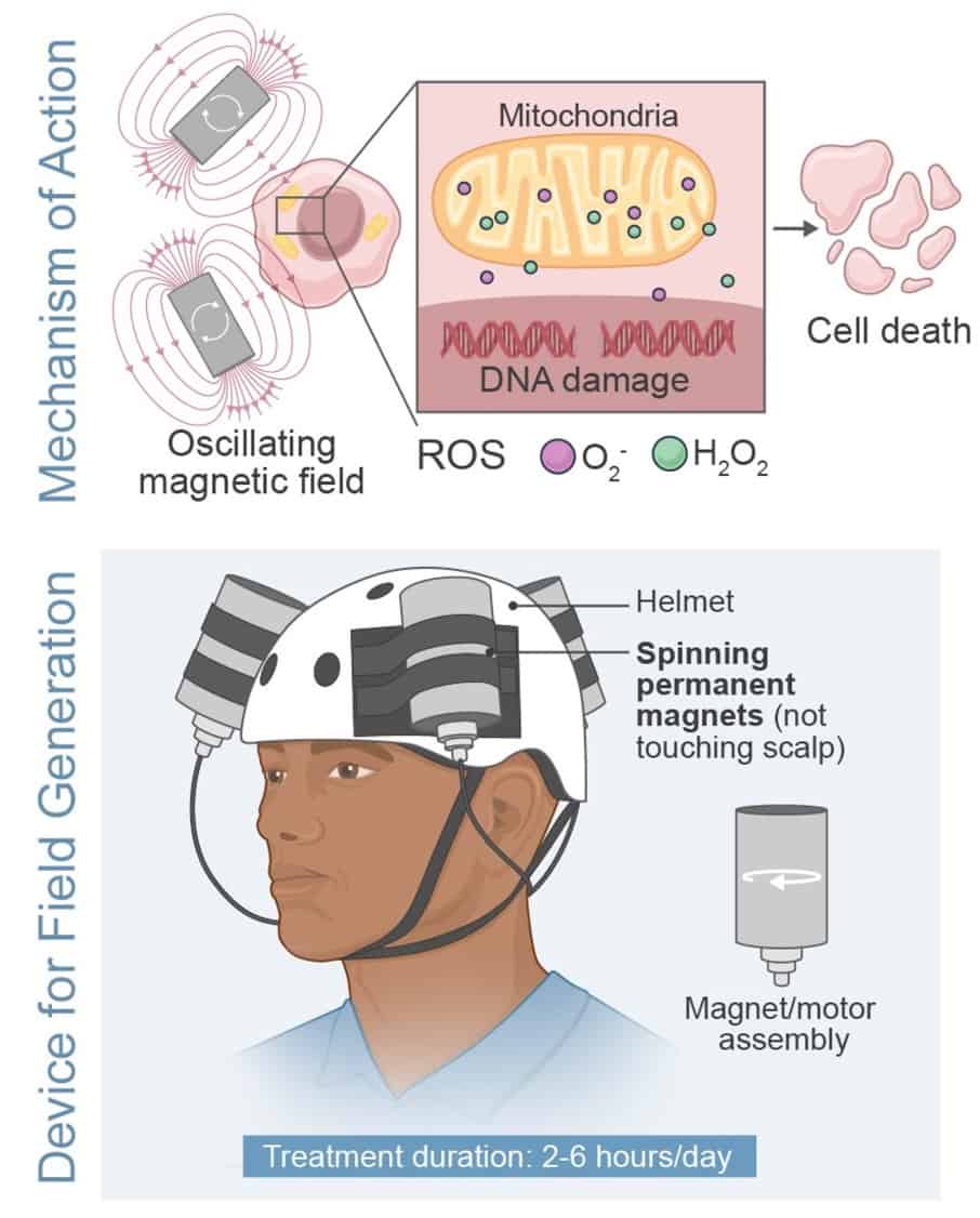
A team of US-based researchers has used an innovative head-mounted device to shrink a brain tumour – potentially paving the way for a powerful new non-invasive therapy for glioblastoma.
In recent studies, the team – which includes researchers based at the Peak Center for Brain and Pituitary Tumor Treatment and Research at Houston Methodist Neurological Institute – found that the oscillating magnetic field-generating device, dubbed an “oncomagnetic device”, was able to rapidly kill glioblastoma cells in culture and shrink human glioblastoma tumours implanted in mice, prolonging their survival. The device was also used to shrink an end-stage recurrent glioblastoma in a patient without access to any other approved treatment option. The researchers describe the results of this case study in Frontiers in Oncology.
As co-author Santosh Helekar from Houston Methodist Research Institute explains, the portable, wearable device consists of “strong permanent magnets rapidly spun by high-speed electric motors, whose rotation and timing are controlled by a programmable microcontroller operated by a rechargeable battery”. The magnet and motor assemblies are housed in vibration-, sound- and heat-insulated enclosures mounted on a helmet worn by the patient. A treatment-specific pattern of rotation frequencies and timings is then used to stimulate the brain to treat glioblastoma.
Glioblastoma is the most common cancer of the brain. Helekar observes that advances in its treatment have prolonged the median survival of patients newly diagnosed with the disease only slightly – from nine months four decades ago to “about 15 to 20 months today”.
Promisingly, this single-patient case report demonstrated that a month-long course of oncomagnetic treatment – during which the oscillating magnetic field was applied for two hours, up to three times a day on weekdays – reduced the volume of end-stage recurrent glioblastoma by more than 30%.
Helekar notes that the new technique is currently being used in both research and clinical settings under the auspices of an ongoing research project supported by the Translational Research Initiative of the Houston Methodist Research Institute.
Cancer cell death: Schematic of the oncomagnetic device and its proposed mechanism of action. (Courtesy: Santosh Helekar)
“Our recently published laboratory research findings show that the oncomagnetic device kills glioblastoma and other cancer cells in culture by increasing reactive oxygen species [ROS] in the mitochondria and cytoplasm of these cells, while sparing non-cancerous cells, such as neurons, astrocytes and bronchial epithelial cells,” Helekar explains.
“We hypothesize that the increase in reactive oxygen species is at least in part due to magnetically induced disruption of the electron flow in the mitochondrial electron transport chain,” he continues. “The rotating magnetic fields influence the spins of unpaired electrons exchanged by free radical intermediates in the chemical reactions taking part in the unmovable transmembrane protein complexes of the electron transport chain. Confirmation of some of the predictions of this hypothesis are going to be published shortly.”
Next steps
One key advantage of the new device is that, in contrast with existing treatments for glioblastoma, it does not have any known serious side effects. Moreover, it does not involve drug treatment nor require shaving of the head.
“The total duration of daily treatment on weekdays is only up to six hours,” Helekar says. “It is likely to be much less expensive because of the low cost and simplicity of the device. The device is very easy and convenient to use because it simply involves the wearing of a helmet up to three times a day.”
Moving forward, the team is currently undertaking laboratory-based preclinical studies of the device to test its biophysical, cellular and molecular mechanisms of action on cells in culture, as well as its safety and efficacy in mouse models of glioblastoma.
“With David Baskin, Peak Center director and vice-chair of neurosurgery as the principal investigator, we are also continuing the FDA-approved compassionate use treatment of patients with end-stage recurrent glioblastoma, like that reported in the recently published case report,” says Helekar. “Our plans are to obtain regulatory approval for a pilot clinical trial to test the safety and efficacy of the device for the treatment of glioblastoma. Furthermore, we plan to collaborate with other institutions nationally and internationally to conduct similar trials in other cancers.”
When the temperature drops, turbulent puffs caused by coughs and sneezes become more buoyant and travel further and last longer, scientists in Japan and Italy have discovered. The researchers say the results of their modelling study could help improve our understanding of the airborne transmission of viruses like SARS-CoV-2.
A turbulent puff occurs when a mass of fluid is ejected from a localized source. In an undisturbed environment, the cloud of fluid – the puff – moves freely and evolves over time. Puffs are important in environmental and health sciences, chemistry, and other fields. How they travel and change over time has implications for the dispersal of pollutants, such as those from chimneys, and the transmission of disease droplets in coughs, for example.
Despite their importance, current theory on puff dynamics is based on work conducted almost 50 years ago, in the 1970s. But that work only focuses on the large-scale dynamics of the puff, such as how fast it moves and its size. It provides scaling laws that explain how a puff increases in size and slows down over time. It does not, however, consider the small-scale dynamics of the turbulent fluctuations inside the puff, and our understanding of these remains elusive.
Complex characteristics
Turbulence in puffs has more complex characteristics than turbulence in continuous ejections of gas or liquid, making it more challenging to study, explains Marco Rosti, an expert in fluid dynamics at the Okinawa Institute of Science and Technology Graduate University in Japan. “But it’s of vital importance – especially right now for understanding airborne transmission of viruses like SARS-CoV-2,” he adds.
To fill in the gaps in the previous theory, Rosti and Andrea Mazzino, a physicist at the University of Genoa in Italy, developed a mathematical model that includes the small-scale structure of turbulence fluctuations in a puff. Their model looked at how both the small-scale and large-scale dynamics of a puff change over time, and how this is affected by temperature, humidity and velocity fluctuations. The researchers used a supercomputer to verify their models against state-of-the-art numerical simulations that could resolve the behaviour of puffs at the large-scale and the small-scale run. They report their results in Physical Review Letters.
Rosti and Mazzino initially found that their results fitted with the previous model of puff dynamics, with turbulence in the puff dictating how it behaved. Over time the puffs expanded and slowed down in a predictable way that was linked to their initial speed, size (radius) and fluid density. But they discovered that if the environment cooled these scaling laws changed, as the temperature difference between the warm puff and the cooler ambient air increased.
Buoyancy matters
At cooler temperatures buoyancy starts to play a role in puff dynamics, their results show. The gas or liquid in the puff is significantly warmer and therefore less dense than the environment. Because of this the puffs rise higher, last longer and travel further. “The effect of buoyancy was initially very unexpected. It’s a completely new addition to the theory of turbulent puffs,” says Rosti.
These findings could improve our understanding and modelling of the airborne transmission of viruses and how it changes with environmental conditions. The researchers write that they expect their results to have “a profound impact on developing evaporation models for virus-containing droplets carried by a turbulent puff, with benefits to the comprehension of the airborne route of virus contagion”.
“How fast the droplets evaporate – and therefore how small they are – depends on turbulence, which in turn is affected by the humidity and temperature of the surroundings,” explains Rosti. “We can now start to take these differences in environmental conditions, and how they affect turbulence, into consideration when studying airborne viral transmission.”
Rosti and Mazzino now plan to study how puffs behave when made of more complex non-Newtonian fluids. “For COVID, this could be useful for studying sneezes, where non-Newtonian fluids like saliva and mucus are forcefully expelled,” explains Rosti.
The British Nobel-prize-winning astronomer Antony Hewish has died at the age of 97. He was awarded one half of the 1974 Nobel Prize for Physics for his research in radio astrophysics and his “decisive role in the discovery of pulsars”. He shared the other half with Martin Ryle who bagged the award for the invention of the “aperture synthesis” – a type of interferometry that mixes signals from a collection of telescopes to produce images having the same angular resolution as an instrument the size of the entire collection.
Hewish was born in Fowey, Cornwall, on 11 May 1924 and went to the University of Cambridge in 1942 before joining the war effort working at the Royal Air force Establishment in Farnborough, although he was seconded to the Telecommunications Research Establishment in Malvern working on airborne radar-countermeasure devices. It was at Malvern where he first met Ryle.
Returning to Cambridge in 1946, he graduated two years later and in 1952 was awarded a PhD in physics. He remained at Cambridge throughout his career, becoming head of the Cambridge radio-astronomy group in 1977 and head of the Mullard Radio Astronomy Observatory from 1982 to 1988.
A ‘scruff signal
As part of the search for mysterious sources of radio waves known as quasars, in the mid-1960s Hewish designed and constructed the Interplanetary Scintillation Array at Mullard. The array was built on a patch of land two-and-a-half times the size of a football pitch a collection of around 4000 dipole antennas that operated at a radio frequency of 81.5 MHz. Taking around two years to build, it was initially operated by Jocelyn Bell Burnell, who was doing a PhD under Hewish’s supervision.
In the autumn of 1967 Bell Burnell noticed a 0.5 cm-long “scruff” signal showing a series of regular peaks in luminosity. Hewish thought that it may have been a radio flare star – or the product of humans or even aliens – and it was dubbed “LGM-1” for Little Green Man. Yet further observations revealed that it had a pulsed nature and turned out to be the first observation of a pulsar – a rotating neutron star that emits a regular ticking signal of radio waves.
In January 1968 the team submitted a paper about the work – “Observation of a rapidly pulsating radio source” – to Nature (217 709). For the discovery, Hewish was awarded the Nobel prize in 1974, although many felt that Bell Burnell should also have been recognized – an omission that Bell Burnell herself attributes to being a student at the time.
Hewish enjoyed listening to music and sailing and during his undergraduate days was a keen rower. He died on 13 September.
The amount of blood flowing through the cerebral arteries and veins can strongly influence brain function, as it impacts both oxygen delivery and removal of waste products from the brain. As such, medical professionals utilize cerebral blood flow (CBF) as a marker of a patient’s cerebrovascular function.
One common technique, diffuse correlation spectroscopy (DCS), utilizes near-infrared light to monitor CBF. In DCS, a near-infrared laser with a long coherence length illuminates the tissue. As the light interacts with moving red blood cells within the arteries and veins, it undergoes scattering. The DCS system detects this scattered light and analyses it to determine the CBF.
Current DCS systems typically use single-photon avalanche photodiodes (SPADs) with a 700–850 nm light source and a source–detector separation of 25 mm. These detectors have a low signal-to-noise ratio (SNR), however, which hinders their ability to accurately determine CBF measurements. This, in turn, precludes the high acquisition rates required to determine dynamic blood flow changes. To improve brain sensitivity and reduce scalp signal contamination, ideal conditions include large source–detector separations, longer wavelengths of light (1000 nm or above) and fast acquisition rates, all of which are difficult to achieve with current SPAD systems.
As an alternative, the authors of a new paper, published in Neurophotonics, propose using superconducting nanowire single-photon detectors (SNSPDs) to improve current DCS methods. SNSPDs consist of a thin film of superconducting material with high single-photon sensitivity and detection efficiency. The authors believe that this method could provide more robust measurements of CBF.
SNSPD advantages
The paper’s first author, Nisan Ozana, alongside a team at Harvard Medical School, Massachusetts General Hospital and MIT Lincoln Labs, has developed DCS systems that use Quantum Opus SNSPDs and a 1064 nm light source. To assess this new approach, they obtained CBF measurements from 11 participants using both SPAD- and SNSPD-based systems, comparing the results obtained from the two systems.
The researchers demonstrated a sixteen-fold improvement in SNR when obtaining CBF measurements using SNSPD-based systems, in comparison with typical SPAD-based DCS systems (operating at 850 nm). This was partly due to a seven to eight times increase in the number of photons available at the detector (operating at 1064 nm) using SNSPD. Additionally, SNSPD demonstrated an increased detection efficiency (88%) compared with SPAD (58%). The significant increase in SNR allowed faster acquisition rates (20 Hz) than SPAD-based systems (1 Hz) at the same source–detector separation, resulting in clear detection of arterial pulses.
The team also found that increasing the source–detector separation to 35 mm improved brain blood flow sensitivity by 31.6% in the CBF measurements. Further, the researchers examined physiological changes during breath-holding and hyperventilation. Measurements obtained during breath-holding displayed the expected relative increase of 69% in CBF. During hyperventilation, they measured a 16.5% relative reduction in CBF, as expected due to the known increase of blood flow into the scalp and decrease of blood flow to the brain. Both results agreed well with previous MRI and PET studies.
The future of SNSPD
The ability to improve the accuracy of CBF measurements using SNSPD-based DCS systems is clear. “While current commercial SNSPDs are expensive, bulky and loud, they may allow for more robust measures of non-invasive cerebral perfusion in an intensive care setting,” say the researchers.
For future implementation, the increased brain sensitivity observed for measurements at larger source–detector distances could be utilized to recover CBF changes more consistently across subjects. “This is needed to provide better accuracy and consistent efficacy when moving to adult clinical applications,” Ozana explains.
Construction will soon be starting on the world’s first national laboratory to be dedicated to quantum computing. With funding of £93m over the next five years, the primary objective of the UK’s National Quantum Computing Centre (NQCC) is to accelerate the scale-up and exploitation of practical quantum computers. The NQCC will be built in Harwell, Oxfordshire, alongside several other top-tier scientific facilities operated by the Science and Technology Facilities Council (STFC), and is due to open in 2023.
One of the NQCC’s key deliverables is to demonstrate a quantum computer with more than 100 qubits by 2025, which means that the NQCC team has already started to commission its first tranche of R&D projects. “The building is important, but we couldn’t wait for it to be finished because the technology is evolving rapidly and our international competitors and collaborators are moving forward at pace,” says the NQCC’s director Michael Cuthbert. “We need to do something tangible, to get started with some development work that we can learn from and that will shape our future technology programme.”
The initial objectives and priorities for the NQCC have emerged from a detailed technology roadmap developed by around 20 of the UK’s leading quantum experts over the last two years. The roadmap highlights current activities in quantum computing, identifies the key strengths of the UK’s quantum community, and evaluates the maturity of different technology platforms and their potential over the next 10 years. Cuthbert and his team have now translated the outcomes of that roadmap into a series of work packages across software, hardware and application development that are now being awarded competitively to both academic and industrial partners.
The NQCC is fortunate to have access to a thriving quantum community of research groups and start-up companies, as well as larger industrial organizations that could become important end-users for future quantum computers. That collaborative ecosystem has been fostered in large part by the UK’s National Programme for Quantum Technologies, which has supported technology hubs in quantum sensing, imaging, communications and computing since 2014. While the UK is traditionally seen as strong in academic research but weaker on commercial exploitation, Cuthbert points out that this co-ordinated activity has already spawned 41 start-up companies that are already capitalizing on the emerging market for quantum technologies. “Between them they have raised more than £135m in investment funding,” he says. “They are developing robust business models and making international connections that could enable them to become the major global players of the future.”
Michael Cuthbert, director of the NQCC: “Quantum computing needs to deliver applications that make a real difference across different economic sectors.” (Courtesy: STFC)
A primary objective for the NQCC will be to accelerate the growth of that quantum economy by speeding up the migration of scientific research into commercial exploitation. “There is often a gap in skills and resources when going from purely academic research into the commercial sector, and the NQCC will be aiming to bridge that gap,” explains Cuthbert. As well as incubating new start-ups and making connections with industry, an important role for the NQCC will be to nurture training and skills development – enabling academics to move into the commercial world and industry professionals with more general engineering and computing backgrounds to gain the knowledge they need to work with quantum technologies.
When the building opens in 2023, it will offer collaborative working spaces along with laboratories for testing devices and building prototype quantum computers. As well as pursuing its own R&D projects, the NQCC will continue to commission external R&D from research groups and industrial partners, and in some cases will co-develop specific technologies or applications. “We want to accelerate our own roadmap, as well as those of the academic and industrial communities,” says Cuthbert. “We don’t want to duplicate work that’s being done very successfully elsewhere.”
Cuthbert is acutely aware that the path towards useful quantum computing will be long and challenging. The initial focus for the centre is to demonstrate a working quantum computer with more than 100 qubits, which will operate in the so-called noisy intermediate-scale quantum (NISQ) regime. Such early machines are vital for demonstrating capability and showing the promise of quantum computers, but they will not be able to challenge the performance of today’s high-performance supercomputers.
“It is a much longer roadmap, perhaps 10 to 15 years, towards large-scale machines that will realize the fully transformative power of quantum computing,” says Cuthbert. “Modest-scale machines are part of the journey to getting there, and that long-term endeavour is one of the reasons we need a national facility.”
A crucial element of that long-term vision is to catalyse a user community that will help to identify useful applications for quantum computers. “Until now we’ve mostly focused on technology development, but ultimately quantum computing needs to deliver applications that make a real difference across different economic sectors,” he says. “The NQCC has an important role to play in providing access to third-party machines, particularly for the research community, and then providing applications support to develop a user community that can really explore the value that can be derived from quantum computing.”
For that reason the NQCC is now commissioning a number of smaller projects to develop use cases for today’s prototype machines. These projects will explore the impact that quantum computing might have in different business sectors and research fields, and attempt to translate complex problems in those different domains into tasks that quantum computing can address. “The outcomes from those projects will go to the heart of whether quantum computing is just a science project, or whether it will really deliver on the potential that everyone is talking about,” comments Cuthbert.
Meanwhile, in its bid to build a prototype machine with more than 100 qubits, the NQCC will initially focus its efforts on two technology platforms – superconducting qubits and trapped-ion systems – that the roadmap identified as having the most chance of early success. However, Cuthbert is quick to point out that other technologies could also play an important role in the future. “We have said all along that it’s far too early to be picking winners. This was about identifying where we should start, rather than saying that this is the one and only technology decision we will ever take,” he says. “We will be continuously assessing that roadmap and the ongoing development of alternative platforms, and figuring out how to bring frontier development work into the NQCC programme.”
The most fundamental challenge for the developers of future quantum computers will be to scale the number of qubits without scaling their inherent noise. Current prototypes incorporate some level of error mitigation to reduce the effects of noise, but many more qubits will be needed to enable full-scale error correction. Some estimates suggest that as many as 10,000 qubits might be needed provide one operational qubit in a general-purpose quantum computer.
Another pressing priority will be to find a way to scale the control system and associated engineering infrastructure along with the devices themselves. The quantum computers that have already been demonstrated by the likes of Google and IBM require thousands of coaxial cables to switch and readout the state of each individual qubit, while trapped-ion systems require optical measurements that become increasingly complex as the machine gets larger.
“We will need some major technology breakthroughs to allow us to address the qubits more quickly and efficiently,” says Cuthbert. “Many groups are already working on technologies to multiplex the signals that are used to control the qubits, and one major step forward would be to integrate some of the control systems into the cryogenic chamber so they are much closer to the physical qubits.”
Overcoming those technology hurdles will require not just fundamental breakthroughs in quantum physics, but also significant innovations in systems engineering and computational science. “We need a whole range of skills and knowledge to deliver the future roadmap for quantum computing,” says Cuthbert, who has just embarked on an ongoing recruitment process that will see 65 people join the NQCC by the time the building opens in 2023. “We need scientists with an academic background in quantum computing who want to play a role in translating the technology, as well engineers and computer scientists who want to work with us to understand and shape the future of quantum computing.”
A new “time lens” that can magnify the difference in arrival times between individual photons within an ultra-short pulse has been developed by researchers in the US. Using an advanced optical setup, Shu-Wei Huang and colleagues at the University of Colorado, Boulder, showed how the arrival times of individual photons within a femtosecond-length pulse could be stretched out while retaining the quantum information that they carry.
Time-correlated single-photon counting (TCSPC) is a long-established technique for obtaining precise 3D images of objects ranging from molecules to geological features. It involves measuring the times taken for individual photons from a pulsed laser to bounce from an object and travel to a detector.
Through additions including photomultiplier tubes and superconducting nanowires, physicists have greatly improved the resolution of this technique in recent years and it can now distinguish between photons separated by times as small as 3 ps. This has proven particularly useful in studying molecules, whereby the laser pulses cause the molecules to emit photons of fluorescent light.
To further improve this resolution, Huang’s team have developed a new time lens, which instead of magnifying physical space, magnifies the time separating the arrival of individual photons.
Late arrivals
The team’s optical setup comprises an off-the-shelf single photon detector, a 30 m spool of nonlinear optical fibre and two pulsed lasers. When a pulse passes through the time lens, Huang and colleagues found that photons at the leading edge of the pulse speeded up, while the photons at the trailing edge slowed down. As a result, the researchers could clearly distinguish between photons arriving earlier and later within the pulse, while retaining the quantum information contained within each photon.
Overall, the setup magnified photon separation times by a factor of 130, to within an accuracy of about 20 fs, and with a 97% photon conversion efficiency. This allowed the team to resolve ultrashort pulses just 130 fs in duration: several orders of magnitude shorter than those resolvable with existing technology.
Although the widespread use of the time lens in TCSPC is still some way off, Huang’s team hope that their new “quantum stopwatch” will pave the way for new advances in lab-based imaging in the future. If achieved, the technology could aid studies of a wide range of ultrafast, molecular scale processes including chemical reactions and metabolic processes taking place within cells.
A structural colour technology that produces concentric rainbows could help autonomous vehicles read road signs, scientists in the US and China claim. As well as exploring the physics of these novel reflective surfaces, the researchers show that they can produce two different image signals at the same time. Autopilot systems that read both signals would be less likely to misinterpret altered road signs, they suggest.
Car autopilot systems use infrared laser-based light detection and ranging (lidar) systems to scan their environment and recognize traffic situations. But these systems cannot recognise traffic signs. To read signs, autonomous vehicles rely on visible cameras and pattern recognition algorithms.
In recent years, researchers have demonstrated that these visual systems can be fooled by physically hacked or damaged road signs. Placing stickers on stop and speed limit signs resulted in some autopilot systems misidentifying them as faster speed limit signs. This could cause cars to accelerate when they should be slowing down.
“The challenge for those artificial intelligent systems is that the signal for them to identify the traffic sign is limited in a conventional traffic sign,” explains optical engineer Qiaoqiang Gan, from the University at Buffalo. “They only rely on the visible pattern.”
Structural colour: White light reflected by microscale concave interfaces produces concentric rainbows. (Courtesy: Jacob Rada)
Gan and his colleagues think they may have a solution. In 2019 they described a new reflective film that consists of a single layer of polymer microspheres on the sticky side of a transparent tape. These structures are known as microscale concave interfaces. When white light is shone on the film it produces multiple concentric rainbows.
“This phenomenon is very interesting and to our knowledge we cannot observe this type of multi rainbow from naturally existing materials,” Gan tells Physics World.
The researchers initially thought that this material could be used to produce hyper-reflective road signs. These could be great at night as they would not need their own light source, cutting energy consumption and light pollution. The colour of the reflection also changes when observed from different distances and angles. The team showed how this could be leveraged to create passive but smart road signs.
For example, if someone was walking along a dark road towards a road sign and a fast-moving car with its headlights on approached from behind them the sign would change colour. This is because the distance and angle between the headlights and the sign changes relative to the pedestrian. For the driver, however, the colour of the sign stays the same, as their position relative to the headlights is fixed. “This is an interesting optical phenomena to two different observers,” Gan says. “One person will see a stable traffic sign; another will see colour changing. In a dark environment this might be helpful to alert this slowly moving pedestrian to pay attention.”
Alternatively, if the sign has its own light source, it appears to change colour to an approaching driver. This could be useful to alert them to particularly important signs.
Improved sign recognition
In their latest work, published in Applied Materials Today, Gan and his colleagues demonstrate how their earlier findings could be exploited to produce different visual and infrared signals. In a series of experiments, they illuminated signs containing microscale concave interfaces with a fixed light source and scanned them with a moving visual camera and a lidar system. Echoing their earlier work, the visual system saw a colour-changing sign, as it shifted position relative to the light source. But as the lidar system moved with the infrared laser, it saw a stable infrared image.
The researchers believe that if autopilot systems could use lidar to map these signs, the simultaneous visible colour change and stable infrared image could significantly improve pattern recognition. Autonomous vehicles could then be less likely to misclassify damaged or altered road signs.
The researchers also probed the physics of the microscale concave interfaces. They found that depending on the angle at which the light enters the spheres, it is reflected internally two or three times before exiting. This means that even a single light source produces beams of light that exit the spheres at a multitude of angles. These then interfere with each other creating the multiple rainbow ring pattern.
Gan tells Physics World that the team is now looking for collaborators to develop and test robotic systems to read the signs.
Few events in history – and even fewer in the history of physics – have generated as much counterfactual speculation as the atomic bombings of Hiroshima and Nagasaki. Did the bombings save lives by forestalling a US invasion of Japan? Could their immense toll of human suffering – an estimated 210,000 deaths, plus hundreds of thousands of survivors injured and sickened by radiation – have been avoided by warning Japan’s generals of the bomb’s power and demanding they surrender? As with all counterfactuals, the answers to these questions are unknowable, but that has not prevented armchair historians from debating them, with diminishing returns (though not, alas, diminishing enthusiasm from book publishers).
Fortunately, Alex Wellerstein is not that kind of historian. He is an expert in science and technology studies at the Stevens Institute of Technology in New Jersey, US, and his book Restricted Data explores a much less examined (and thus far more interesting) counterfactual: What if the secrecy surrounding nuclear weapons had ended with those first atomic bombings?
Today, the existence of a nuclear security state – that is, an institution charged with shielding nuclear know-how not only from a country’s enemies but even from its own citizens – may seem normal. Not so in 1945. Many Manhattan Project physicists, Niels Bohr and Robert Oppenheimer among them, viewed the secrecy involved in building the first atomic bombs as contrary to scientific ideals. Even Edward Teller, the hawkish “father of the H-bomb”, had doubts about limiting the free exchange of nuclear information, albeit for idiosyncratic reasons (he felt it hindered weapons development). In US political circles, prohibitions on what could be said, and by whom, were seen as antithetical to American values – perhaps even contrary to the First Amendment of the US Constitution, which guarantees the right to free speech. How, then, did information about nuclear physics become so verboten that releasing it without US government approval could be punished with fines, prison or even execution?
To answer this question, Wellerstein draws on a voluminous body of documentary research, supplemented by interviews with more than two dozen living or recently deceased participants in nuclear history. One of the great ironies of Restricted Data is that none of this research involved information that is currently secret. Though Wellerstein is clearly well versed in the art of filing Freedom of Information Act requests, he has no security clearance and professes not to want one. That a book of such calibre and depth can nevertheless be written is a testament both to Wellerstein’s scholarship and to one of the book’s central contentions: knowledge, once created, is very hard to keep secret, and nuclear knowledge – stemming as it does from physics principles accessible to anyone with an intelligent mind and the right training – is arguably harder than most.
Plenty of people tried, of course. Between the end of the Second World War and the Soviet Union’s first atomic test in 1949, a vast and unwieldy bureaucracy sprang up from the fertile soil of the Manhattan Project. Its mission, set out in the Atomic Energy Act of 1946, was to keep “all data concerning the manufacture or utilization of atomic weapons, the production of fissionable material, or the use of fissionable material in the production of power” in the hands of a carefully vetted few.
In the context of the early Cold War, this definition of “Restricted Data” might seem sensible. However, Wellerstein points out that it is in fact extremely far-reaching. Among other consequences, he writes, “the statute was open to the interpretation that nuclear weapons information is ‘born secret,’ no matter who or where the new information comes from”. In principle, scientists in other countries, including allies like the UK, could fall foul of it. Certainly, domestic scientists who unwittingly strayed into restricted territory (knowledge of what was useful for weapons was, naturally, also restricted) were likely to have their work declared off-limits by the newly created Atomic Energy Commission (AEC). Worse, the “Restricted” label was soon covertly extended to cover information that “might have an adverse effect on the position of the commission”. Most grievously, this included a series of experiments in the late 1940s in which US scientists injected terminally ill patients with plutonium without their knowledge or consent, to determine how quickly the body excretes radiation.
In the book’s later chapters, Wellerstein chronicles efforts by various individuals to cast light on this dark territory. One such individual, ironically, was US President Richard Nixon, who signed an Executive Order in 1972 disallowing “the use of secrecy to conceal errors or avoid embarrassment”. Another, more satisfyingly, was Hazel O’Leary, who in 1993 became the first woman and (so far) only African American to lead the Department of Energy (DOE), one of the AEC’s successors. Under her tenure, the department declassified more information than it had in the entire previous history of the DOE and its predecessors – including information on the unethical plutonium experiments and their role in setting occupational health limits for radiation exposure.
The more the authorities tried to keep bomb designs out of view, the more attention they drew to them
Ultimately, though, Wellerstein shows that the US nuclear secrecy edifice cracked less through individual efforts than through a high-stakes version of the Streisand effect – where attempts to hide information end up generating more interest in it. Beginning in the cynical 1970s and extending through to the conspiracy-minded present, the more the authorities tried to keep bomb designs, stockpile numbers and so on out of view, the more attention they drew to them. While many secrets still exist, Wellerstein argues convincingly that their preservation is a matter of great tension for the US, which remains in this respect – as in so many others – “a simmering mix of high-minded idealism and ugly, fearful power”.
Have you ever wondered what a black hole “sounds” like? The Belgian mathematician, lecturer, and electronic musician Valery Vermeulen has created an album of electronic music that was composed using data associated with black holes. Collaborating with the cosmologist Thomas Hertog and physicist Matthias Kaminski, Vermeulen selected data from simulation models of astrophysical black holes as well as gravitational-wave observations of the objects.
According to the composer’s studio, Vermeulen has bridged the fields of mathematics and music by designing, “bespoke data sonification systems to create otherworldly expansive soundscapes, that guide us into the higher dimensional realms of deep space and reality”.
The album is called Mikromedas AdS/CFT 001 and it will be released by the London based record label Ash International in December 2021.
Pop-up book
Vermeulen is not the only person listening to black holes. The physicist Mariela Massó Reid and children’s literature expert Dimitra Fimi – both at the University of Glasgow – have teamed up to write a pop-up book called Listen to the Universe.
Aimed at children in Hingoli district of Maharashtra in western India, the book is a conversation between a girl and boy (Lila and Gopu) about the LIGO gravitational-wave detector that is planned for the area. In the book, Lila explains gravitational waves to Gopu, starting with Einstein’s suggestion of their existence in 1915.
“We were incredibly excited when formal approval for the building of LIGO–India came from the Indian Prime Minister in 2016,” says Massó Reid. “However, we were also very conscious that a large observatory was going to be built close to many rural communities. Our initial aim was to introduce and explain the purpose of these large instruments to local people.”
Massó Reid adds that the authors aimed their book at young children “to inspire girls and boys to realize that they can be anything they want – and that girls can be astrophysicists”. They also hope that parents will read the book along with their children and learn about LIGO–India.
The book is illustrated by British illustrator Oliver Dean. It is written in Marathi, the language of the region, and was written in consultation with scientists working on LIGO–India. 1000 copies of the book have been printed initially and there are plans for translations into other languages.