An 1851 experiment in which the French physicist Hippolyte Fizeau showed that light gets “dragged along” when it travels through a moving medium has been given a 21st-century update by researchers in the US, China and Japan. Working in two independent teams, the researchers observed an analogous effect whereby plasmon-polaritons – hybrid quasiparticles made of photons and oscillating electrons – get dragged by drifting electrons within graphene (a one-atom-thick sheet of carbon). The new effect, which is much more pronounced than the one Fizeau found for light, provides an additional tool with which to study nonequilibrium effects in electron fluids and could also lead to improvements in photonics devices.
Any moving medium can drag a wave that passes through it. If the medium is travelling in the same direction as the wave, the wave’s speed will increase; if the medium’s motion is in the opposite direction, the wave’s speed will decrease. In 1818, Augustin-Jean Fresnel predicted that light waves would experience the same effect. However, since light normally travels so quickly, the magnitude of the drag effect is extremely small and it can only be detected using highly sensitive techniques.
Fizeau’s success in doing just that helped to inspire Einstein’s special theory of relativity. It also brought him a unique honour. Of all the notables whose names were inscribed on the base of Paris’ Eiffel Tower during its construction in 1889, Fizeau was only one who was still alive at the time, and several streets in France are named after him (see image).
Quasiparticle flow
In the new studies, two teams of researchers – one led by Dmitri Basov of Columbia University and the other by Feng Wang of the University of California at Berkeley – chose to study Fizeau drag in graphene because the electrons and plasmon-polaritons in this material travel at similar speeds: the electrons drift at reasonably large velocities and the long-lifetime plasmon-polaritons propagate much slower than light. The fast-moving electrons therefore make a more efficient drag medium for the plasmon-polaritons than the moving medium in Fizeau’s experiments did for light.
Both teams of researchers began their experiments by firing a beam of infrared light at a gold nanobar to “launch” plasmon-polaritons in a two-terminal device made of graphene. The researchers then used a unique near-field nanoscopy technique to take a snapshot of these quasiparticles as they propagated along and against the flow of electrons. Both groups found that plasmons travelling in the direction opposite to that of the electrons’ flow have a shorter wavelength (and thus lower speed) than those travelling in the same direction. The difference between the two speeds is between 3 and 4%.
Massless “Dirac” particles
Denis Bandurin, a member of Basov’s group, explains that the Fizeau drag effect in graphene can be described by laws similar to those of special relativity. This fact sets graphene’s plasmon-polaritons apart from plasmons in conventional materials, which obey the rules of classical physics. In conventional materials, for example, a plasmon’s final velocity is simply the sum of its initial velocity and the electron drift velocity. In graphene, however, electrons behave like massless “Dirac” particles, and must therefore be treated using a quasi-relativistic approach.
By demonstrating that it is possible to carry out relativistic experiments in a simple tabletop setting, the two teams’ results should open the door to further studies of non-equilibrium light-matter interaction effects at the nanoscale. Their findings may also have applications for photonics devices thanks to a nonlinear property known as non-reciprocity. This property is normally very difficult to achieve in optical experiments, but it is induced in graphene by the drag effect, and its presence means that graphene’s physical properties change if the direction of time is reversed.
Usually, a strong external magnetic field or chiral optical pumping (which requires intense laser light) is needed to break this so-called time-reversal symmetry. Such strong fields and intense light cannot be applied to a real-world device because they would affect all the individual components in it. However, both teams have shown that the flow of drifting electrons might offer an alternative way to break time-reversal symmetry in a graphene-based device. Being able to do this could bring improved control of photonics devices, and perhaps new functionalities as well.
Bandurin and his colleague Yinan Dong are now searching for ways to accelerate the electrons in graphene to even higher velocities so that their flow matches the speed of the plasmon-polaritons in the material much more closely. “We will also be looking into Fizeau drag at the technologically important terahertz frequencies, where the effect is expected to be even stronger,” Bandurin tells Physics World.
The two groups describe their experiments in back-to-back papers in Nature.
In a new blind survey of gravitational microlensing, an international team of astronomers has detected likely evidence for four Earth-sized planets wandering freely through interstellar space. Using observations from the aging Kepler Space Telescope, researchers led by Iain McDonald at the University of Manchester picked out key signs of microlensing by the planets in a crowded and noisy field of stars. Their success in the face of challenging circumstances clearly demonstrates the feasibility of blind, space-based microlensing surveys in future missions.
In some star systems, astronomers predict that the strong gravitational tug of large planets could have thrown their smaller planetary neighbours out into interstellar space. Without any host star, these roughly Earth-sized “free-floating planets” (FFPs) would be virtually impossible to detect using conventional exoplanet searching techniques – but should be detectable through the effect of gravitational microlensing.
First predicted by Einstein as part of his theory of general relativity, this effect occurs when a massive object passes in front of a more distant star in our line of sight. As the object’s gravitational field bends more of the star’s light towards us, it causes a brief burst in its observed brightness. In the case of Earth-sized FFPs, these bursts would be extremely faint, and last for little more than an hour.
To search for these signals, McDonald’s team analysed observations taken by NASA’s Kepler Space Telescope as part of its later K2 mission. Between April and July 2016, the telescope surveyed a region of densely packed stars, close to the centre of the Milky Way. Since Kepler was designed to detect exoplanets transiting their host stars, and was also reaching the end of its lifetime, it was far from optimized for the task of microlensing detection. In addition, many stars in the crowded field had far more variable brightness than the subtle signals associated with microlensing.
Despite these challenging factors, the latest advances in photometric techniques allowed McDonald’s team to identify 27 short-duration microlensing candidate signals in a blind survey of Kepler’s data. Out of these, four events were not only entirely new, they also had durations lasting less than 0.1 days, and weren’t accompanied by any stronger signal, which might indicate a host star. Both of these factors were key attributes of microlensing by an Earth-sized FFP.
With such a poorly-optimized setup, the success of the team’s results provide reassurance that future missions, which are actually intended to search for microlensing signals, will be a resounding success. McDonald and colleagues will now await the first observations of two upcoming missions: the ESA’s Euclid mission, and NASA’s Nancy Grace Roman Space Telescope – now scheduled for launch in 2022 and 2025, respectively. Through these observations, astronomers could finally determine how common these wandering interstellar exoplanets really are, and shed new light on their turbulent origins.
“In the absence of any other proof,” Isaac Newton is once said to have proclaimed, “the thumb alone would convince me of God’s existence.” With 29 bones, 123 ligaments and 34 muscles pulling the strings, the human hand is indeed a feat of nature’s engineering. It lets us write, touch, hold, feel and interact in exquisite detail with the world around us.
To replicate the wonders of the human hand, researchers in the field of “soft robotics” are trying to design artificial structures made from flexible, compliant materials that can be controlled and programmed by computers. Trouble is, the hand is such a complex structure that it needs lots of computing power to be properly controlled. That’s a problem when developing prosthetic hands for people who have lost an arm in, say, an accident or surgery.
Designers seeking to make their structures move are, however, finding inspiration from a surprising source: the study of movement in plants. Now if you’ve ever observed the slow movement of a leaf as it turns towards the Sun, a plant might seem an unlikely choice given that such motion occurs at just a few microns per second. But plants can also act surprisingly quickly. They disperse seeds, for example, at tens of metres per second, which is an astonishing seven orders of magnitude faster than the speed at which leaves turn to the Sun.
Quite how plants make an array of movements over such varying timescales has long fascinated scientists. What’s more, lacking any of the muscles or joints found in animals, plants have to exploit other – often ingenious – methods to induce controlled, reliable motion. And rather than being controlled by a central brain, these movements are usually the result of an external stimulus, such as gravity, light or even touch.
In trying to work out how plants move, biologists have naturally focused on the underlying biochemical signalling that triggers the motion. But what about the mechanics of the motion itself? How, in other words, does a plant move in such a precise, controlled and quick way? It’s a question that researchers have only recently started to consider from a physics points of view. As it turns out, the motions are often built into the architecture of plants themselves.
Seeds of success
Consider how a plant disperses seeds. It needs to spread them far and wide to maximize the chances of its potential descendants finding fertile ground, while also boosting the species’ resistance to disease and predators. Some, like dandelions, do this via seeds that are so light they get carried by currents of air. Others, such as burdock, have seeds with hooks that attach themselves to animals, including humans, who unwittingly transport them to new ground. Yet others, like the fern, catapult their seeds.
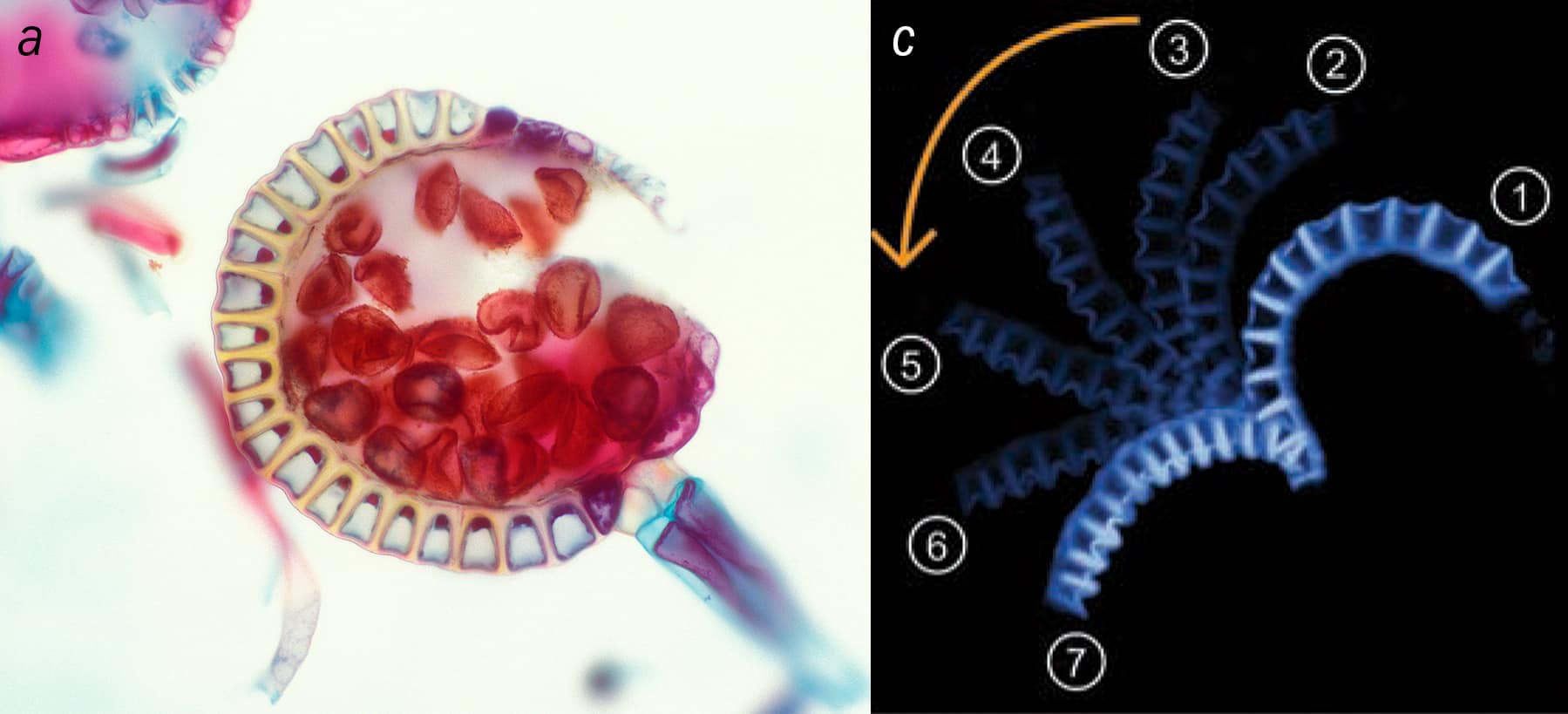
1 Catapulting to successa Leptosporangiate ferns disperse their spores using an ingenious mechanism, in which the spores (coloured red) are initially held in a small spherical basket, known as a “sporangium”. One side of the basket is ringed with U-shaped cells (yellow) that curve round to form a semi-circular annulus. b When liquid in the cells evaporates, the annulus, which starts in position 1, straightens out (position 4) and then bends over backwards (position 7), with the spores held at the outermost tip. The annulus then straightens out again (not shown), firing the spores far and wide like a catapult. (Courtesy: Herve Conge, ISM/Science Photo Library; Science/AAAS)
The most striking example of a catapult is found on leptosporangiate ferns, which hold the spores on the underside of leaves in tiny spherical baskets, about 0.2 mm in diameter. Each basket, known as a “sporangium”, is ringed on one side by a series of cells arranged in a semi-circle. In summer, as the weather gets warmer and drier, water between the cells in this “annulus” starts to evaporate and it stretches out to form a straight arm, with the spores held at the tip. The arm then gets bent backwards, like a catapult primed for operation (figure 1).
This dramatic release of energy accelerates the seeds up to 105g, flinging them from their basket into the surrounding countryside at speeds of 10 m/s
A taste for animals
But plants don’t only need to ensure their offspring survive. They also have to find nutrition to support themselves, with most plants taking minerals from the ground and energy from the Sun. However, a few plants – roughly 0.2% of all flowering species – have an extra source of nutrition: they eat animals. The most famous example of a carnivorous plant is the Venus flytrap (Dionaea muscipula), which is native to the subtropical wetlands of the eastern US.
Each of the leaves on this beautiful plant ends in a pair of lobes, hinged at the middle like a clam. When a fly or other insect lands on the lobes, tiny trigger hairs make the lobes swing shut, creating a cavity that holds the prey inside. Enzyme-containing secretions from the leaf dissolve the animal’s soft tissue, allowing the plant to absorb vital nutrients before the leaf re-opens. What remains of the fly is blown away, ready for the trap to snare its next victim.
The Venus flytrap can’t afford to be slow, given that a fly will respond to movement in barely 400 ms. In fact, the flytrap snaps shut in about a quarter of that time, well before the animal has time to get away. But how does the plant act so fast? For many plants that move, the secret lies in a kind of hydraulics. By changing the concentration of ions in cells in different areas of a leaf, a plant can shift water around internally. Cells with less water shrink while those with more fluid get bigger, allowing a plant to, for example, lengthen one side of a leaf but shorten the other. The leaf can therefore move, just as contracting the bicep in your upper arm can lift your hand up.
The amount of liquid that must be moved increases with the size of the plant, but there’s a limit to how fast the liquid can travel. So without some kind of clever trickery, a plant of a given size can’t move any faster than a certain speed. What this means is that if the Venus flytrap were to shut using hydraulics alone, by the time the leaves had closed, the fly would have long flown off to safety. To exceed the speed limit imposed by the slow motion of liquids in the plant tissue, this plant uses the same physics that occurs when an umbrella blows inside out.
As described by Charles Darwin in his 1875 book Insectivorous Plants, the leaves on a Venus flytrap are convex (outwardly curved) when open, but concave (inwardly curved) when shut. Although the open state is a mechanically stable configuration, the leaves lie very near to an elastic instability. So when a fly stimulates the trigger hairs, one side of the leaf expands in volume.
Using high-speed video, a team led by L Mahadevan from Harvard University found in 2005 that the expansion pushes the leaf beyond its stability limit. It buckles, snapping shut in a few tenths of a second – just as a gust of wind can blow an umbrella inside out so it flips from one stable geometrical configuration to another (Nature433 421). Known as the “snap-through buckling instability”, engineers exploit the effect to change the shape of carefully designed structures, but it looks like the Venus flytrap got there well before we did.
Wheels of fortune
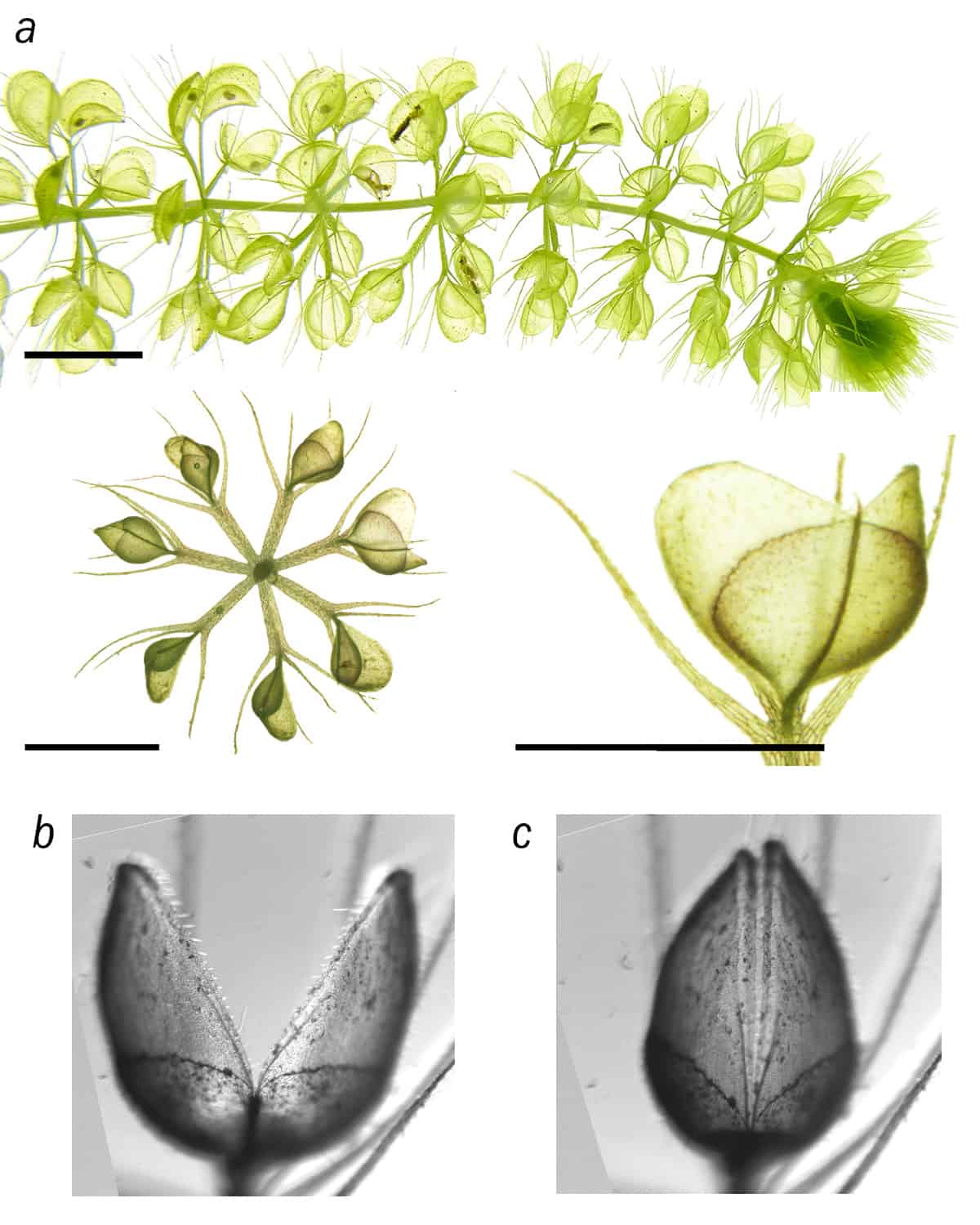
A less-well-known carnivorous plant with interesting physics is the aquatic waterwheel (Aldrovanda vesiculosa). Native to Asia, Australia, Europe and Africa, it is an invasive, underwater plant that takes its name from the series of curved leaves arranged in a circle around the plant’s long, central stems. Though lacking a physical hinge, each leaf has two lobes, making it look like the main character in the Pac-Man video game (figure 2).
2 Wonderful waterwheela The aquatic waterwheel plant lives underwater and consists of a series of leaves arranged in a circle around a stem. Each leaf has two lobes, forming a trap that can be either open (b) or closed (c), snapping shut when a fly or other insect lands on it. The region linking the two “jaws” deforms slightly causing a much bigger displacement at the leaf tips. The trap is also aided by being a constant state of pre-stress, which helps it to further speed up the motion. (Courtesy: Taken from Proc. Royal Society B 10.1098/rspb.2018.0012; reused with permission; courtesy Plant Biomechanics Group)
Just like the Venus flytrap, the leaves have a set of trigger hairs on their surface, snapping the lobes together when an insect lands on them. And for this plant too, motion powered by hydraulics alone would not be fast enough for it to catch a meal. So to capture its prey, which includes water mites and mosquito larvae, the waterwheel uses the principle of “kinematic amplification”. This is where a small, controlled input motion in one part of a structure creates a larger displacement elsewhere. You see this principle in action with a door, which you can easily swing wide open at the handle, or push open by nudging it near the hinge (albeit by applying a much bigger force).
As a team led by Anna Westermeier from the University of Freiburg, Germany, observed in 2018, the waterwheel uses the same approach by creating a tiny, hydraulically induced deformation that widens the base of the trap by a few hundredths of a millimetre. Thanks to kinetic amplification, this is enough to move the walls of the trap by about 200 times that amount, allowing the plant to close and trap prey inside – with no hinges in sight.
The rapid motion is also helped by the traps being in a constant state of “pre-stress”. Like a spring confined to a small space, the leaf has stored elastic energy, which – when motion is initiated – can be released, speeding up the movement (Proc. Roy. Soc. B205 20180012). Provided the prey can’t pry the leaves apart, this cunning design ensures that the waterwheel doesn’t go hungry.
Sticky subject
Another fascinating carnivorous plant is the Cape sundew (Drosera capensis). Native to South Africa, this beautiful plant has long, thin leaves covered with hair-like tentacles. They secrete a sticky fluid that forms a drop at the tip of each tentacle. When an insect lands on the leaf, it gets stuck on the sticky surface, which curls over, trapping the unfortunate animal. This process brings more of the plant’s digestive glands into contact with the prey, maximizing its nutritional intake (figure 3).
3 A sticky ending The leaves of the Cape sundew are covered in tentacles with a drop of sticky fluid at the tips. When a fly lands on the leaves, they curl round the insect, bringing digestive cells into contact with the prey, which can therefore be more easily eaten. The asymmetric configuration means that a simple, symmetric stimulus (a fly landing on the leaf) creates an asymmetric deformation. This principle could be useful in designing robotic replacements for the human hand. (Courtesy: iStock/Tailex)
This genera of plant so fascinated Darwin that he once claimed to “care more about Drosera than the origin of all the species in the world”. However, it was only in 2019 that an interdisciplinary research collaboration led by Caterina La Porta and Stefano Zapperi at the University of Milan, of which I was a part, worked out the full mechanical details of this strange motion and the biochemical signalling that triggers it.
Using an optical microscope, the upper and lower layers of cells in the Cape sundew leaf were found to have different shapes. The lower layer has elongated cells running down the length of the leaf, while the upper layer has more circular cells. When the plant is stimulated, either by an insect landing on its surface or a drop of milk being placed on its surface (as we did in our experiments), the plant alters the internal pressure of its cells, which change size in response.
Much like a balloon, where the inflated shape depends a lot on its initial, deflated shape, the upper and lower cells respond differently to the pressure change, with the long cells on the bottom of the leaf extending much more than the rounder cells on the upper layer. This differential rate of growth changes the overall geometry of the leaf, which curls up around its prey like a forefinger on a hand beckoning inwards (Proc. Nat. Acad. Sci.116 18777).
The bending, in other words, is literally encoded in the cellular structure of Cape sundew’s leaf, which converts a uniform biochemical signal into an asymmetric response. Inspired by this behaviour, our collaboration realized that the Cape sundew could also be useful for soft robotics. That’s because an artificial hand, say, has to respond in a predictable way to a particular input: it needs a pre-determined motion from a given stimulus. We therefore began planning new artificially structured metamaterials that could do just that.
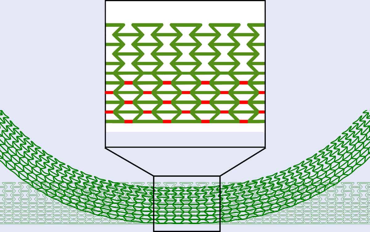
Using an ordinary plastic, we built an artificial, bi-layer structure that copied the behaviour of the Cape sundew plant (figure 4). The upper layer is made of hexagons where two of the sides are pulled in, which is a geometry well known to get thicker when stretched (i.e. has a negative Poisson’s ratio). The lower layer had the same structure, but with a single extra link added to each lattice unit cell. This geometry acts more conventionally by getting thinner when stretched (i.e. has a positive Poisson’s ratio).
4 All curled up Inspired by the way the leaves of the Cape sundew plants curls up around a prey, the author and his colleagues created this artificial bilayer lattice. As the inset shows, the structure has a deformed hexagonal lattice on the upper half and a set of tessellating trapeziums on the lower half, with additional links in red. When compressed by a symmetric force on the upper and lower edges, the structure curls up just like the plant. The design could be useful for those working in soft robotics who want to design prosthetic hands that can function just like the real thing. (Courtesy: Daniel Rayneau-Kirkhope)
This bilayer structure was easy to manufacture and had far-reaching mechanical consequences. Computer simulations showed that our material would curl up into a circle if air were pumped into the voids in the material – just like the plant that inspired it. What’s more, we found we could also make this material deform from its initial, flat configuration into a circle by compressing it from its sides. In other words, there was more than one way to trigger this hingeless behaviour.
It’s not too fanciful to imagine a soft material like this, where the motion is built into the architecture, being used to make robotic fingers or limbs that can curl up or straighten out on demand. By replacing conventional rigid hinges, such materials could allow robots to grasp fragile objects without damaging them, such as plates, glasses and cups from a dishwasher. They would need less computational power and none of the complicated feedback loops that are normally required to stop robots from unintentionally smashing objects in their grasp.
Robotic future
From self-cleaning windows inspired by superhydrophobic lotus leaves to surgical staples inspired by porcupine quills, our work is just part of a growing trend for novel, practical applications inspired by mechanisms found in plants and animals. But rather than simply copying animals or plants to create robots, researchers are seeking to tease out the basic principles and mechanics, marrying them with modern engineering materials to create an entirely new generation of machines.
What is wonderful about following nature’s lead is that we know that the mechanics work. Living things we see all around us have been tested over thousands and millions of years, with faulty designs long since weeded out. If a plant seed dispersal mechanism does not work, its species will become extinct; if a plant cannot get food reliably, it will die. Newton may have seen God’s presence in a thumb, but one wonders what he would have made of a robotic plant-inspired hand.
An adaptive optics OCT image (top) and the WeakGCSeg algorithm result (bottom), which identified and traced the shapes of individual retinal ganglion cells. (Courtesy: Sina Farsiu, Duke University)
The eyes are sometimes referred to as a window to one’s soul, a phrase with unclear origin, but filled with truth. In fact, our eyes provide a literal window into our brains. This is because neurodegenerative conditions such as Parkinson’s disease and Alzheimer’s disease, as well as eye diseases such as glaucoma, can directly affect a patient’s optic nerve, retinal cells and surrounding visual structures. The eye, or more specifically, the retina, is often known as an extension to our central nervous system (CNS).
One type of cell, the retinal ganglion cell (GC), is a neuronal cell whose axons form the optic nerve. These cells are key to one’s vision as they process and relay optical information to the brain. In neurodegenerative diseases, GCs often degenerate and disappear, eventually leading to blindness. For this reason, being able to quickly identify the extent of such degeneration has proven to be an important biomarker for both diagnosis and treatment monitoring of neurodegenerative diseases. In a study published in Optica, researchers in the US present a new method to visualize and quantify individual GCs.
A weakly supervised segmentation network…
Ophthalmic imaging systems, such as optical coherence tomography (OCT), are clinically used to visualize the different layers of eye tissue, including the GC layer, in order to diagnose and track the progression of eye diseases. However, as these traditional methods are limited to low resolution, they can only measure the thickness of these layers, without providing much information on individual cells.
A state-of-the-art technology, called adaptive optics OCT (AO-OCT), is sensitive enough to image individual retinal GCs. However, the current standard approach for quantification involves manual marking of each AO-OCT volume, which is not only subjective, but also time consuming and impractical for large datasets.
This method consists of a three-step process. First, the group uses an automatic algorithm to pre-process an entire AO-OCT volume, to extract the retinal layer containing the GC somas. Second, the researchers pass this volume through a localization network that is trained to produce a probability map indicating the locations of potential somas. Interestingly, the network is not trained using ground-truth segmentation maps, but rather using “weakly” annotated labels in the form of small spheres (with a radius of 2 μm) at each manually annotated cell location. In the final step, the researchers perform post-processing to transform the network’s predictions into individually segmented somas.
… accurately localizes individual ganglion cells
To test their method, the researchers analysed both healthy subjects and glaucoma patients, and showed that their proposed framework was able to accurately segment GC somas of both groups. By using WeakGCSeg’s output, they could differentiate between the two cohorts based on the number and size of the predicted GCs. Not only that, but this framework was able to achieve high detection performance regardless of the imaging device used, while being on par with or exceeding the performance of human expert graders.
In the future, the group hopes to extend this work using larger datasets and looking at different retinal diseases to fully capture the framework’s generalizability. The researchers note that WeakGCSeg also has the potential to be used with other neurodegenerative disorders, such as Alzheimer’s or Parkinson’s disease.
Stroke – a life-threatening condition arising from restriction of the blood supply to the brain – is a leading cause of long-term disability. In Europe, there are over 1.5 million new cases each year, with less than 15% of patients achieving full recovery. A key challenge in stroke rehabilitation is that recovery varies widely between patients: roughly two thirds of stroke patients recover naturally from their initial impairments, the remainder do not. Critically, natural recovery is especially variable in severely impaired patients.
The ability to forecast an individual’s potential recovery could help doctors select personalized therapy and maximize treatment outcome, particularly for those with severe impairments. With this aim, an international research team headed up at the Swiss Federal Institute of Technology (EPFL) has developed a machine-learning system that makes high-accuracy predictions of recovery outcome for stroke patients.
“The key is to find the optimal neuro-rehabilitative strategy to maximize individual treatment outcome,” explains EPFL’s Friedhelm Hummel in a press statement. “This tool can support the prediction of individual courses of recovery early on and will have an important impact on clinical management, translational research and treatment choice.”
Personalized predictions
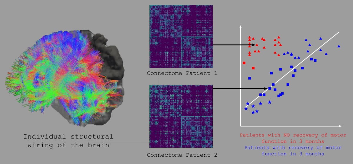
Hummel and collaborators developed the predictive tool based on analysis of the connectome – a map of the whole brain’s wiring generated from multiple MR images. Their goal was to use information from whole-brain structural connectomes as a potential prognostic factor to determine individual outcome after stroke (recovery versus no recovery), using a machine learning approach based on support vector machines (SVMs).
To train their prediction model, the researchers analysed whole-brain structural connectomes of 63 patients two weeks after stroke onset, while simultaneously assessing the patients’ motor impairment. Within this dataset, 39.7% of the patients did not show natural recovery; in a subgroup with severe motor impairment, 63.9% did not naturally recover. The team also tracked connection changes in the patients’ brains up to three months later.
The SVMs were trained to separate patients who recovered naturally from those who did not, based on their connectomes. The SVMs then defined the underlying brain network patterns for each patient to make predictions about their recovery potential, with a particular focus on those who were severely impaired.
Internal validation of the prediction model revealed that features extracted from the connectomes two weeks after stroke could segregate patients with recovery from those without. Internal validation of the model demonstrated high classification performance, with a prediction accuracy of 83% for all patients. Importantly, the classification accuracy was even higher – 92% – in the difficult-to-predict group of severely impaired patients.
MRI-based techniques are used to determine the individual structural wiring of the brain and the underlying connectome. Features from this complex information can classify patients with high precision into those who show natural recovery after stroke or those who do not. (Courtesy: F Hummel, EPFL)
The researchers further validated the results in a smaller independent dataset. Here, the classification across all subjects, including severely impaired patients, also achieved high accuracy. They note, however, that these results should be taken with caution, as the external validation data sample was small.
The areas that matter
The team also used the whole-brain connectomes to identify relevant brain areas and connections that may support or hamper motor recovery, determining network nodes of particular importance for recovery in severely impaired patients. The identified brain areas are likely to underlie the processes of spontaneous biological recovery of motor functions and could influence the design of new therapies.
Feature extraction revealed that connectivity within the core parietofrontal motor network is crucial for favourable outcomes, with secondary motor areas especially important in patients with severe initial impairment. In addition, functional systems such as the attentional, somatosensory or multimodal areas clearly contribute to motor function recovery and improve the classification.
The researchers, who report their findings in Brain, conclude that computational analyses of whole-brain connectomes have high potential for predicting patients’ degree and course of recovery at an early stage following stroke. Hummel notes that for routine clinical use, the prediction tool will require further development and, ideally, automation.
“The next steps we are planning are to further evaluate this approach in other data sets and develop the analytical approaches towards a simpler application for direct clinical use,” he tells Physics World. Furthermore, we continue to analyse structural and functional connectomics and its impact on stroke recovery to use this information towards personalized treatment strategies.”
Stephen Hawking’s 40-year-old theorem about the area of a black hole’s event horizon has been confirmed thanks to data from the first burst of gravitational waves detected by LIGO. Known as Hawking’s area theorem, it states that the entropy of a black hole should not decrease. Because a black hole’s entropy is proportional to the area of its event horizon, that means the event horizon area should not decrease if two black holes merge, as they did in the cosmic cataclysm that produced the gravitational-wave signal dubbed GW150914.
To test Hawking’s area theorem, astronomers led by Maximiliano Isi of the Massachusetts Institute of Technology (MIT) re-examined the GW150914 signal, which was picked up by LIGO’s detectors in 2015 and announced in February of the following year. These ripples in space-time developed when black holes of 36 and 29 solar masses merged to form a new black hole of about 62 solar masses, with the three remaining solar masses converted into gravitational-wave energy.
If Hawking’s area theorem holds, the event horizon area of the newly merged “daughter” black hole should not be less than the combined area of the event horizons of the two parent black holes. Instead, Isi explains, “The combined changes in black hole masses and spins should conspire to result in an area increase – or, strictly speaking, to prevent an area decrease.”
Putting Hawking to the test
Gravitational-wave signals from merging black holes display a distinct sequence. At first, the in-spiraling black holes produce gravitational waves that increase in frequency and amplitude. A “ring-down” period then follows immediately after the merger, when the daughter black hole is in a distorted state and produces gravitational-wave vibrations somewhat analogous to the sound waves from ringing a bell.
By analysing the in-spiral and ring-down phases of GW150914, Isi’s team calculated the area of the two black holes’ event horizons based on their masses and rates of spin. They found that the combined area of the parent black holes’ event horizons was approximately 235 000 km2, whereas the area of the daughter black hole’s event horizon was approximately 367 000 km2. The total area had indeed increased, proving Hawking’s theorem to 95% confidence.
Deviations into exotic physics
Gaurav Khanna, a physicist at the University of Rhode Island and the University of Massachusetts, US, who was not involved in the research, calls the MIT study a “truly impressive work” that offers “the clearest such result” that Hawking’s theorem is true. “It’s really cool when gravitational-wave data is able to help test fundamental theorems of black-hole physics,” Khanna says.
Isi’s team now plan to study more black-hole mergers, searching for deviations from the theorem that may offer clues to new kinds of object, or even new physics. “Theorists have come up with more or less plausible models that could result in mixed populations of compact objects that could resemble black holes,” Isi tells Physics World. As examples, he cites quark stars and gravastars, which are hypothetical alternatives to black holes that contain a gravity-repelling area of space that prevents further collapse. Such extreme forms of matter could, he says, “support very compact objects that look like black holes from afar but have other properties as you get close to where the event horizon would be”.
Isi also points out that Einstein’s general theory of relativity doesn’t mesh well with quantum physics, and may eventually need to be corrected. “If so, it is likely that black holes would have additional features beyond what we expect,” he says. “More precise measurements in the future might allow us to place interesting constraints on these models.”
This article was amended on 14 July 2021 to correct Maximiliano Isi’s institutional affiliation; he did his PhD research at the California Institute of Technology but is now at MIT.
Monoglot culture Should we let a language hailing from a tiny island in north-western Europe determine the integrity of the global scientific discipline? (Courtesy: Shutterstock/Aine)
English is the lingua franca of science. An estimated 98% of all scientific publications are written in English and the vast majority of talks at international and most local conferences are given in the language. To disseminate their results as widely as possible, students who are non-native English speakers must therefore not only learn their field of expertise but also what is in effect a new “dialect”. While English’s ascendency is widely accepted and unchallenged, the situation leads to a gatekeeping of scientific information. It is a way of doing science that is unfair and needs to be undone.
English’s dominance was not always the case. In 1880 only 36% of scientific publications were written in English, and centuries prior to this Arabic dominated medical, mathematical and astrophysical texts. The “Islamic golden age” was a time when the foundations of the scientific method were laid, when innovations and improvements to many works from Aristotelian physics were made.
Indeed, many vital texts in physics were first written in Arabic. These include al-Khāzini’s The Balance of Wisdom as well as al-Haytham’s Book of Optics (Kitāb al-Mānazir), which was the first text to postulate that light is reflected off different surfaces. This idea was in stark contrast to the prior Greek Euclidean and Ptolemaic theories that light rays were emitted from the eyes and back again. In fact, al-Khwārizmī’s The Compendious Book on Calculation by Completion and Balancing – influenced by the Indian text Āryabhatīya in 499 CE – was the first work to teach algebra and one of the most transformative and important works in the history of mathematics.
However, due to the colonial nature of the Western science curriculum that I encountered first at school and then as a physics undergraduate, I learnt plenty about the scientific virtues of Aristotelian physics but nothing about their non-Western counterparts. That’s a shame as the importance of teaching and translating the works of the Islamic golden age was somthing that European scientists of that era understood well.
Al-Khwārizmī’s books on algebra, for example, were used until the 16th century as the principal mathematical literature at European universities – a lot of mileage for texts that were written in 820 AD. The ability of science to be disseminated throughout all languages has been vital for its survival and continuation and was instrumental to the revival of learning science in the West. Academics at the time knew that science should be a multilingual enterprise.
Transformative work
It is unfair now to demand that scientists all over the world treat science as a monoglot exercise – something that should remain firmly within the constraints of the English language. Science is difficult enough without adding the need for non-native English speakers to be able to fluently present and publish only in English. How many talented non-English speaking students and academics feel as though their ability to survive in academia depends on their capacity to speak and write in a language that is not their own? How many scientists have non-English scientific works that have been ignored simply because they were not written in the “right” language? Why isn’t more being done to aid them?
Many scientists have already spoken out about the struggle of not having English as their mother tongue. In 2019 Adriana Romero-Olivares, who was then a biology postdoc at the University of New Hampshire, wrote in Science about the harsh, undeserved criticism that was given to her and her collaborators about the way their paper was written. She cited one reviewer who criticized her very first paper by saying that: “The authors need a native English-speaking co-author to thoroughly revise the grammar of this manuscript.”
Romero-Olivares says that her “heart sank” when she received such feedback, especially given that the reviewer did not say anything about the underlying science (Science 10.1126/science.caredit.aaz7179). There are many other similar accounts that indicate attitudes that contribute to the systematic bias in science – not overtly racist but an inbuilt injustice due to the centring and framing of the West as a global scientific powerhouse.
Hoping for a different dominant language or multiple languages seems unlikely to happen, at least in my lifetime. But publications and journals that are not written in English should be regarded as equally valuable as those that are, and efforts should be made to translate between all of them. At the very least, we need to make sure those in academia who are not fluent in English feel adequately supported. This could mean having translation services and language support built into grants for non-English speaking academics. Another is the inclusion of language-editing services such as that offered by IOP Publishing, which publishes Physics World. It could also mean ensuring that there is free, easy-to-access English-language training services for all who require it.
We need to do a lot of introspection. Every native English-speaking scientist – especially those who do not know another language – should try to understand how isolating it must be to carry out research and present and publish in a dialect that is not their own. We should not let a language hailing from a tiny island in north-western Europe determine the integrity of a discipline that is carried out globally. By doing so we are inhibiting the progress that scientists of the past tried so hard to protect.
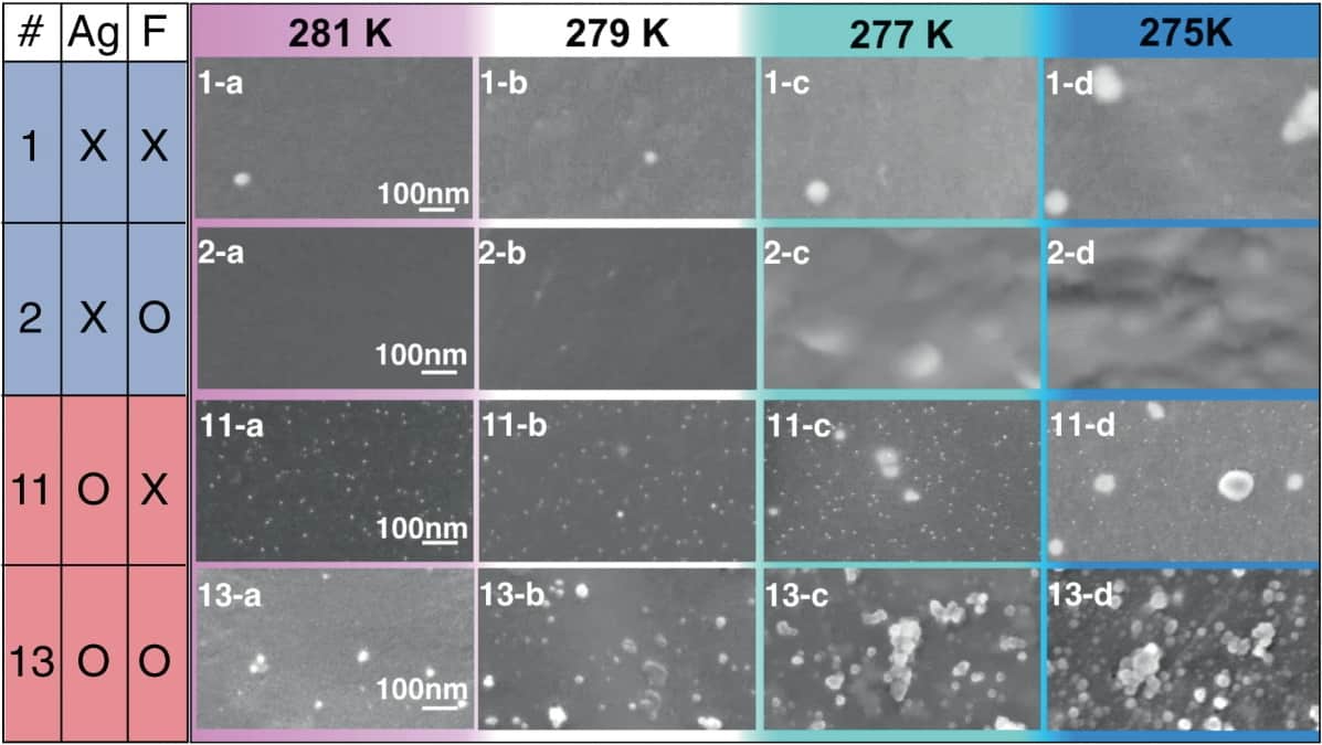
Films prepared from aqueous semiclathrate hydrate solutions with and without silver nanoparticles. In sample 13, which included both silver nanoparticles and fluoride anions, 10–30 nm clusters formed even at 281 K. As the temperature decreased further, the number and density of the clusters increased, and then the material crystallized with a small degree of supercooling. (Courtesy: T Sugahara/Osaka University and H Machida/Panasonic Corporation)
The rate at which water molecules cluster together to form ice crystals can vary depending on which “nucleation seeds” are introduced, researchers in Japan have discovered. Through high-resolution imaging experiments, a team from Panasonic Corporation, Osaka University and Waseda University found that crystallization in water-based materials is accelerated by the addition of silver nanoparticles – suppressing the often-inconvenient effect of supercooling. The discoveries could pave the way for improvements in heat-storing materials.
When water is cooled below its freezing point, molecules will quickly start to cluster around impurities in the liquid. Through the process of nucleation, these clusters become larger and more frequent, before eventually amalgamating to form solid ice crystals. Nucleation can be inhibited when water contains few impurities, and is left undisturbed during cooling – allowing it to maintain its liquid state even below its freezing point.
This supercooling effect can impair the performance of latent heat storage materials. In these systems, heat can be extracted or stored in frozen water-based solids, without changing their temperature – but this cannot be done efficiently if the materials are still liquids below their regular freezing points. This inconvenience is now driving research into how supercooling can be supressed, allowing nucleation clusters to form more readily. Currently, however, the mechanisms by which these clusters form around impurity seeds are still poorly understood.
To study the effect, the team constructed experiments involving clathrate hydrates: water-based solids that resemble ice, in which polar or hydrophobic molecules are trapped within “cages” of hydrogen-bonded water molecules. After introducing different nanoparticle seeds to clathrate hydrates in liquid form, the researchers used a combination of scanning electron microscopy and X-ray absorption spectroscopy to directly image the formation of nucleation clusters.
When they added silver nanoparticles measuring between 5 and 10 nm across, the images revealed that clusters up to 30 nm in diameter would readily form around them. With some clusters even remaining at temperatures as high as 8°C, these nanoparticles clearly accelerated the crystallization process. In contrast, nanoparticles of unreactive noble metals – including palladium, gold and iridium – didn’t promote crystallization at all.
The discovery has shed new light on the nucleation mechanism, and the researchers anticipate that their results will provide a starting point for enhanced control over supercooling. By introducing the appropriate nanoparticles to clathrate hydrates, they envisage that it should be possible to develop more advanced latent heat storage materials. If achieved, these systems could be applied to technologies including solar energy and heat recovery systems for buildings.
The smallest source capable of delivering high-intensity pulses of extreme-ultraviolet (XUV) light has been claimed by researchers at ELI-ALPS in Hungary, INCDTIM in Romania, and the Max Born Institute in Germany. The new optical technique could make high-intensity XUV pulses far more accessible to labs worldwide, opening up new possibilities for high-speed nanoscale imaging.
Although intense pulses of lower-frequency UV light are routinely generated in many research facilities, comparable intensities at higher XUV frequencies had proven far more difficult to produce. Currently, the most compact sources use a technique called high harmonic generation (HHG), whereby a target material is placed at the focus of intense near-infrared (NIR) femtosecond laser pulses. This causes the target to strongly emit light at higher harmonic frequencies to the NIR light, which conveniently lie in the XUV range.
To generate suitably intense XUV pulses, the NIR pulses must be focussed onto large target areas using large spherical mirrors, requiring an apparatus that exceeded 10 m in size. So far, these bulky and expensive setups can only be implemented at a limited number of research facilities.
Highly pressurized gas jet
Now, Balázs Major and colleagues have developed a more advanced HHG technique. This has a highly pressurized gas jet acting as the HHG target, but crucially the target positioned some distance away from the focus of the NIR light. Since the NIR beam is spread out as it encounters the the gas, it interacts more and generates more XUV photons. As an added bonus, the resultant XUV pulses have a large divergence, which means that they can be focussed to a very small spot for nanoscale imaging.
The NIR light is removed from the beam using an aluminium filter and the XUV pulses are focussed onto a spot just 600 nm across using a small spherical mirror. The entire setup fits on a 2 m tabletop and the team was able to create XUV pulses with intensities as high as 2×10¹⁴ W/cm2. This already exceeds the performance of existing, far bulkier XUV sources. Simulations done by the team suggest that further improvements could boost this intensity by a factor of 1000.
To demonstrate their laser’s capabilities, the researchers used it to induce both two- and four-photon absorption in argon atoms. These nonlinear processes produce doubly and triply ionized states of argon respectively, with a likelihood that scales with the square of XUV intensity – making the team’s setup the most compact apparatus to date that can trigger the process reliably.
The researchers hope that the compactness and affordability of the laser could increase access to intense XUV pulses for universities, research facilities and industries worldwide. In particular, it could allow researchers to easily carry out femtosecond, or even attosecond imaging of systems ranging from electron dynamics to biomolecular reactions.
Quantum computers can now simulate much larger quantum systems than was previously thought possible thanks to algorithms developed by researchers in the UK and Germany. The new algorithms divide up quantum computational resources according to which parts of the simulation require them most, making it possible to extract information about a large quantum system from many smaller, more manageable calculations – in effect, running the simulation in parallel. The result should boost the capabilities of the current generation of so-called noisy intermediate scale quantum (NISQ) computers, which lack the computational resources required to perform useful algorithms in materials science or drug discovery, both of which depend heavily on a deep understanding of quantum effects.
Quantum computers promise to perform complex calculations today’s classical supercomputers cannot. A bottleneck for achieving such a “quantum advantage” is that it is very difficult to engineer a large, error-free quantum computer. While upgrading the quantum technologies themselves might seem like the obvious solution, it is also possible to improve the algorithms that run on such computers – for example, by changing the way thinformation.
Representing quantum states efficiently
In the latest work, published in npj Quantum Information, researchers at University College London (UCL), the Technical University of Munich, King’s College London and the biotech company Kuano (formerly a quantum start-up called GTN) took inspiration from a set of mathematical tools known as tensor networks. Tensor network methods were originally developed to simulate quantum systems classically, and their chief selling point is that they are very efficient at storing the information needed to describe certain classes of large quantum states. They achieve this efficiency by focusing on the quantum effects that are most important (like entanglement), purposefully allocating fewer computational resources to those that are not.
By translating these methods to quantum computing algorithms, the team showed that similar advantages apply even when the processors are quantum. The approach allows computational resources to be divided, so that the separate components of the algorithms can be run in parallel on different quantum processors and their results combined at the end. This may be useful in simulating large molecules that have complicated quantum interactions only in certain regions. These regions could each be assigned a processor, with the simpler interactions between them relegated to a separate computation of their own.
Extra advantages
While the team’s approach emulates what tensor networks already do on classical computers, running such computations on quantum computers has potential advantages that could never be attained classically. This is largely because of something called the bond dimension, which is a way of modelling the amount of entanglement in a quantum state. For highly entangled quantum systems, this bond dimension becomes large and makes the computation inefficient. On a quantum computer, however, the resources required may be greatly reduced. “Tensor networks are the very best way to simulate many quantum systems on classical computers,” says Andrew Green, a condensed-matter physicist at UCL and a co-author of the study. “We expect that translating them to quantum computers will unlock a quantum advantage.”
To test their approach, Green and colleagues simulated an Ising spin chain, which is a large one-dimensional quantum system that is often used to model interesting optimization problems. The scale of this system is far beyond what has previously been achieved on a quantum computer, indicating that it is possible to extract useful information about large quantum systems from many small-scale calculations on current quantum computing hardware.
According to the researchers, a possible follow-up would be to develop algorithms to simulate two- or three-dimensional quantum systems, as well as systems that may be far less ordered than those that scientists have investigated so far. They hope that while such systems – like those with complicated forms of entanglement – are proving challenging for classical tensor network approaches, translating them to quantum computers may finally lead to a breakthrough.