Creating a quantum computer that integrates a large number of components is a huge challenge for many reasons. One is that most quantum bits (qubits) used today must be chilled to near absolute zero and therefore isolated from the room-temperature electronic components used to control them. This makes it extremely difficult to have large numbers of qubits and controllers packed into a small space. But researchers in the Netherlands may have a solution, as the science writer and educator Karmela Padovic-Callaghan explains in this episode of the Physics World Weekly podcast.
The “wonder material” graphene is a sheet of carbon just one atom thick that has extraordinary physical properties. One promising use for graphene is to make products more sustainable. On hand to talk about how graphene can do its bit to improve the environment is Andrew Pollard, who specializes in the metrology of 2D materials at the UK’s National Physical Laboratory.
Water is another substance that has wonderous properties that have proven very useful for sustaining life on Earth. Recently, researchers used a laser technique to study supercooled water in a previously inaccessible temperature regime. Katherine Skipper, a PhD student contributor to Physics World joins us to chat about this breakthrough. Skipper also talks about her research at the University of Bristol, which is focussed on active matter that comprises two-faced Janus particles.
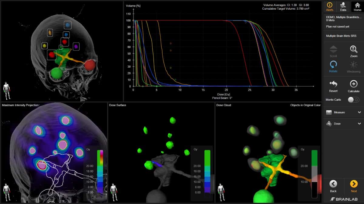
Indication-specific: Elements Multiple Brain Mets SRS dose planning enables the delivery of highly conformal single-session treatments for multiple metastases. (Courtesy: Brainlab)
Simplicity, automation, efficiency and dose targeting with sub-mm accuracy: these are the operational reference points for the radiation oncology team running the stereotactic radiosurgery (SRS) programme at Scripps MD Anderson Cancer Center in San Diego, California. Underpinning that stereotactic clinical workflow is a portfolio of enabling technologies from Brainlab, a German healthcare equipment maker specializing in hardware and software products for SRS, surgical navigation and the next-generation “digital operating room”.
“Think automated treatment planning, high-precision patient positioning plus online imaging and thermal surface guidance,” says Steve Kirsner, director of medical physics at Scripps MD Anderson. “Our established suite of Brainlab products makes it easy for physicists to do what they have to do, allowing us to deliver high-quality care to our cancer patients every time. You can’t ask for more than that.”
Kirsner, for his part, heads up a 16-strong multidisciplinary clinical team (eight medical physicists and eight dosimetrists) working across three radiotherapy centres in the greater San Diego area. Right now, those three clinics treat around 160 patients a day, with a fourth facility being commissioned ahead of coming online for initial patient treatments in August. “We run a busy SRS linac schedule for diverse brain indications,” adds Kirsner. “That’s complemented by an extensive programme of stereotactic body radiotherapy (SBRT) to treat a range of cancer types, including prostate, lung, spine, liver, pancreas and oligometastasis.”
End-to-end workflows
In developing its SRS programme, the Scripps MD Anderson team has worked closely with Brainlab engineers to implement indication-specific, end-to-end workflows for a range of brain stereotactic treatments. That specificity means the algorithms in Elements, Brainlab’s à la carte treatment planning software, are able to address the unique clinical challenges surrounding a given disease indication. Elements Cranial SRS, for example, automatically creates SRS treatment plans for complex cranial indications – including, but not limited to, vestibular schwannomas, meningiomas and large metastases – while Elements Multiple Brain Mets SRS dose planning enables the delivery of highly conformal single-session treatments for multiple metastases, minimizing dose spread to adjacent healthy tissue and critical structures.
“Elements is one of the most user-friendly platforms for SRS treatment planning,” explains Kirsner. That ease of use is particularly important when running a medical physics service across multiple sites and multiple teams. It’s straightforward, for example, to create a set of standard planning templates for a given clinical indication. Those templates can then be shared across every facility to give treatment planners a common baseline and reference point from which to develop individualized SRS plans on a patient-by-patient basis. “Put simply,” adds Kirsner, “Elements has enabled us to create a unified framework for treatment planning, driving standardization and best practice across multiple Scripps MD Anderson treatment facilities.”
It helps, of course, that Elements offers a streamlined, automated process to arrive at a high-quality SRS treatment plan, while verification of the plan and translation to the treatment machine are equally friction-free. All of which equates to a win-win for healthcare providers and patients. “Efficiency and patient throughput are key metrics for any oncology centre,” says Kirsner, “while there’s a growing demand from patients for the benefits associated with SRS treatment schedules – not least fewer visits to the clinic and a faster return to family and friends.”
Alongside Elements for treatment planning, Kirsner’s team is also taking advantage of Brainlab’s in-room X-ray-based ExacTrac positioning and monitoring system. ExacTrac X Ray enables robust verification of patient positioning – to isocentre with sub-mm accuracy – as well as intrafractional monitoring of patient motion and internal anatomical shifts during treatment. The latest iteration of the platform, ExacTrac Dynamic, is able to track patient position along an additional coordinate – using a 4D thermal camera to create a hybrid thermal image that correlates the patient’s heat signature to their reconstructed 3D surface structure.
Multiple perspectives: in addition to X-ray-based patient imaging, ExacTrac Dynamic (shown here in relation to the treatment couch) uses a thermal imaging camera to correlate the patient’s heat signature to their reconstructed 3D surface structure. (Courtesy: Brainlab)
ExacTrac Dynamic is currently being installed as part of the commissioning of the fourth Scripps MD Anderson radiotherapy clinic, with the three established treatment centres all due to upgrade to the new imaging system by the end of this year. “We like the intuitive user experience with ExacTrac,” says Kirsner, “and the fact that it allows us to image what we treat at all times, regardless of couch angle or gantry position.” In addition, the new thermal surface guidance capability will allow Kirsner and his team to automatically track both internal and external motion throughout the entire treatment of the patient. “ExacTrac imaging also integrates seamlessly with our Varian machines,” Kirsner adds. “This will allow treatment to be paused if either the X-ray imaging or surface guidance are deemed out of tolerance – so we’re excited to see the clinical impact of the latest surface-tracking functionality in ExacTrac Dynamic.”
Clinical validation
While the near-term priority for Kirsner and colleagues is the commissioning and go-live of their new radiotherapy facility, the team is also focused on achieving Novalis-Certified status by year-end. Novalis Certification is an independent accreditation programme that offers formal recognition of the highest clinical standards in SRS/SBRT treatment delivery. As such, a key outcome for cancer treatment centres is the implementation of standardized protocols for continual self-assessment and quality improvement, further reinforcing the access that Brainlab customers get to Novalis Circle, a global network of oncology centres dedicated to best practice, knowledge-sharing and collaboration on all aspects of SRS/SBRT.
“I see the independent audit of Novalis certification as a great way to strengthen the SRS/SBRT programme at Scripps MD Anderson,” concludes Kirsner. “We are all doing the best we can for our patients, but you can always learn something new from colleagues running their own radiotherapy programmes. It’s my belief that our practice here is already Novalis-compliant, so the next step is to formally document those processes and workflows for external review.”
Researchers in the US have created a new solid-state lithium-metal battery that can charge and discharge over a record-breaking number of cycles at a high current density. The proof-of-concept device, which is fundamentally different from existing liquid electrolyte lithium-ion batteries, could extend the lifespan of electric vehicle batteries to 10–15 years, similar to that of petrol and diesel cars.
The ideal battery for an electric vehicle would last a long time and store up a lot of charge very quickly. While lithium-metal batteries score well on the second of these criteria thanks to their high capacity and high energy density, their longevity leaves something to be desired. This is because, during charging, lithium ions move from the cathode to the anode. When this anode is made of lithium metal, needle-like structures called dendrites form on the electrode surface and grow into the electrolyte. Eventually, these unwanted structures pierce the barrier separating the anode and cathode, causing the battery to short or even ignite.
To overcome this problem, researchers have previously tried replacing the liquid electrolyte in these devices with a solid-state one that is more difficult for the dendrites to grow through. In practice, however, lithium dendrites can still burrow through the barrier via micron- or submicron-sized cracks produced when the battery is assembled.
Sandwich effect
Researchers led by Xin Li, a professor of materials science at Harvard University’s John A Paulson School of Engineering and Applied Sciences (SEAS), have now developed a solid-state battery in which lithium dendrite penetration is no longer a problem. Rather than stopping the dendrites dead in their tracks with a single barrier, their new battery design takes a multi-layered approach. The design, which is detailed in Nature, incorporates a less-stable electrolyte sandwiched between layers of more-stable solid electrolytes. Together, these layers keep dendrite growth under control.
The full battery “sandwich” consists of six layers: a lithium-metal anode, a graphite coating, the first electrolyte, the second electrolyte, another layer of the first electrolyte, and finally the cathode. The first electrolyte, which has the chemical formula Li5.5PS4.5Cl1.5 (LPSCI), is prone to dendrite penetration. The second electrolyte, Li10Ge1P2S12(LGPS), is more robust. The idea is that dendrites are permitted to grow through the graphite and electrolyte no. 1 but stop when they reach electrolyte no. 2 – and, crucially, before they short out the battery.
“Incorporating instability to stabilize the battery might sound counterintuitive,” says team member Luhan Ye. “But just like an (expansion) anchor can guide and control a screw going into a drywall, so too can our multilayer design guide and control the growth of dendrites. The difference is that our anchor (the LPSCI) quickly becomes too tight for the dendrite to drill through, so the dendrite growth is stopped.”
Superior cycling performance
The researchers found that when they paired their lithium metal anode with a LiNi0.8Mn0.1Co0.1O2 cathode, the cycling performance was very stable, with the battery maintaining 82% of its capacity after 10,000 cycles at a 20C rate (8.6 mA/cm2) and 81.3 % of its capacity after 2000 cycles at a 1.5C rate (0.64 mA/cm2). The design allows for a specific power of 110.6 kW/kg and a specific energy of up to 631.1 Wh/kg from the cathode material.
The test device cycled for 1800 hours at 0.25 mA/cm2, which is substantially better than the single-electrode-type batteries the researchers tested. It can also cycle at an extremely high current density of 20 mA/cm2 with a low “overpotential” (that is, the potential difference between a theoretical or thermodynamically determined voltage and the actual voltage under operating conditions) of ~0.5 V without significant signs of short-circuiting, even at a temperature of 55 °C.
Longer lifetime and faster charging
According to the researchers, the new battery technology could make the lifetime of electric vehicles comparable to that of petrol- or diesel-powered ones – 10 to 15 years – without the need to replace the battery. With its high current density, the design could also “pave the way for electric vehicles that can fully charge within 10–20 minutes,” they say.
“This proof-of-concept design shows that lithium-metal solid-state batteries could be competitive with commercial lithium-ion batteries,” Li adds. “And the flexibility and versatility of our multilayer design makes it potentially compatible with mass production procedures in the battery industry.”
The researchers say they now plan to scale up their device to a pouch cell the size of an ID card. “We expect that the attractive performance we have presented in our work will hold, since our innovation focuses on the scalable factors of materials chemistry and their combinations,” Li tells Physics World. “The design also opens the door to many new fundamental studies in solid-state battery research that were hitherto not possible without a stable lithium-metal anode.”
As the push toward stronger and faster MRI scanners continues, so does concern over magnet safety, according to Filiz Yetisir, who discussed the potential effects MRI has on patients at the recent International Society for Magnetic Resonance in Medicine virtual meeting (ISMRM 2021).
There are three main components of MRI scanners – the main magnet, gradient coils and radiofrequency (RF) coils – that are essential to a machine’s function. However, each of these components carries unique risks for patients and operators, namely the projectile effect, nerve stimulation, hearing damage and tissue heating.
Yetisir, a postdoctoral graduate student at Harvard University School of Medicine, discussed these potential effects and provided tips for reducing their impact during procedures in a May 16 session at ISMRM 2021.
The main magnet
Consider that junkyard magnets capable of lifting cars have strengths of 1 T. Most of the MRI scanners used in clinical settings today have strengths of 1.5 T and 3 T, while scanners used mainly in research have strengths of 7 T – and in some cases up to 11 T. Recently, 7 T scanners, such as the Magnetom Terra scanner by Siemens Healthineers and the Signa 7.0T scanner from GE Healthcare, for instance, have been cleared for clinical marketing by the US Food and Drug Administration.
The main safety risk associated with the main magnet is the projectile effect, or the “missile effect,” according to Yetisir. MRI magnets attract iron particles with tremendous force. This can include other ferromagnetic objects in the scanning room, such as chairs or oxygen tanks.
An office chair caught in an MRI scanner. (Courtesy: of Frank Shellock, MRISafety.com)
“If there’s a person inside the scanner or in the path of the flying object, the projectile could result in serious injuries or even death,” she said.
In 2001 in New York, for instance, 6-year-old Michael Colombini died after being hit in the head with an oxygen tank. “The most important thing is to remember that the magnet is always on,” Yetisir said.
Gradient coils
The magnetic field produced by the gradient coils in the MR scanner can induce an electric field in the patient’s body due to Faraday’s Law of Induction. This electric field may form “hot spots”, which can create enough electric potential to stimulate large nerves. This phenomenon is known as peripheral nerve stimulation (PNS).
PNS causes discomfort for subjects due to a tingling or tapping sensation, and its severity depends on several factors, including the gradient coil type and wire pattern, the patient’s body size, shape and tissue distribution, and nerve orientation and diameter, for instance.
“Similar to PNS, the electric fields induced by the gradient coils can stimulate the heart, leading to cardiac arrhythmia,” Yetisir said.
Acoustic noise due to mechanical vibration is another safety concern associated with gradient coils because it can be discomforting for the patient and may damage hearing, especially with fast sequences such as echo-planar imaging.
Yetisir recommended communicating with patients regarding tingling or tapping sensations and monitoring the subject closely. If the patient experiences discomfort, one solution is to switch from first-level mode to normal mode or to select a slow gradient mode, if it is available.
Earplugs should be used by anyone in the scan room, extra protection should be used for vulnerable subjects, such as neonatal or paediatric patients, and for mechanical vibrations, table pads can be used for vulnerable patients, Yetisir suggested.
“If the patient is still too uncomfortable, the scan should be stopped,” she said.
Radiofrequency coils
The radiofrequency coils (the “antennae” of the MRI system) have a magnetic field that can induce an electric current in conductive human tissue. These currents can lead to power deposition, which eventually leads to temperature elevation and may cause heat stress or burns.
Filiz Yetisir.
While vendors control specific absorption rate limits in the scanner itself, patients need to be carefully screened for metals and tattoos, as well as permanent makeup, which can increase the risks. No cables should touch the patient, Yetisir said.
Moreover, avoid “limb loops,” which are caused by hands touching hips, or by clasped hands, for instance, because they may increase local RF heating by up to a factor of three, Yetisir said.
In addition to the potential effects associated with the three main components of MR scanners, patient implants may pose additional safety risks, Yetisir said. Implants interact with all three magnetic fields produced by the MR scanner’s components. Metallic implants have higher conductivity than human tissue and can lead to increased nerve stimulation, heating and vibration, and ultimately malfunction of the implant.
Check the MRI labelling from the manufacturer on the patient’s implant and pay specific attention to the main field strength and gradient limits, gradient field amplitude and slew rate limits, exposure time limits and exclusion zones, Yetisir suggested.
“New implant designs are being developed to address some of the limitations in MRI due to implant safety,” she concluded.
Astrophysicist Catherine Heymans has become the first woman to be appointed Astronomer Royal for Scotland since it was created almost 200 years ago. Heymans, who is based at University of Edinburgh, will become the eleventh person to hold the role after it became vacant in 2019 following the death of the incumbent John Campbell Brown, who held the position since 1995.
Heymans has a DPhil in astrophysics from the University of Oxford and, following postdoctoral positions in Germany and Canada, moved to the University of Edinburgh in 2008. Since 2019 she has held a joint position at Ruhr-University Bochum as director of its German Centre for Cosmological Lensing.
I hope to be able to demonstrate that science is for everyone
Catherine Heymans
Created in 1834, the position of Astronomer Royal for Scotland was originally held by the director of the Royal Observatory in Edinburgh. Since 1995, however, it has been awarded as an honorary title. Heymans was recommended to the Queen for the role by an international panel that was organized by the Royal Society of Edinburgh. She is expected to officially begin the position in the coming days following the signing of the official Royal Warrant.
Astronomy for all
Heymans told Physics World that it is an “honour” to have been appointed not only the first female Astronomer Royal for Scotland but also the first female for all the Astronomer Royal positions in the UK. Heymans adds that she wants to use the position to show girls that science is an “amazing, creative and innovative career, and absolutely for them”.
Heymans, who has given numerous popular talks about her research including in 2013 to celebrate the 25th anniversary of Physics World, says that John Campbell Brown especially used the role to engage with the public, something that she would like to carry on. “Science is such a thought-provoking subject, yet so many people are scared of it,” she says. “I hope to be able to demonstrate that science is for everyone, irrespective of gender, age, (dis)ability, race, religion or belief, or sexual orientation – it doesn’t matter who you are or where you come from – the universe is for everyone.”
Indeed, one of Heymans’ first initiatives will be to install telescopes at all of Scotland’s remote outdoor learning centres that are visited by school pupils. “My hope is that once that spark and connection with the universe is made, children will carry that excitement home with them and develop a life-long passion for astronomy or, even better, science as a whole,” she says.
UK Astronomer Royal Martin Rees says that Heymans’ appointment is “excellent news”. “She is a distinguished successor to her predecessors in this role and I wish her a long and successful tenure,” he adds.
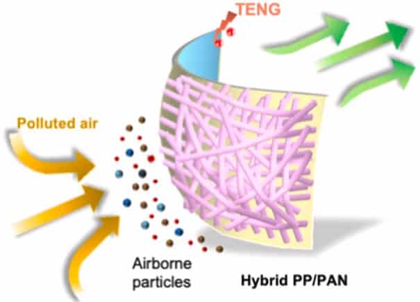
More effective: diagram of the mask design showing the polymer fibres and the TENG. (Courtesy: LingyunWang et al./Nano Energy)
A new design for a self-powered air filter mask with enhanced removal efficiency for nano and microparticles and an extended lifetime of at least two days of continuous service has been unveiled by researchers in China.
Filter masks play an important role in our wellbeing as the presence of airborne particulate pollutants is widespread and has a negative impact on human health. Most recently, the COVID-19 pandemic has shown us the importance of using face masks as protective equipment, to prevent the respiration of hazardous particles in the environment. However, current disposable masks are unable to remove extremely small particles effectively. Another shortcoming is that they can only be used for a few hours, after which, the efficiency of filtration decreases.
In this latest research, Lingyun Wang and colleagues at City University of Hong Kong and the Chinese University of Hong Kong have discovered that the inclusion of polymeric fibres with a small diameter within the range of nanometres are the key to enhancing a mask’s ability to eliminate suspended particles. They have created a hybrid air filter using the fibres that has a porous structure, with varied pore sizes to increase the amount of filtered material.
Charge separation
However, the capture efficiency of these fibres alone is still relatively low. This is because filtration relies on the passive accumulation of particles due to the physical impact of the particles on the fibres and Brownian motion. To boost the efficiency, Wang and colleagues took advantage of the fact the polymers have a marked separation of positive and negative charges due to their large dipole moment, which can attract more particles through electrostatic interactions.
To boost the long-range attraction of the filters to negatively charged and dipolar particles in air, the team applied an external electrical field to the hybrid air filter using a triboelectric nanogenerator (TENG). Today’s N95 masks exploit a similar strategy, using charged polypropylene to attract airborne particles. However, N95 masks can only achieve this attraction for a few hours, after which their ability to attract particles decreases. By actively applying a voltage using triboelectricity, the team enhanced these electrostatic forces and increased lifespan of their filter masks to up to two days.
The researchers also harnessed the TENG to give the filter antiseptic properties by incorporating silver nanowires with the fibres. Silver has well-known antibacterial properties because it releases ions that damage bacterial proteins. This effect can be enhanced using an electric field and this allowed the team to achieve a 35% sterilization rate.
Low-cost and green energy
The TENG provides low-cost and green energy, which is harvested from the movement between two materials that generate opposite charges by contact friction. One of the materials gains electrons while the other loses them, generating an electrical current that is used to power the filter masks. Therefore, a mask can be self-powered by the wearer’s movement only, without the need for batteries or additional energy sources.
In the new device, the team connected the polymer fibres to the TENG via an electrically conductive substrate. The hybrid filtering material was tested using a custom-made set-up that produced a continuous air flow while monitoring the numbers of particles in air before and after the contact with the filtration masks.
The researchers found that the filters can operate for two days under a continuous airflow, while maintaining a high removal efficiency even for small particles. They now hope to incorporate their technology into practical lightweight facemasks that are charged using human motion.
The findings are reported in a paper in Nano Energy.
Chemical reactions are complex. Even if only a few molecules are involved, the final configuration will depend on a huge number of parameters – including, in principle, all the possible locations each electron in each atom can occupy as the reaction takes place. Calculating these trajectories is beyond the power of today’s best computers, but researchers led by Kang-Kuen Ni of Harvard University in the US have now demonstrated an alternative. By cooling molecules of potassium and rubidium down to a fraction of a degree above absolute zero, they reduced the number of possible reaction outcomes to just 57. They then probed all these outcomes down to the level of individual quantum states, paving the way for a better understanding of chemistry.
When molecules are cooled close to 0 K, they enter their absolute quantum mechanical ground state, with the lowest possible levels of electronic, vibrational and rotational energy. Additionally, their kinetic energy, which governs their movement through space, becomes vanishingly small. Ultracold molecules are therefore an ideal test bed for validating quantum-dynamical models of reactions.
Ultracold KRb reactions
In their study, which is published in Nature, Ni and her colleagues focused on the reaction that occurs when ultracold potassium-rubidium (KRb) molecules exchange atoms to form K2 and Rb2. This reaction releases energy (it is exoergic), but the energy released is not sufficient to excite vibrational motion in the product molecules. This means that all the energy must instead go into rotational and translational motion (recoil energy).
Under these conditions, a quantum statistical model that includes conservation of energy conservation and angular momentum predicts that the number of possible joint quantum state pairs in the product molecules is relatively small, at 57. However, measuring these states is still no easy task and requires special techniques.
The researchers detect the rotational states of their K2 and Rb2 molecules by ionizing them with a laser pulse. The ionization process is selective, occurring only when the laser frequency matches the gap in the molecules’ energy levels, which is different for each rotational state. The ionized molecules then fly through a series of circular electrodes (all integrated into the reaction chamber), at which point electric fields propel them towards a position-sensitive detector. Once the researchers mapped how often a particular rotational state originated from the reaction, they changed the laser frequency to match the energy levels of the next rotational state and repeated the process until the survey was complete.
Ni and colleagues also needed to determine whether the molecules they detected were produced in the same reaction event. For this, they applied the principle of conservation of linear momentum. When two KRb molecules of similar momenta collide, the resulting K2 and Rb2 molecules fly away from each other with equal but opposite momenta. This means the product molecules arrive at the detector at different locations, but their arrival can be correlated. This correlation enabled the researchers to distinguish reaction products from molecules that originated in other processes.
Quantum-dynamical model
In a series of experiments (most of which were run remotely to comply with COVID restrictions), the Harvard team measured the scattering probabilities for all 57 allowed rotational state-pairs of the products. They found that while 50 of these probabilities matched theoretical predictions, the remaining seven did not. In one of these non-matching outcomes, the reaction took place close to the threshold of the exoergic energy, where most of the energy couples only to rotational motion. This leaves very little recoil energy for the reaction products to fly apart and get detected, meaning that the lack of theoretical agreement could be an artefact of the team’s measurement system. The other deviations, however, have no straightforward explanation, and contradict the prediction that all product states should have an equal probability of forming.
“For many physical chemists it’s been a long-term dream to be able to follow a chemical reaction from start to finish at the quantum state resolved level,” Ni says. “In this work, we achieved that.” Ni notes that the deviations they observed “may not be understood for another 10-20 years, until perhaps a quantum computer solves the quantum reaction dynamics calculations”. However, she concludes, “the data is there for future benchmarking of theory for when that becomes available”.
“A significant achievement”
Balakrishnan Naduvalath, a physical chemist at the University of Nevada in Las Vegas, US, who was not involved in the study, says that precisely mapping the quantum states of reaction products is “a significant achievement that no other research group has accomplished for a bimolecular chemical reaction in the ultracold regime”. The experiment, he adds, “provides much deeper insights into chemistry and at the same time challenges theory. The availability of experimental results surely would motivate theorists to develop new methodologies (and benchmark them) to study this class of four-atom reactions that might also benefit other areas of chemistry.”
As a next step, members of the Harvard team plan to put their system to work on molecule-to-atom reactions, which are less complex, and for which the available theoretical predictions are more accurate. They also hope to leverage such reactions to generate quantum entanglement resources.
Researchers in Italy have developed organic detectors that can quantitatively and reliably measure proton radiation dose, both in real time and integration mode. The organic devices, based on semiconductor thin films, demonstrated direct detection of 5 MeV protons. The team suggest that this new class of material has the potential to create flexible, portable and tissue-equivalent proton detectors for use in applications such as proton therapy.
Organic semiconductors have been demonstrated previously to be reliable detectors of ionizing radiation; but the multi-institutional team, led by Beatrice Fraboni of the University of Bologna, believes that this is the first study to evaluate the detector’s responsivity to proton beams.
Organic detection devices have unique advantageous features for developing flexible, large-area, direct proton dosimeters, according to the researchers. Organic semiconductors can be deposited from solution using low-cost techniques that are easily scalable onto large areas. Low-temperature fabrication processes (below 180°C) allow for fabrication of flexible devices onto plastic substrates. The devices operate at very low bias (less than 1 V), are portable and wearable. Finally, their composition and density make them human-tissue equivalent in terms of proton absorption. Thus they can be employed as medical dosimeters without requiring complex calibration procedures.
The detectors fabricated by the team have a photoconductor structure in which the active semiconducting layer is an organic thin film of microcrystalline TIPGe-Pn. This 150 nm film is deposited from solution onto two interdigitated gold electrodes on a plastic substrate, which ensures the mechanical flexibility of the system.
The researchers tested the detector’s response to proton irradiation, both in real time and in integration mode, reporting their findings in Science Advances. They irradiated the detectors using a 5 MeV proton beam from the 3 MV Tandetron accelerator at the LABEC ion beam centre in Firenze.
The best sensitivity obtained by the detectors was 5.15±0.13 pC/Gy, with a calculated limit of detection of down to 30±6 cGy/s. The sensors demonstrated a stable and reproducible response to proton beams with fluences between 3.5 x10⁹ and 8.7×10¹¹ protons/cm2, and maintained a linear response up to a total dose of 28 kGy.
The researchers note that while the energy of therapeutic beams is commonly above 70 MeV, the proton energy tested in this work is similar to end-of-range values, in particular the energies of scattered protons reaching internal tissues surrounding the target. The detector could therefore be employed to monitor dose to healthy tissues during treatments, such as the dose delivered to the rectal wall during proton therapy of prostate cancer, for example. The organic sensors could fit into a rectal balloon to help ensure the safety of surrounding healthy tissues.
Another potential application is as a medical dosimeter to measure radiation absorbed by astronauts during long-duration space missions.
“Our work demonstrates the possibility to operate simultaneously the same sensor in real-time mode and in integration mode, exploiting the interface coupling of the organic semiconductor with the plastic substrate,” first author Ilaria Fratelli tells Physics World. “While the energy released by the proton beam in the organic semiconductor is registered by an instantaneous increase of current, the energy released in the plastic substrate generates an accumulation of trapped charges, which induces an increase of the device conductivity proportional to the total radiation dose absorbed by the system.”
The team is now planning further tests in proton therapy centres to investigate the effect of higher energy proton beams. “We are also continuing our research through two parallel pathways,” says Fratelli. “We are studying the fundamental mechanism of interaction between protons and the materials forming the device, to reach full control and optimization of the sensing system. And we are working on the geometry and architecture of the device, on the interface effects that rule the integrative response of the detector, and on the semiconducting material employed as the real-time active layer.”
Physics is more stimulating than ever and the world increasingly depends on it. Yet, sadly, many women still do not feel inclined to pursue the subject. My own motivation for studying physics at university was my inquisitive nature: I simply enjoy delving into mathematics to understand how the world works. However, studying a subject like physics, where men dominate, can be intimidating. Sometimes I feel overwhelmed and question whether I even belong.
I have experienced biases during my time in physics, such as being told that I am too “girly” to be a physics student. Gender bias can also have a big impact on women when it comes to looking for jobs. Women who want to pursue a research career in physics, for example, must publish more papers in top-tier journals than men just to be accepted for the same job position. The gender gap can leave aspiring female physicists feeling inadequate and isolated.
Yet this is a problem not exclusive to physics: women in other sciences go through similar issues. We as individuals feel as though we are carrying the hopes, aspirations and responsibilities for all women in our field. Hiding from the spotlight or “blending in” can only make matters worse. It just means that examples of female success, such as the African American female mathematicians and computer scientists who worked at NASA in the 1970s, end up being ignored or forgotten.
While studies have revealed no difference in the capabilities of girls at physics – they perform as well if not better in exams than boys – physics stands out as the second most popular A-level subject for boys in the UK but only the 18th most popular for girls. This means that science, technology, engineering and mathematics (STEM) subjects at university and occupations in these fields are dominated by men. Between 2017 and 2018, for example, women accounted for just 35% of STEM graduates from UK universities, while, according to the UK Higher Education Statistics Agency, the yearly increase in women studying core STEM subjects is only around 1000 students. Despite the many recent efforts to inspire women to pursue STEM subjects, it seems that no significant change has occurred. So, will there ever be an even division between males and females in science or is the divide firmly engraved in us from ancestral societal roles?
Changing perceptions
Stereotypes – whether of gender, race or culture – will always exist. They emerge from a fundamental human desire to use our cognitive skills to classify information. We might know nothing about a person, but we subconsciously make assumptions about them based on those stereotypes. Women, for example, are perceived as affectionate and caring towards others while men are considered to be assertive and wanting to dominate. The problem with these generalizations is that they result in two key variations between male and female roles in society. First, women are more likely to hold low-authority positions and men are more likely to hold high-level positions. Second, women are therefore more likely to be homemakers and men are more likely to be employed in a paid business.
When it comes to physics, we portray physicists as hard-working, clever, socially inept…and male. However, there is a point when such stereotypes morph into discrimination, such as gender bias. Many wrongly correlate girls’ loss of interest in mathematics and physics to a fear of being “unfeminine” or to them being unable to cope with the perceived difficulty of the subject. Those assumptions then underscore even more firmly traditional beliefs of men’s competitive instincts, durability and motivation to master hard topics. The net result is a subconscious barrier to physics in young girls’ minds.
I believe that changing the public’s perception of what a physicist is will be key to redefining the cultural barriers between physics and wider society. It will take time, but the focus needs to be on children, who are scientists by nature, in that they show a curiosity for how the world works. Indeed, it has been shown that girls only begin to develop negative opinions about science once they reach the age of 10, especially when they become aware of female societal roles. Children gather their perspective on gender “norms” from people around them and increasingly from the media and social media too – so the more they see diversity in the appearance of scientists the better. Sadly, some secondary-school physics textbooks do not include a single female physicist.
Women pursuing a career in any STEM field should be looked after. Finding other women who have been through similar challenges will help to remind us that we are not alone in doubting ourselves in STEM. For now, we need to see these thoughts as a compromise for the reward of an accomplished life. Only in time can we change the perception of what it is to be a physicist, but that change will come.
A ground-breaking R&D collaboration between clinical physicists at MedAustron and their industry partner IBA Dosimetry, a German supplier of independent QA solutions and services to radiation oncology clinics, is rewriting the rulebook on patient-specific QA for proton therapy. A case study in clinical translation, the partnership is focused on practical implementation of myQA iON, IBA Dosimetry’s patient QA dose-verification software, yielding operational insights and technical innovations that will enable proton therapy clinics to increase their workflow efficiency while simultaneously enhancing patient safety and treatment outcomes.
From a commercial perspective, IBA Dosimetry is positioning myQA iON as a “game-changer” in patient QA – a software-as-a-service solution that supports the planning, delivery and management of proton therapy while ensuring interoperability with the proton treatment systems of all leading radiotherapy equipment manufacturers. As such, myQA iON gives physicists and dosimetrists the flexibility to combine Monte Carlo dose recalculation, QA based on irradiation log files, plus real-world detector measurements within a unified, automated and web-based software verification system that enables users to access their QA on-campus or remotely from any device that connects to the hospital network.
A division of labour
Over the past 18 months, MedAustron, a cancer treatment centre specializing in proton and carbon-ion therapy and related research, has emerged as one of IBA Dosimetry’s flagship customer sites supporting the clinical roll-out of myQA iON. Joint activities have spanned beta testing, customer training as well as acceptance and commissioning, while subsequent physics and clinical validation by the MedAustron team enabled efficient implementation of myQA iON into the patient QA workflow. “Having the chance to collaborate with a company like IBA Dosimetry provides us with a long-term QA solution, including service and maintenance,” explains Loïc Grevillot, beam delivery and Monte Carlo simulation group leader at MedAustron.
That division of labour on QA is also driven by operational necessity, given that the MedAustron clinic is still work-in-progress. The facility currently has two clinical treatment rooms with fixed proton beam lines plus one treatment room set aside for clinical and preclinical research studies. A fourth treatment room with a proton gantry will come online next year, extending patient treatment hours across the site to full capacity. As such, measurement-based patient-specific QA (with set-up and beam time) is in direct competition for beam-time needed to support the commissioning effort at MedAustron. Beyond the commissioning phase, of course, patient-specific QA will need to be streamlined further in order to maximize the beam-time allocated for patient treatment. “That’s why MedAustron wanted to be a pioneer in Monte Carlo-based independent dose calculation,” Grevillot adds. “We’re now using myQA iON second-check calculations for a preselected subset of plans embedded in an extensive machine-based QA programme.”
So how did the MedAustron team set about integrating myQA iON into routine clinical practice? According to Grevillot’s colleague Ralf Dreindl, the first step is to identify the relevant commissioning tasks – covering beam-model aspects, CT calibration and clinical testing. “Our strategy involved moving from simple geometries in water and air phantoms to complex clinical cases in patient geometry in order to get the precise overall picture,” he explains. Other considerations include the clinical simulation settings – in terms of the trade-off between dosimetric accuracy and simulation times – as well as benchmarking the implemented gamma index for 3D dose verification. “Our clinical workflow foresees post-processing tasks that start immediately after the clinical approval of a treatment plan,” explains Dreindl. “One of these tasks is the independent dose calculation of the approved plan or beam set.”
Operationally, however, the rework of established clinical routines is always a delicate and nuanced undertaking. The MedAustron team therefore defined a two-month transition period for clinical implementation, with plan complexity being the main driver of dosimetric approval via myQA iON’s independent Monte Carlo dose calculations or the usual patient-specific QA measurements. “Since the transition period ended,” Dreindl adds, “we are now using myQA iON for all normofractionated proton treatments in the horizontal beam lines.”
Benefits realization
In-house analysis indicates significant – and immediate – efficiency gains since the MedAustron team implemented myQA iON clinically on the centre’s two horizontal proton beam lines. During the first two months of operation (starting in February 2021), Dreindl and colleagues noted an average 24% reduction in patient-specific QA measurements for single-field optimized proton beams (in which the spot positions and weights of each proton field are optimized individually, yielding uniform dose distribution over the tumour target). Multiple-field optimized beams (with highly conformal dose distributions to the target volume) are also now part of the mix for independent dose check, yielding up to a 50% reduction in patient-specific QA measurements since the beginning of April. For the near term, says Dreindl, hypofractionated treatment schedules will always be measured manually in addition to the independent dose check provided by myQA iON.
Game-changer: myQA iON combines Monte Carlo dose recalculation (shown here for a prostate case), QA based on irradiation log files, plus real-world detector measurements within a unified, automated and web-based software verification system. (Courtesy: MedAustron)
More broadly, the reduction in patient-specific QA measurements is strongly dependent on the “patient mix”. For the moment, MedAustron can only simulate horizontal proton beam lines, but there are plans to start with the vertical proton beam line in spring 2022 after commissioning is complete on the new treatment room with proton gantry. Other joint lines of enquiry with IBA Dosimetry include the implementation of irradiation log-based QA at MedAustron – to provide fraction-by-fraction monitoring and evaluation of treatment delivery accuracy – as well as the integration of the GATE-RTion/IDEAL Monte Carlo dose calculation engine (developed by MedAustron) into myQA iON to support the clinical implementation of independent dose check for carbon-ion therapy. “We cannot fully replace the patient-specific QA measurements with independent dose check,” notes Dreindl, “though the integration of log-file-based QA may be an interesting route to further reduce QA measurements beyond the current 50% threshold.”
Better together
Just 18 months after they started working together, the collaboration between MedAustron and IBA Dosimetry is going from strength to strength. “As a clinical user, it’s essential for us to have a comprehensive tool like myQA iON which has undergone a certification process,” notes Markus Stock, head of the medical physics division at MedAustron. “Working as part of a collaboration, we always have a hot-line to the team at IBA Dosimetry for assistance on installation, commissioning and the testing of new functionality.”
One thing is certain: as cancer care providers seek continuous improvements in treatment efficacy, next-generation particle therapy systems will be pushed to the limits – of physics and engineering – when it comes to targeting accuracy, dose distribution accuracy and the sparing of healthy tissue. All of which translates into evolving and increasingly complex demands on patient, machine and workflow QA. “That’s why it’s essential for industry and clinical users to work hand-in-hand to deliver user-friendly, patient-centric QA technologies that support clinical decision-making and workflow efficiency,” concludes Stock.