Do not linger after using a public toilet is the advice from researchers at Florida Atlantic University (FAU), who have done a comprehensive study of how aerosols with the potential to carry disease are created and dispersed by flushing toilets and urinals. Siddhartha Verma and colleagues studied three scenarios – toilet flushing, covered toilet flushing and urinal flushing – in a medium-sized public restroom on the FAU campus.
“After about three hours of tests involving more than 100 flushes, we found a substantial increase in the measured aerosol levels in the ambient environment with the total number of droplets generated in each flushing test ranging up to the tens of thousands,” reports Verma.
“Both the toilet and urinal generated large quantities of droplets smaller than 3 micron in size, posing a significant transmission risk if they contain infectious microorganisms. Due to their small size, these droplets can remain suspended [in air] for a long time,” he adds.
Lid makes little difference
The team detected droplets at a height of 1.5 m above a toilet or urinal (face height for many people) and found that the droplets persisted at this height for more than 20 s after the flush. Unfortunately, closing the toilet lid before flushing did not result in a significant reduction in the number of particles detected – which suggests that aerosol particles can easily escape through gaps around the seat and lid.
The observed build-up of aerosols over time suggests that the ventilation in that particular facility was not adequate. “The study suggests that incorporation of adequate ventilation in the design and operation of public spaces would help prevent aerosol accumulation in high occupancy areas such as public restrooms,” says team member Manhar Dhanak.
The team describes its work in Physics of Fluids and they have made the above video to show how aerosols build up after a flush.
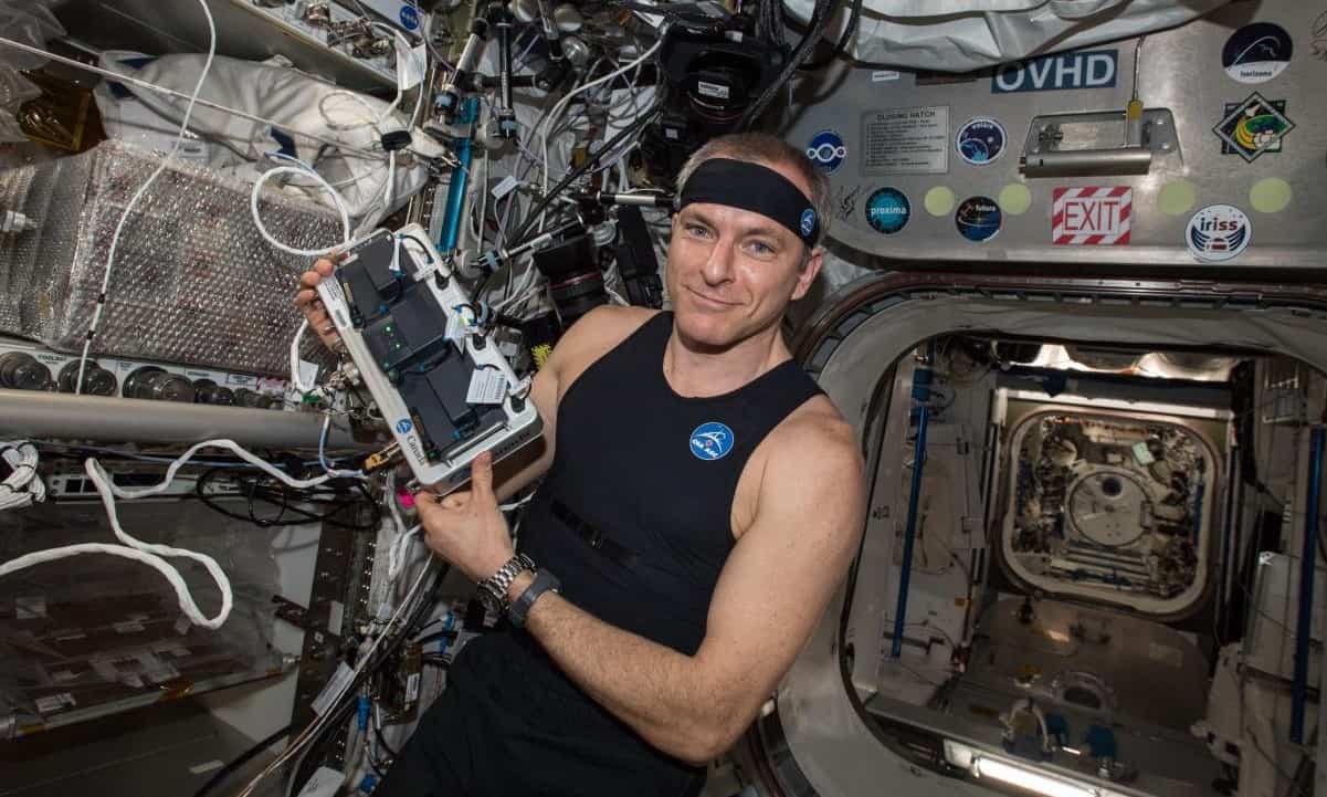
What to wear while floating around the International Space Station (ISS)? How about a “technology-packed tank top” – which the Canadian astronaut David Saint-Jacques models in this photo?
The Bio-Monitor shirt was created for the Canadian Space Agency by Montreal-based Carré Technologies, which produces wearable sensors. Saint-Jacques and fellow astronauts helped researchers at the Schlegel-University of Waterloo Research Institute for Aging evaluate the performance of the shirt in 2019 and now the results have been released.
“The Bio-Monitor shirt allows simultaneous and continuous direct measurements of heart rate, breathing rate, oxygen saturation in the blood, physical activity and skin temperature, and provides a continuous estimate of arterial systolic blood pressure,” explains Waterloo’s Carmelo Mastrandrea.
According to Mastrandrea, the shirt removes need for astronauts to stop what they are doing to make the measurements at regular intervals. It also means that the astronauts can be monitored while doing a range of activities. The crew also wore the shirts on Earth before heading into space and the researchers were able to confirm that astronauts experience a large reduction in physical activity when they are in space. And if astronauts continue to wear the shirt after they have returned to Earth, it could provide early warning of any problems they have re-adapting to gravity back on Earth.
You don’t need to be an astronaut to have a Bio-Monitor shirt – they are available to the public and can be used to monitor sporting performance and health.
Mastrandrea describes the research in a poster at the recent Experimental Biology 2021 conference.