A team of researchers from the Ecole Polytechnique Federale de Lausanne has developed a retinal implant that transposes images acquired by camera-equipped smart glasses into a simplified, black and white image made from 10,500 pixels. Although it has not been approved for human trial yet, the team has tested the implant in both a mouse model and a dedicated virtual reality programme, reporting the findings in Communications Materials.
For many patients suffering from retinitis pigmentosa – an inherited disease where progressive loss of retinal photoreceptors eventually leads to blindness – current retinal implants do not provide clear benefits. In fact, three years after surgery, most patients have stopped using them.
Two limiting parameters are often cited as the reason for the interruption: a small field vision angle (usually limited to 20°) and coarse visual resolution (less than 100 pixels in the most commonly used implant). These require the patient to constantly scan their environment to recreate a mental map of their surroundings, which is impractical and cognitively exhausting.
One electrode: one pixel
To tackle these limitations, Diego Ghezzi and his team developed POLYRETINA, a wide-field high-density epiretinal prosthesis that can be implanted at the back of the retina, close to the optic nerve. The implant contains 10,498 photovoltaic pixels (80-µm diameter, 120-µm pitch) distributed in a tiled fashion over a 13 mm-diameter active area, and provides a 43° vision angle.
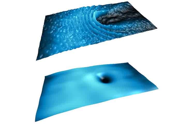
A camera embedded in the smart glasses captures images in the wearer’s field-of-vision and sends the data to a microcomputer placed in one of the glasses’ end-pieces. The data are then turned into light signals that are transmitted to the 10,498 electrodes of the retinal implants, creating a star-spangled-sky-like version of the image.
The team conducted a battery of tests to ensure that the implant was fit for purpose. Combining conjugated polymers and less rigid substrates, for example, allowed for a wider coverage of the retinal surface. However, the main question was how many electrodes the prosthesis should contain: a small number would not significantly improve resolution compared with existing implants; a large number increases risks of crosstalk with neighbouring pixels.
By firing combinations of pixels of increasing pattern complexity, the researchers confirmed that even when using 10,498 electrodes, the voltage generated by each pixel is sharply discriminated from its neighbouring pixels and does not show a voltage summation effect. This was observed even in the most extremes cases where a central pixel is off while the surrounding eighteen pixels are on.

Virtual reality while waiting for human trials
The researchers performed further experiments ex vivo on a mouse model of retinitis pigmentosa and showed that each electrode could reliably produce a dot of light in the retina.
“We wanted to make sure that two electrodes don’t stimulate the same part of the retina. So we carried out electrophysiological tests that involved recording the activity of retinal ganglion cells [a type of neuron at the inner surface of the retina]. And the results confirmed that each electrode does indeed activate a different part of the retina,” explains Ghezzi.
Currently, the team is awaiting approval to test their prosthesis in humans. Meanwhile, to continue testing the implant, they have developed a virtual reality programme that recreates what the patient would see using their prosthetic. The simulations confirmed the ability of the current setup to generate perceptible images and the implant’s readiness for clinical trials.
 Florian Stieler, PhD, is the senior medical physicist at the University Medical Center Mannheim, Department of Radiation Oncology and Medical Faculty Mannheim, University of Heidelberg, Germany.
 Manon Spaniol is a medical physicist trainee and PhD student at the University Medical Center Mannheim, Department of Radiation Oncology, University of Heidelberg, Germany.
 Eun Young Han is an associate professor of radiation oncology at the University of Texas MD Anderson Cancer Center at Houston, USA. She works as a clinical medical physicist. She earned her PhD at the University of Florida focusing on Monte Carlo-based internal dosimetry using pediatric and adult anthropometric phantoms. Today, her clinical and research interests are in the areas of end-to-end test and dosimetric comparison of brain stereotactic radiosurgery using Gamma Knife ICON and spine SBRT using MR-guided Linac.
 Jinzhong Yang is an assistant professor of radiation physics at the University of Texas MD Anderson Cancer Center, USA. He is the physics lead of the MR-Linac programme at MD Anderson. He earned his PhD in electrical engineering from Lehigh University in 2006. Dr Yang has more than 12 years of research experience in medical image registration and image segmentation, with a focus on translating novel imaging computing technologies into clinical radiation oncology practice. His current research interest focuses on MR-guided online adaptive radiotherapy.
 Jan Würfel studied physics at Karlsruhe Institute of Technology (KIT) and holds a PhD in molecular electronics. He currently works as a research scientist at PTW Freiburg with a key focus on improving detector performance and investigating new detector materials. In addition, Jan frequently serves as a speaker on a variety of dosimetry topics for the PTW Dosimetry School. His research and lecturing interests include small field dosimetry, detector physics and reference dosimetry.