An international team of researchers has used Google’s Sycamore quantum computer to power an online Zoom meeting for the first time. The US tech giant’s device, which consists of 53 programmable superconducting quantum bits, has already been shown to outperform classical computers at certain tasks. The new discovery could allow meeting participants to appear in more than one break-out room at the same time – a phenomenon that the team has dubbed “quantum Zoom advantage”.
Conventional, classical computers store and process information as bits that can have one of two states – “0” or “1”. But quantum computers like Sycamore exploit the ability of quantum particles to be in “superposition” of two or more states at the same time. N such qubits can therefore be combined or “entangled” to represent 2N values at once, allowing quantum devices to process information in parallel on a massive scale.
This unprecedented power has now been exploited for the first time in a video call when Benedetta Brassard – a quantum physicist at the the University of Waterloo in Canada – accidentally installed Zoom on Sycamore during an online meeting. Brassard is part of the International Fault Tolerant Benchmarking Team (FiT/BiT), which she set up to diversify participation in measuring the performance of quantum computers.
Distracting meme
“I was in a FiT/BiT board meeting and just thought I would have a quick check of the Sycamore dashboard to see how my quantum calculation was going,” Brassard told Physics World. But after being distracted by an amusing Shor’s algorithm meme, she somehow ported the Zoom meeting to Google’s quantum processor.
The 11 participants became encoded in Sycamore’s 53 superconducting qubits and found themselves in confusing quantum superpositions of Zoom settings. “Some colleagues were telling me that I was on mute, while others could hear me,” recalls Brassard.
“I knew something was really wrong when multiple versions of the meeting kept popping up on my screen”. Brassard now believes that Sycamore was using the “many worlds” interpretation of quantum mechanics while running Zoom. “The only way to steer it back to the classical world was to keep making measurements – which meant that I actually had to pay attention to what other people were saying”.
Is Google’s quantum supremacy not so supreme after all?

Fortunately for her fellow FiT/BiT members, Brassard had supervised a PhD student on the implementation of Instagram on D-Wave’s 2000Q quantum annealer. “The problem was to work out the optimum time of the day for influencers to post pet-related images – which we discovered is an NP hard problem,” she explained. As a result, Brassard already knew how to transform an app from a quantum to a classical state.
Brassard and colleagues have published a paper describing the Zoom incident in the journal Quantum Advances in Computing and Correlation (QUACC). They now hope to develop a quantum formalism to allow meeting participants to exist in multiple break-out rooms at once. “This could lead to the real quantum advantage of making online meetings shorter and more bearable,” she said.


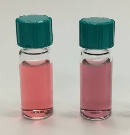
 Electrolyte development is a critical component in the quest for higher-performing energy storage devices. Ionic electrolytes such as ionic liquids, plastic crystals and their polymer composites can offer important safety and performance advantages over traditional molecular-solvent based systems, particularly for devices utilizing reactive metals such as lithium or sodium.
 Prof. Jenny Pringle works at the Institute for Frontier Materials at Deakin University, Australia. She is a chief investigator in the ARC Centre of Excellence for Electromaterials Science (ACES) and the ARC Industrial Transformation Training Centre “StorEnergy”. She received her degree and PhD at The University of Edinburgh in Scotland, UK, before moving to Monash University, Australia, in 2002. From 2008–2012 she held an ARC QEII Fellowship, investigating the use of ionic electrolytes for dye-sensitized solar cells. Pringle moved to Deakin University in 2013. There she leads research into the development of new ionic liquids and organic ionic plastic crystals for applications including thermal energy harvesting, gas separation membranes, and lithium and sodium batteries.