New techniques for imaging paediatric patients, including a hybrid imaging sequence that may make abdominal MRI exams easier for children to undergo, were among the topics presented in a virtual scientific session at the recent European Congress of Radiology (ECR 2021).
Conventional abdominal MRI exams can be challenging to perform on children. The procedure involves a series of differently weighted imaging sequences executed during repeated breath-hold manoeuvres. Because of this, the exam is complex, time-inefficient and sensitive to artefacts related to incomplete suspension of respiration.
A team at UKBB, the University Children’s Hospital of Basel, is currently evaluating a new radial volumetric encoding (RAVE) hybrid T2/T1 imaging technique that enables free-breathing abdominal MRI scans. The sequence follows a multi-parametric approach that enables the acquisition of both a T2-weighted and a T1-weighted image in a single scan.
Speaking at ECR 2021, Katja Glutig of the Universitäts Klinikum Jena described an initial feasibility study to determine whether this sequence is feasible for use in paediatric patients. She believes that the sequence will be particularly favourable for children and adolescents with cystic fibrosis.
The study included 15 patients aged between one and 19 years who had abdominal MRI scans at UKBB during 2019. Patients underwent a standard MRI exam using routine sequences on a 3T scanner, followed by an axial RAVE T2/T1 hybrid sequence at the end of the exam. The sequence, developed at New York University School of Medicine’s Center for Advanced Imaging Innovation and Research (CAI2R) in 2017, is described in detail in Magnetic Resonance in Medicine.
Two paediatric radiologists independently assessed images acquired from basic T2- and T1-weighted MR images and those of the hybrid sequence. They ranked the overall image quality, respiratory motion artefacts, clarity of portal vein wall delineation, sharpness of the hepatic margin and the quality of fat suppression.
“Our results indicate that the quality of the T2-weighted component of the RAVE-T2/T1 aspect was significantly higher than the standard T2 HASTE FS sequence in all categories except the quality of fat suppression,” Glutig reported. “Similarly, the quality of the T1 RAVE acquired aspect compared to the standard T1 DIXON sequence aspect was significantly higher for respiratory motion artefacts, clarity of portal vein wall delineation and sharpness of hepatic margin.”
“There will be several advantages to this sequence when it can be implemented in clinical practice,” Glutig tells Physics World. “It will help improve analysis of very small lesions, such as those in the kidney. The radial acquisition allows a patient to breathe freely during the measurement. In spite of free breathing, the MR images are almost free of artefacts and are of good diagnostic quality, corresponding to a standard sequence or better. This will be beneficial when imaging cystic fibrosis patients, who often suffer from constant coughing, causing motion during a scan.”
Glutig advises that data from a pilot study being conducted in Jena evaluating a RAVE hybrid sequence for cystic fibrosis patients are currently being analysed, and that the researchers hope to publish their findings later in 2021.
Decreasing the radiation dose
Speaking in the same session, Lütfiye Özlem Atay from Gazi University Faculty of Medicine described a technique to reduce radiation exposure when scanning paediatric cancer patients, who may require multiple ionizing radiation-based imaging exams during their course of treatment. Replacing PET/CT with PET/MRI, using hybrid PET/MRI scanners, can reduce radiation exposure by up to 70%. Atay and colleagues are working to reduce radiation exposure even more, by decreasing the injected radiotracer dose.

Atay presented a study suggesting that a third-dose of radiotracer can produce diagnostic-quality images in paediatric oncologic PET/MRI, with only a small relative percentage change in quantitative parameters. The study included 54 patients, aged between two and 18 years, with 12 different types of cancer. The patients underwent whole-body PET/MRI scans performed on a 3T scanner with a time-of-flight PET detector, roughly one hour after injection of 1.9 MBq/kg of 18F-FDG – half of the recommended tracer dose, and currently used as standard at Gazi University Hospital.
To investigate whether the injected tracer activity could be reduced further, the researchers used the acquired list-mode data sets from 77 PET/MRI exams to retrospectively simulate images acquired at one third (1.2 MBq/kg) and one quarter (0.9 MBq/kg) dose. They placed volumes-of-interest within organs and around FDG-avid lesions to examine the influence of dose reduction on quantification.
Atay reported that signal-to-noise ratios differed significantly among PET data sets, showing gradually increasing image noise with decreased tracer dose. However, image quality and lesion detectability were comparable, for both visual assessment and quantitative contrast-to-noise analysis.
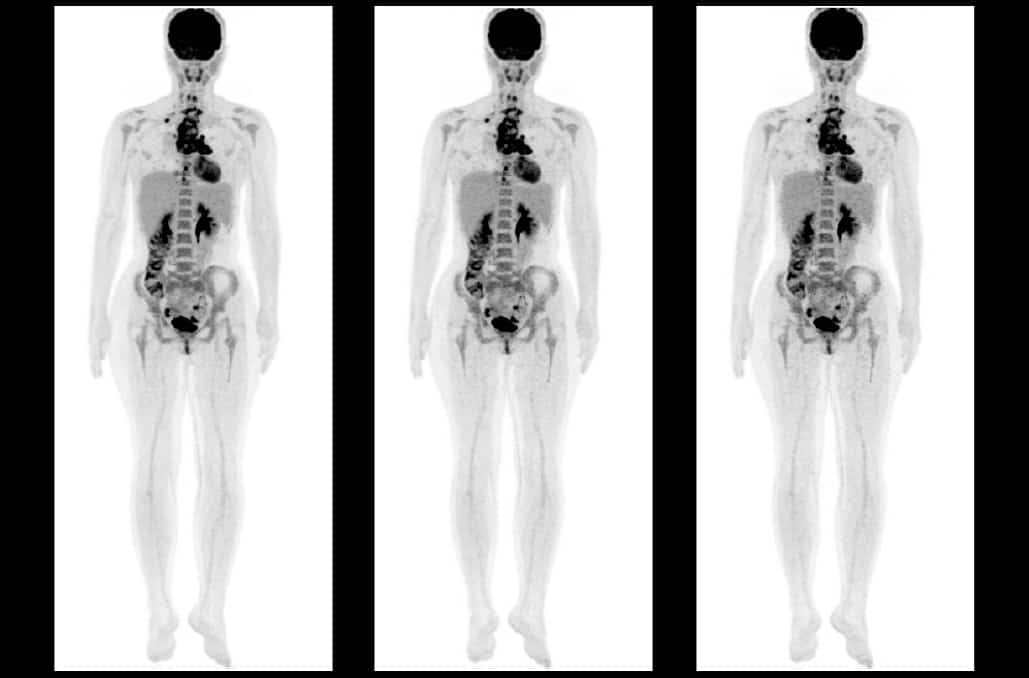
“We’ve been using half-dose tracer since September 2017, and wanted to determine the effect of even lower tracer doses,” explained Atay. “When considered with the elimination of CT-related radiation dose, using injected radiotracer activity of third dose allows a radiation dose reduction of more than 80% in PET/MRI exams compared to PET/CT. This situation provides a significant reduction in the cumulative ionizing radiation dose, especially in paediatric patients who require repeated PET imaging during treatment and follow-up.”
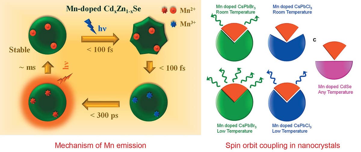
 Dr Ranjani Viswanatha is an associate professor at the Jawaharlal Nehru Centre for Advanced Scientific Research (JNCASR), Bangalore, India. She graduated from the Indian Institute of Science, Bangalore, with MS and PhD degrees. After several postdoctoral stints at the University of Arkansas, US, and Los Alamos National Lab, she joined JNCASR as an assistant professor. Dr Viswanatha’s research interests include optical, magnetic, magneto-optical, and electronic structure studies of nanocrystals with an emphasis on II-VI semiconductors and perovskite nanomaterials. Her research has been published and cited in international journals, and she holds several patents in the field. She is a member of the editorial advisory board of ChemPhotoChem. Her work has been recognized through several awards and honours.

 The QUASAR™ Multi-Purpose Body Phantom is a flexible QA tool designed to perform comprehensive testing recommended by AAPM TG 53/66/76 and IAEA TECDOC- 1583.
 Joanne Tang is an application specialist at Modus QA. Having joined Modus QA after completing her BSc and MSc in medical biophysics at the University of Western Ontario, she is currently involved in customer application support of the QUASAR™ Multi-Purpose Body Phantom.