Wearable supercapacitor stores energy using human sweat

A wearable supercapacitor that stores energy using human sweat has been developed and demonstrated in the UK by Ravinder Dahiya and colleagues at the University of Glasgow’s Bendable Electronics and Sensing Technologies Group. The team created the device by depositing a specialized polymer onto a highly absorbent cloth. Their design addresses many of the problems facing designers of flexible energy storage systems and could lead to a diverse range of new technological applications.

Creating wearable electronic technologies poses a unique set of challenges. Researchers must ensure that a high density of self-powered devices including sensors, displays, and circuits can be seamlessly integrated into flexible, durable, and comfortable materials. This is extremely difficult to do with conventional batteries, which are inflexible, prone to overheating, and rely on toxic, environmentally unfriendly electrolytes that could harm a wearer.

To address these issues Dahiya and colleagues have made a high-performance supercapacitor from easily wearable materials that used the wearer’s sweat as a biocompatible electrolyte. Like a rechargeable battery, a supercapacitor stores and releases electrical energy.

Sweat trap

The system traps sweat in highly absorbent cloth made from mixed fibres of polyester and cellulose. Each side of the cloth is coated with thin layers of the polymer PEDOT:PSS, which were doped with impurities to improve their conductivity. A solar cell provides the electrical input that causes positive and negative ions contained in sweat to be absorbed and diffused into the surfaces of each oppositely charged polymer coating – thereby storing electrical energy

Dahiya’s team tested the performance of their supercapacitor by asking volunteers to run while wearing the device as a small patch attached to their clothes. They found that the device became fully charged with just 20 μl of sweat, and generated 10 mW of power until the running stopped. This sufficient to operate a small bank of LEDs.

Bendy and washable

They showed that the device continued to work while subject to different amounts of bending. It operated after 4000 cycles of charging and discharging and did not deteriorate during washing.

The researchers also used their supercapacitor to power a cloth-based sensor of runners’ sweat salinity – offering a safe and sustainable route towards meeting the power requirements of such a system.

Since polymers bond well with textiles, Dahiya’s team envisions a wide range of applications for their device, from patient monitoring and self-health management, to improving energy efficiency in homes. They now hope to explore how sweat power could be integrated into other rapidly developing technologies including the Internet of things, augmented and virtual Reality, and robotics; potentially allowing for a better degree of connectedness between humans and technology.

The device is described in Advanced Materials.

Planned NASA space telescope renamed after astronomer Nancy Grace Roman

NASA’s troubled space telescope, WFIRST, has been renamed the Nancy Grace Roman Space Telescope in honour of NASA’s former chief of astronomy.

The telescope has had a difficult existence so far, having twice been given zero funding by the Trump administration only for the US Congress to reinstate its budget.

In March this year, NASA approved construction of the $4bn space telescope, and the naming of the Roman Space Telescope has now cemented NASA’s commitment to the project, which ranked highest for astrophysics at the last US National Academy of Science Decadal Survey.

“It’s a mark of how far the mission has come that we are being renamed,” says Julie McEnery, who is deputy project scientist for the mission. 

Astronomer Nancy Grace Roman (1925–2018) studied astronomy at Swarthmore College in Pennsylvania and in 1949 completed a PhD at the University of Chicago, where she worked at the Yerkes Observatory. She joined NASA in 1958, where she became one of the chief architects of NASA’s space telescope programme.

“She imagined the Hubble Space Telescope before it was even a concept,” says NASA associate administrator for science, Thomas Zurbuchen. “She really was one of the pioneers.”

The Roman Space Telescope is currently funded through to September 2020 with a planned launch for the mid-2020s. However, NASA has not requested funding for the telescope for 2021 as the agency seeks to devote its resources to completing the James Webb Space Telescope, which is planned for launch next year.

Far-ultraviolet light could fight the next pandemic, assessing the physics in a sci-fi blockbuster, how to detect a black hole in the solar system

In this episode of the Physics World Weekly podcast the science journalist Jon Cartwright explains how ultraviolet light at a specific wavelength could help in the fight against the next pandemic.

Physics World’s science fiction aficionado Tushna Commissariat is on hand to talk about the plausibility of the physics in the Chinese blockbuster film Wandering Earth.

We also chat about two recent proposals to use fleets of tiny spacecraft to find out if a primordial black hole is lurking in the outer solar system.

This podcast is sponsored by Teledyne Hastings Instruments.

Prompt gammas could measure body composition during particle therapy

Prompt gamma imaging enables real-time beam range measurements during particle therapy, by detecting the prompt gamma rays produced when the treatment beam interacts with atomic nuclei within the patient. Such in vivo range verification should improve the accuracy and effectiveness of proton and ion-beam therapies, which are highly sensitive to even slight targeting inaccuracies.

A recent variant of this technique, prompt gamma spectroscopy (PGS), uses time- and energy-resolved detection of prompt gamma rays for range verification. And because PGS measures the energy of the emitted prompt gammas, it can also be used to determine the elemental concentrations of irradiated tissues. This ability to measure the composition of treatment targets in vivo could enable assessment of tumour hypoxia across several particle therapy fractions, for example, or tracking of calcifications in brain metastases.

A research team headed up by Joao Seco at the German Cancer Research Center (DKFZ) has now demonstrated a technique that uses PGS to determine the elemental composition of irradiated tissues. The approach – called proton and ion beam spectroscopy (PIBS) – uses a new generation of CeBr3 scintillation detectors that measure the entire spectrum of the ion-beam-induced prompt gamma rays.

“The CeBr3 scintillation detectors have similar performance as the LaBr3 counterparts used in the prototype PGS system at Massachusetts General Hospital. Both perform very well in terms of energy and time resolution, which is key for PGS,” explains first author Paulo Martins. “LaBr3 detectors have a slightly better energy resolution. However, they are intrinsically radioactive. Therefore, for prompt gammas below 3 MeV, CeBr3 scintillators may outperform LaBr3 scintillators by about a factor 50 in noise reduction.”

Oxygen levels

Martins and collaborators, also from the Heidelberg Ion-Beam Therapy Center (HIT) and the Max Planck Institute for Nuclear Physics, used beams of protons, helium ions and carbon ions at HIT to perform PIBS. They first investigated sugar-in-water solutions with different oxygen concentrations, irradiating the samples with 90.7 MeV protons.

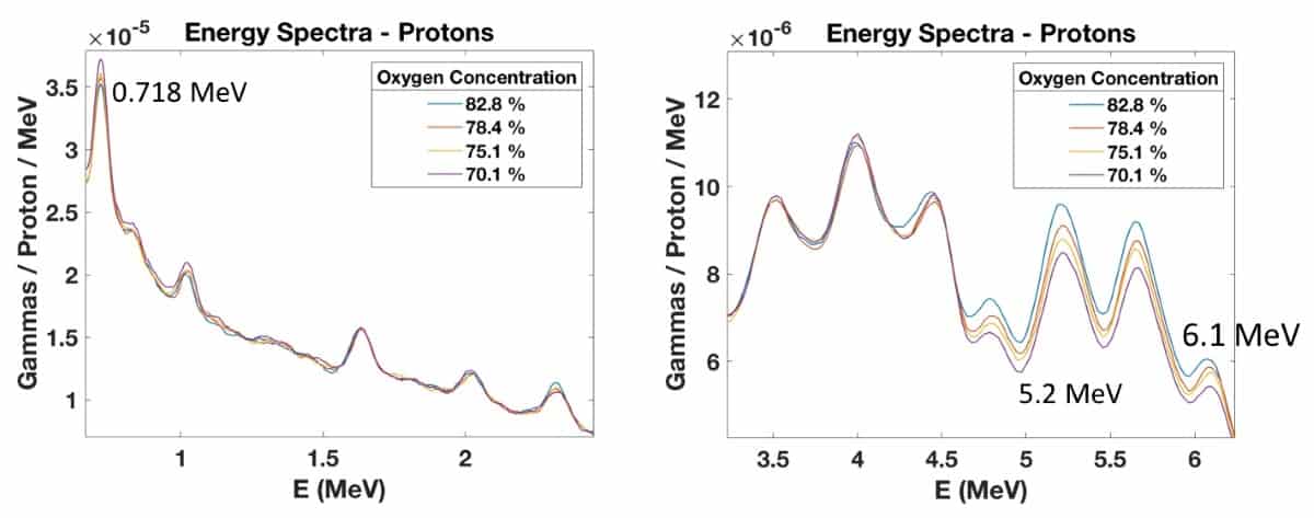
In the low-energy region, they observed prompt gamma emission at 0.718 MeV that increased with decreasing oxygen concentration or increasing carbon concentration. This energy line results from the excitation of carbon nuclei followed by prompt gamma emission. The high energy region showed peaks at 5.2 and 6.1 MeV, which increased with increasing oxygen concentration, due to the excitation of oxygen nuclei. Irradiating the samples with 92 MeV/u helium ion beams produced similar behaviour.

Energy spectra

To determine the mass of oxygen irradiated, the researchers placed EBT dosimetry films within the targets along the beam direction. They observed – as seen in previous studies – that the total prompt gammas detected within the 5.2 MeV energy peak increased linearly with the mass of oxygen irradiated.

To confirm the ability of PGS to measure oxygen concentrations deeper in water phantoms, they also irradiated five water/sugar samples with 113.6 MeV protons through 7 cm of water (two water flasks). They observed a clear relationship between increased oxygen concentration and prompt gamma production at 5.2 and 6.1 MeV. The PIBS setup could detect 3% changes in oxygen concentration in the samples.

Tissue surrogates

Next, the team irradiated six water/sugar samples and 12 tissue surrogate inserts with 88.1 MeV/u helium ion and 161.5 MeV/u carbon ion beams. The tissue surrogates contained varying concentrations of oxygen, carbon and calcium, and included five types of bone surrogate with increased calcium concentration, as well as breast, solid-water, muscle and liver surrogates with very low calcium concentrations.

The data from water/sugar samples with higher oxygen concentrations (67.1–88.9%) and tissue surrogate inserts with lower oxygen concentrations (14.9–36.5%) exhibited a logarithmic trend between prompt gamma production at 5.2 MeV and oxygen concentration.

Prompt gamma production

The researchers also evaluated whether this relationship held true for other elements, such as calcium. In the low-energy window, irradiating the tissue surrogates with 88.1 MeV/u helium ion beams generated prompt gamma peaks at 1.66 MeV, resulting from calcium reactions. Again, they observed a clear relationship between calcium concentration and prompt gamma production.

The team reported that PIBS could clearly identify calcium concentration changes of 1% between adipose and breast surrogates, and 2% oxygen variations between the various tissue surrogates.

“Conversely to PGS, the aim of PIBS is not range verification, but rather determination of the physical and chemical properties of the irradiated targets,” Martins tells Physics World. “This feature is, however, linked to the range in the patient as the body composition will ultimately affect the location where the particles will stop, thus influencing the spatial resolution. By combining PGS with PIBS, other modalities such as CT or PET may be suppressed in future treatment planning, with strong impact on treatment workflows.”

Martins says that future studies will involve in vivo measurements in mice and benchmarking against other state-of-the-art techniques, such as dual-energy CT, PET, nuclear magnetic resonance spectroscopy and dual-energy X-ray absorptiometry. “Such outcomes may be the key to monitor tumour hypoxia in the course of therapy and detect calcifications,” he adds.

The research is published in Scientific Reports.

Retinal imaging goes high resolution

Age-related macular degeneration (AMD) affects 26% of Europeans over the age of 60, but because its early signs are not easily detected, many patients only learn that they have it after their vision has already deteriorated. A team of researchers at the Ecole Polytechnique Fédérale de Lausanne (EPFL) in Switzerland has now invented a technique that makes it possible to observe the retinal cells involved in the disease in vivo. These observations, which take just a few seconds to perform, could help clinicians diagnose AMD earlier and bring about better follow-up treatment for patients, says team leader Christophe Moser.

According to the World Health Organization, 80% of the vision impairment linked to AMD could be avoided if the condition were diagnosed and treated early enough, notes study lead author Timothé Laforest. Unfortunately, standard ophthalmic exams based on visual acuity tests – the familiar wall charts – are not accurate enough. Even advanced imaging tools like optical coherence tomography (OCT) cannot provide a detailed view of the retina, which is a complex tissue composed of many layers, including photoreceptor segments, the retinal pigment epithelium (RPE) and the nerve fibre layer.

Imaging individual retinal cells within this complex tissue is difficult. Eye-motion artefacts; ocular aberrations (which reduce the lateral resolution); and the fact that retinal cells are transparent (and so have low contrast compared to surrounding cells) all contribute to the challenge. OCT, in particular, is limited because it involves sending a beam of light through the pupil. Most of this light is either absorbed or reflected at the interface between photoreceptor segments, meaning that the comparatively weak signal of light backscattered from the RPE – the layer researchers would like to study in AMD – gets overwhelmed. “The result is that these techniques cannot provided a detailed view of the minute changes occurring in the early stages of AMD,” Laforest says. “Patients are thus diagnosed when significant damage to the retina has already occurred.”

Radically different method

The EPFL team developed a radically different technique for imaging retinal cells with high contrast and high resolution. Rather than sending light through the pupil, the new technique, which they have dubbed transscleral optical phase imaging (TOPI), illuminates the white part of the eye (the sclera). This tactic greatly increases the contrast of many retinal structures, as team member Mathieu Künzi explains.

One reason for the increased contrast is that although photoreceptor cells strongly reflect light that enters through the centre of the pupil, their reflectivity reduces sharply for light entering via the pupil’s edge. This angular-dependent reflection of the retina is known as the optical Stiles-Crawford effect, and it ensures that very little high-angle transscleral light couples to the photoreceptor cells – meaning that a large fraction of this light instead reaches the crucial RPE layer.

Another advantage is that the angle of the light in transscleral illumination is much larger than would be possible if the light were sent in through the pupil. This high angle means that different retinal layers are not excited in a uniform way, enhancing the contrast of these transparent structures.

A third advantage stems from the fact that the team image the reflected light from the retina using a conventional transpupillary advanced optics (AO) camera system. This system corrects eye aberrations in real time, enabling them to obtain cellular-level resolution images. Importantly, it also avoids collecting any directly-backscattered light because such light does not overlap with the light collection path passing through the pupil. Instead, only light that is multiply scattered by the different retina layers enters the optical system and reaches the camera.

Crucial information for detecting AMD’s first signs

“By imaging the RPE layer in this way, we can extract morphological parameters such as cell density and area,” explains Moser. “According to ophthalmologists, this information is crucial for detecting the first signs of AMD, monitoring patients and investigating the efficacy of new treatments.”

The researchers tested their technique on ex vivo retinas of humans, rats and pigs before obtaining approval from the Canton de Vaud Ethical Commission to apply it to 11 healthy human subjects. The results of these last-stage proof-of-concept experiments show that TOPI can quantify retinal cells from the RPE depth down to the nerve fibre layer in just 19 seconds. “Being so fast makes TOPI compliant with routine ophthalmologic examinations in hospitals or doctors’ surgeries,” Laforest and Künzi say.

The researchers will soon begin a larger-scale clinical trial at the Jules Gonin Eye Hospital in Lausanne. “The goal of this first pilot study, which will involve 100 people, is to image healthy volunteers of different ages,” Laforest tells Physics World. “We will also image AMD patients to qualitatively and quantitatively describe how RPE cells evolve in the process of normal aging as opposed to how they degenerate in AMD.”

The TOPI technique is detailed in Nature Photonics.

Colourful bilayer paint can cool buildings by reflecting sunlight

A bilayer paint that can dramatically cool sunlit surfaces yet comes in a range of colours has been developed by scientists in the US and China. The researchers say that the solar-scattering underlayer of their coating can cool surfaces in strong sunlight by almost 16 °C, compared with conventional paint. Such coatings could potentially be applied to buildings, cars and even textiles to increase radiative cooling in the summer, the team claims.

According to the US Energy Information Administration, around 15% of household electricity usage in the US is for cooling indoor spaces. As well as being energy intensive, cooling generally requires the use of coolants that can damage the environment. As a result, there is a lot of interest in developing low energy, eco-friendly alternative approaches to cooling buildings.

One area that has had a lot of attention is using surfaces that are very good at reflecting sunlight. “If you can enhance the reflectance of the wall or roof you can reduce the solar heating, and reduce the electricity consumption,” explains Yuan Yang, a materials scientist at Columbia University in New York City.

A glaring problem

Achieving this can be as simple as painting surfaces white or covering them in mirrors or highly reflective metallic surfaces. Researchers have also looked at other man-made and natural photonic structures that are highly reflective. The problem with these ideas is that they reflect all or most of the sunlight, making the buildings glaringly bright. And they offer limited options for adding colour to buildings.

To tackle these issues, Yang and his colleagues focused on reflecting infrared light in the 0.74–2.5 μm wavelength range (near-to-short wavelengths). This light accounts for about half of all solar energy, so reflecting this part of the spectrum reduces solar heating considerably. To achieve this, the researchers developed a coating that consists of two layers. The outer layer is commercial paint that provides colour and the inner layer reflects near-to-short infrared wavelengths to reduce solar heating.

Blue, red, yellow and black

In tests, blue, red, yellow and black hues of the paint reflected radiation more effectively than commercial, single layer paints of the same colour. Under a midday summer Sun, the black version kept an object 15.6 °C cooler than a standard black paint.

Yang told Physics World that while the overall effect on the internal temperatures of a building depends on many factors, such as building size and the ratio of wall to windows, the coating can reduce solar heating on a wall by around 10–20%.

While the outer layer absorbs visible light to produce the desired colour, the inner layer of the coating is made of a porous polymer that scatters and reflects near-to-short infrared light that passes through the outer layer. The researchers say that this strategy allows them to create coatings with near-identical colours and visible reflectance to standard commercial paints, but with significantly higher reflectance of infrared light.

“Our strategy is to enhance the reflectance in the infrared light so we can keep the same colour and also reflect more sunlight, to keep the building cool,” Yang explains. He adds that the pore size of the bottom layer is optimized to be in the range of infrared light, which makes it highly effective at reflecting those wavelengths.

Different shades

In addition to using different colour paints, the researchers found that by changing the thickness of the top layer of the coating they could achieve different shades of the same colour. For example, a thin top layer of Sudan blue produced a whitish blue, while a thick coat created a darker blue. They also demonstrated the stability and durability of the coating, with no change in the colour or reflectance of the bilayer after being placed outdoors or in an oven at 60 °C for 30 days.

According to the researchers, the bilayer design increases the versatility of the coating, making it suitable for many applications. The top layer can be changed to achieve specific attributes – such as weather proofing, while the infrared reflectance of the bottom layer is maintained. This could be particularly useful for the automotive industry, where a range of photonic designs and surface finishes are required, they add.

And they even claim that fibres can be dipped and coloured with the coating to produce fabrics and clothes with high solar reflectance, to help keep you cool in the summer.

The new bilayer is described in Science Advances.

How do high radiotherapy dose rates affect implanted cardiac devices?

When a patient with a pacemaker or other type of cardiac implantable electronic device (CIED) requires radiotherapy, the treatment plan is designed to avoid direct irradiation of the device, to prevent malfunctioning or even permanent damage. However, temporary direct irradiation of a CIED can occur unintentionally due to sudden patient movement during treatment.

The impact of radiation dose rates of 8 Gy/min or less on the functioning of CIEDs has been thoroughly investigated in prior research. But stereotactic radiotherapy treatments of tumours in the head-and-neck, cervical spine and lung can deliver higher dose rates – to targets that are in close proximity to CIEDs. As such, researchers from Japan have tested the impact of higher dose rates, of up to 24 Gy/min, on four implantable devices. They found that all malfunctioned temporarily when exposed to dose rates greater than 8 Gy/min.

Implantable cardiac pacemakers continuously sense the intervals of cardiac rhythm. They emit electrical pulses to prompt the normal heart rate, or pace, when programmed limits of intervals are exceeded. Another type of CIED, implantable cardioverter-defibrillators (ICDs), also monitor cardiac rhythm, but deliver a high-voltage shock to the heart when needed to stop the abnormal rhythm.

Both of these CIEDs have a battery-powered pulse generator and encased electronics consisting of a semiconductor element and a control circuit. When the control circuit is exposed to radiation, it can cause overcurrent, recognized as false cardiac activity. This may then cause pacing inhibition in patients with pacemakers, or inappropriate shock in patients with defibrillators, causing palpitations, loss of consciousness and/or dizziness.

Lead author Kazuhiko Nakamura, of the Aichi Medical University Hospital, and colleagues tested two pacemakers and two cardiac resynchronization therapy devices. Each was placed on a 20-cm stack of tissue-equivalent phantoms on a treatment couch top, in the centre of a radiation field. Each device was irradiated at dose rates of between 4 and 14 Gy/min with a 6 MV flattening filter-free (FFF) beam, and between 4 and 24 Gy/min with a 10 MV FFF beam. During each irradiation, the team evaluated CIED function using an electrocardiogram.

Transient malfunctions caused by CIED irradiation have different effects depending upon the patient’s CIED dependency. The researchers performed pacing inhibition tests to determine the effects of transient malfunctions in patients with high CIED dependency. They report that for 6 MV beams, pacing inhibition occurred during irradiation in three of four devices at dose rates of 4–12 Gy/min, and in all four at 14 Gy/min. For 10 MV beams, dose rates of 8–24 Gy/min impacted all four CIEDs, and 4 Gy/min affected all but one.

For low CIED dependency patients, the team evaluated the occurrence of asynchronous pacing, the presence of pacing pulses unrelated to those of the device. With 6 MV irradiation, they observed asynchronous pacing in one of the CIEDs at dose rates of 4–8 Gy/min, and in all four at 10 Gy/min or greater. For 10 MV beams, two CIEDS exhibited asynchronous pacing with dose rates of 4–24 Gy/min.

Each CIED malfunctioned under different conditions, and the authors caution that other CIED models might be affected differently. All malfunctions in the four CIEDs tested were transient and reversible, returning to normal function when the irradiation was stopped. No device had permanent damage, and none had significant malfunctions requiring a power-on reset or loss of pacing, even at dose rates of 24 Gy/min.

The authors believe that CIED malfunction has a low impact on patients, and that the risk of irradiation is low because patients’ positions are constantly monitored during radiotherapy, enabling irradiation to be halted immediately if abnormal movement is detected. They caution, however, that clinicians administering radiotherapy should be aware of the risk of malfunction from direct exposure of the CIEDs to X-ray dose rates greater than 8 Gy/min, and should closely monitor and manage patient movement.

This study is described in the Journal of Radiation Research.

Cotton yarn flexes its muscles

Materials that expand and contract in response to changes in their environment have attracted considerable interest as building blocks for actuators or “artificial muscles” in miniaturized medical devices, robotics and smart textiles. Usually, these artificial muscle fibres contain exotic, non-textile materials such as graphene, carbon nanotubes or shape-memory alloys. Now, however, a team of researchers in China has succeeded in making them from ordinary cotton using a twisting and plying technique. The cotton muscles contract when exposed to water moisture, and the degree to which they twist and unwind compares well to muscles made from more complex materials.

Until now, most such actuators have been based on electrochemically-stimulated carbon-nanotube yarns, polymers that change shape when an electric field is applied, or shape-memory materials such as metals or polymers that exist in two phases and can therefore suddenly contract and expand at a given temperature. Researchers led by Zunfeng Liu of Nankai University in Tianjin had previously explored silk as a natural alternative, making moisture-sensitive artificial muscles and smart textiles from the protein-rich fibre of commercial silkworms. But since silk makes up less than 0.20% of the global textile market, they decided to investigate more widely-used natural fibres as well.

A logical choice

In many ways, cotton was a logical choice. It is widely available, accounting for nearly 40% of the world’s textile production in 2018. Cotton yarn is mechanically strong, with excellent hygroscopic (moisture-absorbing) properties, and its composition – mainly cellulose – makes it biocompatible with the human body. It is pleasant to wear, especially when the weather is warm and humid. It also softens when it absorbs moisture, is alkaline-resistant and tolerates a wide range of temperatures. All these properties make it a good candidate for crafting moisture-responsive smart textiles.

cotton muscle

Liu and colleagues started out with ordinary shop-bought cotton from a commercial 17-ply 35-tex yarn. To make their muscle, they hung a load at the bottom of a strand of yarn and twisted it from the top using a motor (at a twist speed of 50 turns/minute). They then folded the twisted yarn in the middle and hung a load at this middle point to form a two-plied structure.

When exposed to water moisture, the cotton yarn expands in volume and untwists rapidly, providing 42.55°/mm of rotation at speeds of up to 720 rpm (as measured with a high-speed camera). Removing the water moisture reverses this process.

Smart window made of cotton

To demonstrate the practical nature of their technique, Liu’s team designed a smart window from their yarn that spontaneously closes when it is wet and opens when it dries. The researchers say they would like to explore other real-world textile technologies applications for their cotton yarn muscle by improving its durability and sensitivity to moisture. “We are also looking into developing a technique to mass produce the muscle,” Liu tells Physics World.

The work is detailed in Chin. Phys. B, which is published by the Institute of Physics Publishing (IOPP).

From Post-it Notes to microwaves – why serendipity lies at the heart of innovation

Writing in this column two years ago, I quoted Geoffrey Nicholson – the father of the Post-it Note. While working at the US adhesives giant 3M, he is alleged to have said that “research is the transformation of money into knowledge; innovation is the transformation of knowledge into money”. But don’t imagine that innovation is a planned, formulaic process. It’s not a case of pouring the funds in and waiting for new technology and innovative products to pop out.

The invention of the Post-it Note, for example, was entirely accidental. Spencer Silver, a scientist at 3M, had been studying strong adhesives when he came across one that was seemingly useless. As he later recalled, it “stuck lightly to surfaces but didn’t bond tightly to them”. Silver initially had no idea what to do with his discovery, but years later another 3M scientist, Art Fry, suggested creating a bookmark that could stick to paper without damaging it.

Thanks to Nicholson’s vision, that bookmark eventually became the Post-it Note and is now a classic example of an invention or new technology looking for an application. Another is penicillin, which Alexander Fleming discovered in 1928 after leaving out some old bacterial cultures and noticing a few weeks later that some mold had killed the bacteria.

There are so many examples of serendipitous inventions that I wonder if the only way to discover things is by accident

Then there’s vulcanized rubber. The American chemist Charles Goodyear had been trying to create a weatherproof rubber for years, but succeeded in 1839 only when he accidentally dropped some regular rubber mixed with sulphur onto a hot stove and found that it still maintained its structure.

In fact, there are so many examples of serendipitous inventions – take your pick from superglue, Coca Cola, Teflon, champagne and chewing gum – that I wonder if the only way to discover things is by accident.

The X-factor

The first accidental physics-based discovery that I can think of occurred on 8 November 1895 when the physicist Wilhelm Conrad Röntgen was experimenting in his laboratory in Würzburg, Germany. Studying a vacuum tube covered in cardboard, he suddenly noticed a mysterious glow emanating from a chemically coated screen nearby.

After playing around some more, Röntgen discovered that when he put his wife’s hand between the glow and the screen, he was able to see her bones. This observation led to the world’s first X-ray photographs, with Röntgen going on to win the inaugural Nobel Prize for Physics in 1901. His finding revolutionized diagnostic medicine.

Perhaps my favourite example involved Percy Le Baron Spencer. Employed as a physicist at Raytheon in the US, in 1945 he was studying the high-powered microwaves emitted by an active radar set when he noticed that a chocolate bar in his pocket had melted.

Seeking to verify his accidental discovery, Spencer created a high-density electromagnetic field by feeding the microwave power from the magnetron into a metal box from which it had no way to escape.

When he placed popcorn in the box and fired up the magnetron, Spencer discovered that the temperature rose rapidly and the corn popped. On 8 October 1945 Raytheon filed a US patent application for Spencer’s microwave cooking process, and an oven that heated food using microwave energy from a magnetron was soon placed in a Boston restaurant for testing.

When he placed popcorn in the box and fired up the magnetron, Spencer discovered that the corn popped

By 1947 Raytheon had built the “Radarange” – the world’s first commercially available microwave oven. Almost 1.8 m tall and weighing 340 kg, this colossus cost about $5000 (roughly $57,000 in today’s money). It consumed 3 kW of power, about three times as much as today’s microwave ovens, and was water-cooled. Unsurprisingly, it wasn’t an overnight success and only sold into niche commercial applications where the speed of cooking mattered. One early example was installed in the galley of the first nuclear-powered merchant ship the NS Savannah.

Things didn’t progress much until the late 1970s when Japanese companies such as Sharp Corporation figured out how to make microwave ovens small and cheap enough for people to use at home. The market boomed and I can remember my own family buying one of these early cookers in the 1980s. My father thought it was amazing (in fact, I suspect he bought it himself) but my mother wasn’t impressed. Still, what was wonderful was you could bake a potato in it in under four minutes.

Do try this at home

All the discoveries I’ve mentioned occurred more by accident than design, but don’t assume that scientific discoveries automatically lead to technological innovation and overnight command sucess. Bringing real, commercial products to market takes significant time, effort and know-how.

Nevertheless, I hope we don’t end up in a risk-free world where ad-hoc experimentation is stifled by worries over process or by overzealous health and safety concerns. After all, who knows what we might be missing out on.

Realizing that many of you may still be self-isolating or stuck indoors by the time you read this article, let me finish by giving you a couple of great (and safe) physics experiments you can do with your own microwave oven. One is to cut a grape nearly in half and place it inside, with the cut side facing up. Turn the oven on for 10 seconds and, boom, you’ll create some fantastic plasma balls. The other is to use your microwave to measure the speed of light with a chocolate bar. I’ll leave it to you to figure out exactly how.

In fact, perhaps the current period of enforced isolation will get the entire physics community thinking great thoughts and making amazing discoveries. After all, remember what happened when the plague forced Isaac Newton to flee Cambridge for the family home in Lincolnshire – he only went and devised the universal law of gravitation. So who knows what fantastic new technologies will emerge from the current lockdown in years to come?

Hydrogen flames spread by breaking into fractal-like patterns

Hydrogen flame fronts can spread efficiently in adverse combustion conditions, researchers in Spain and Germany have shown. The team, led by Mario Sánchez-Sanz at the Carlos III University of Madrid, made the discovery by observing how hydrogen flames can efficiently access new fuel by breaking into fractal-like patterns. Their results provide important new information for those designing storage cells for hydrogen fuel.

Hydrogen is rapidly emerging as a desirable alternative to fossil fuels because it simply creates water when burned. However, the safe storage of hydrogen also poses significant challenges. Storage systems are prone to leakage and the gas is extremely difficult to detect in industrial settings – creating an ever-present danger of explosion.

Currently, it is widely believed that these dangers can be avoid by storing hydrogen in confined spaces, and in low-concentration mixtures with air. The idea being that these poor combustion conditions should quench any flames before they can reach a large proportion of the gas.

Mind the gap

Sánchez-Sanz’s team investigated this storage scenario through a setup involving two transparent vertical plates, separated by a narrow gap. After filling this gap with a low-concentration mixture of hydrogen in air, they ignited the gas from either the top or the bottom. Then they tracked the path of the resulting flame fronts by imaging the trails of condensed water they left behind.

For gaps less than 6 mm wide, and with hydrogen concentrations at around 5%, these flames did not quench as the team had expected, but broke into smaller propagating cells that were separated by cold, unburnt gas. Two distinct propagation modes were revealed in their observations. In one the flame fronts split into fractal-like patterns resembling the leaves of a fern. The movements of the fronts resembled the spread of starving colonies of fungi and bacteria as they search for nutrients. In the second propagation mode, the fronts broke into a smaller number of stable flame fronts that moved in straight, steady trajectories like the smouldering patterns that occur during the combustion of thin, solid materials.

Through computer simulations, Sánchez-Sanz and colleagues determined that each of these patterns emerge due to the high diffusivity of hydrogen, combined with extreme heat loss at the two plates. They also repeated their experiments with two far less diffusive hydrocarbon fuels, in which flames were quickly quenched by heat loss.

The research is described in Physical Review Letters.

Copyright © 2025 by IOP Publishing Ltd and individual contributors