The effect of rapid rotation on a single quantum spin in a piece of diamond has been measured for the first time. Alexander Wood of the University of Melbourne and colleagues rotated the diamond at 200,000 rpm and used laser light and microwaves to measure the effect on the spin. The technique could be further developed to measure rotation on the nanoscale, say the researchers.
Diamonds contain impurities called nitrogen-vacancy (NV) centres that comprise a single electron-like spin that is very well isolated from the surrounding environment. The spin can be measured and manipulated using light and microwaves. As a result, NV centres have proven to be very useful in a wide range of applications from quantum sensors to quantum-information storage.
Spin echoes
Spin is quantized intrinsic angular momentum, and this means that an NV spin should be affected if the diamond is rotated. To study this effect, Wood and colleagues used a technique called optically detected spin-echo magnetic resonance – a technique that has been developed to detect and image small numbers of electron or nuclear spins in samples.
In the experiment, a piece of diamond is mounted onto a rotating cylinder and a magnetic field is applied along the axis of rotation of the spin. The measurement technique involves first putting the NV spin into a lower energy state by firing a laser pulse at the diamond. Then the diamond is subjected to a series of microwave pulses, which rotate the direction of the NV spin. Finally, the energy state of the NV spin is read-out by observing the fluorescent light that it emits.
The team found that the probability that the NV spin ended-up in a higher-energy state depended upon the angle between the diamond’s axis of rotation and the polarization of the applied microwave signal. This is just as predicted by theory and the team say that the technique could be developed as a way of “probing rapid rotation and motion on quantum-relevant timescales”. Potential applications include sensitive torque detectors and studies of the fundamentals of quantum mechanics.
Delivery as planned: the ClearView 3D Dosimeter enables clinical users to visualize and verify the detail of complex SRS dose distributions. (Courtesy: Modus QA)
The growing clinical application of stereotactic radiosurgery (SRS) for the treatment of metastatic tumours in the brain presents a significant dosimetric and quality assurance (QA) challenge for medical physicists and their clinical colleagues. Put simply, the precision targeting inherent to SRS necessitates all manner of patient, machine and process-level checks to verify that conformal, high-dose radiation is delivered to the patient as intended – usually in one or a few fractions – while minimizing damage to surrounding healthy tissue and organs.
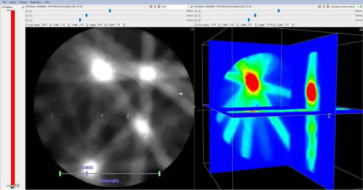
With this in mind, Modus QA, a Canadian supplier of QA products and services to radiation oncology clinics, is stepping up the commercial roll-out of its ClearView 3D Dosimeter, a non-diffusing, radiochromic hydrogel dosimeter designed specifically to support advanced radiotherapy techniques like SRS. Working in tandem with the vendor’s VISTA optical CT scanner, the dosimeter helps users to confirm that planned treatments are delivered accurately, visualizing the intricate detail of complex dose distributions for multiple-lesion dosimetry.
John Miller: “The goal is an integrated 3D dosimetry system that gives users more accurate measurements of dose and dose distribution.” (Courtesy: Modus QA)
“Gel dosimetry can be used to measure any 3D dose distribution,” explains John Miller, founder and co-owner of Modus QA, “though the most important application we see is for SRS treatment of metastatic tumours in the brain – a truly complex problem with multifoci targets distributed in 3D space.” Fundamentally, ClearView is a tool that enables medical physicists to meaningfully compare 3D dose distributions – measured versus calculated – and get them to a pass or fail ahead of SRS treatment. “It’s a necessary and sufficient test,” adds Miller.
Joined-up thinking
If that’s the back-story, what of the specifics? The ClearView 3D Dosimeter itself comprises an optically clear, low-scattering and colourless hydrogel matrix suffused with a radiochromic indicator dye. The dye turns purple after irradiation, with the change in optical attenuation throughout the gel directly proportional to the absorbed radiation dose.
Spatially, while the radiochromic dose response of the ClearView gel is accurate at the submicron scale, the resolution of the dose image is ultimately determined by the spatial accuracy of the optical CT scanner used for readout. In this case, the latest iteration of the Modus optical CT scanner (VISTA 16) is capable of imaging 0.25 mm isotropic voxel sizes, though in practice the more commonly used spatial resolution of 0.5 mm will be down-sampled to match the treatment plan.
Also worth noting is the joined-up approach that Modus has taken to ClearView and VISTA product development. VISTA, for example, is built with a convergent green light source (530 ± 10 nm) to minimize the scatter associated with cone-beam optical CT, while ClearView has been optimized for use with VISTA by increasing the clarity and reducing the scatter of the hydrogel. “The goal is an integrated 3D dosimetry system,” Miller explains. “The combination of the two products ultimately gives users more accurate measurements of dose and dose distribution.”
Into the workflow
When it comes to deploying ClearView and VISTA into the SRS QA workflow, Miller reckons an experienced user will need about 60 minutes to measure a dose distribution and compare it to the calculated dose distribution. The stepwise process includes acquisition of an optical CT scan of the gel before irradiation (the reference scan); irradiation of the gel with the planned treatment dose; a 45-minute wait for the gel chemistry to develop; followed by another optical CT scan (the data scan). Each scan takes less than five minutes, while the CT reconstruction is automatic.
“It’s the comparisons at this point that are crucial,” says Miller. Users can do a gamma distribution to see a gamma map in 3D; also a conformity index between planned isodose surfaces and measured isodose surfaces. In a multimet treatment, meanwhile, users are able to look at the centre of mass of each target and compare with the plan to ensure the centre of each target is hit with the centre of each dose distribution. “You can then look at contours around that to make sure the shape of the dose distribution is correct,” he adds.
The ClearView roadmap
Looking ahead, the priorities for 2020 are already nailed down for the Modus product development team. With the ClearView roll-out under way to new clinical customers, the emphasis now shifts to commercial release – slated for the autumn – of the work-in-progress VistaACE analysis software. “The software will effectively complete the product launch,” notes Miller, “enabling medical physics teams to use ClearView, VISTA and VistaACE on site as an integrated solution for 3D gel dosimetry.”
Beyond that, intriguing possibilities are coming into view on the ClearView development roadmap. A case in point is a concept called 3D dosimetry as a service (3DDaaS). “There’s no timeline for the launch of 3DDaaS as yet,” Miller admits, “but the idea is that this will be a remote accreditation service for QA of clinical procedures – for example, to support the commissioning of new radiotherapy equipment or new treatment techniques in the clinic.”
Right now, Modus is focused on accelerating clinical uptake and validation of ClearView beyond its current “early-adopter” institutions, which include the London Regional Cancer Program in Ontario and the University of Michigan. A key opportunity to engage with end-users directly will come in June at the International Conference on 3D and Advanced Dosimetry in Quebec City.
“Watch this space,” Miller concludes. “We’ll be there with at least the work-in-progress VistaACE.”
Using the ClearView 3D Dosimeter
Selling the benefits of gel dosimetry to the medical physics community is all about openness, claims John Miller of Modus QA. “Transparency is key,” he explains. “We’ve tested ClearView rigorously and are being upfront and open with all our specifications – acknowledging that radiochromic gels have their limitations with respect to linearity, range and energy and dose-rate effects.”
Benefits of the Modus QA ClearView 3D Dosimeter include:
Stability: chemistry is stable for more than 60 days prior to irradiation under recommended storage conditions. The signal is geometrically stable any time after irradiation (though for best dosimetric results, Modus recommends optical scanning between 45 min and 24 hr after irradiation).
Linearity: dose response is linear within the range 0 to 80 Gy, making ClearView suitable for SRS QA.
Spatial resolution: the gel is accurate to the submicron level.
Material properties: near-tissue equivalence provides a patient-like testing environment, while low-scatter substrate is ideal for optical cone-beam CT scanning.
A full list of ClearView specifications is available here.
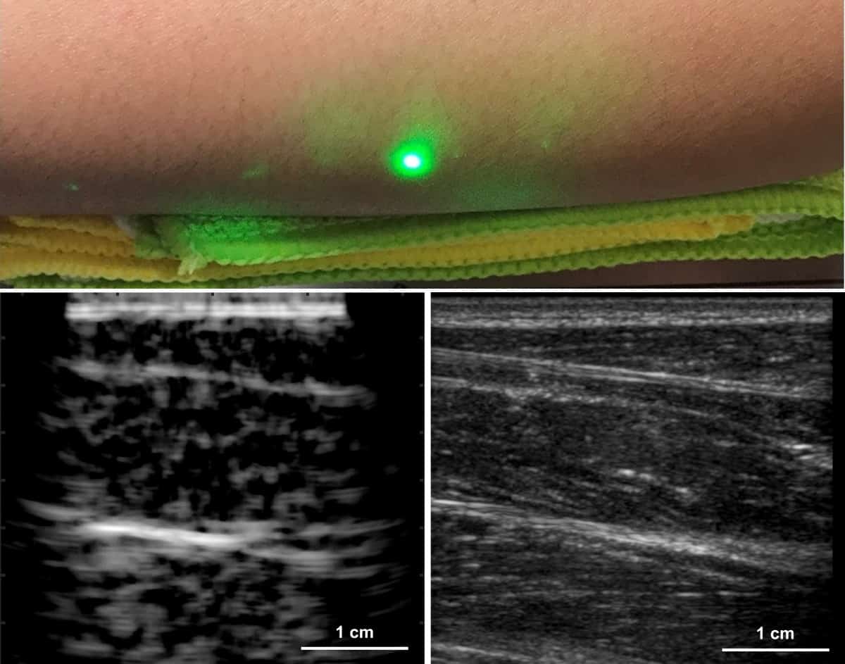
The new laser ultrasound technique was used to produce an image (left) of a human forearm (above), which was also imaged using conventional ultrasound (right). (Courtesy: Xiang Zhang et al)
Fully contact-free laser ultrasound (LUS) imaging has been demonstrated in humans by researchers at Massachusetts Institute of Technology (MIT), in collaboration with MIT Lincoln Laboratory. Xiang Zhang and colleagues used an infrared laser to generate sound waves at the tissue surface of volunteers’ forearms. A second beam detected the propagating sound waves by measuring how the subjects’ skin vibrated in response. The technique could be especially useful for imaging where physical contact is not tolerated, such as over wounds and on other sensitive areas (Light Sci. Appl. 10.1038/s41377-019-0229-8).
In conventional ultrasound imaging, an array of transducers is pressed against the skin directly or with a coupling gel to help transmit the acoustic waves into the tissue. The method is inexpensive, convenient and produces images in real time, but it has some disadvantages.
One significant limitation is the pressure that is typically required to maintain acoustic contact between the device and the target tissue. This limitation is exacerbated for contact-sensitive applications where such pressure would be too painful, such as for burn victims or trauma patients.
Another problem is the low degree of reproducibility, due to the fact that the operator usually defines the image orientation and field-of-view by manipulating the transducers manually. This means that patient images acquired at different times are difficult to compare, and treatment or disease progression cannot easily be tracked.
A variation on the technique – ultrasound tomography – addresses the issue of reproducibility, but the solution comes at the expense of convenience, as it involves part of the patient being immersed in a water tank. The problem of physical contact, meanwhile, is partly dealt with by photoacoustic imaging. In this method, the ultrasound pulses are generated within the tissue remotely by a laser, but the reflected signal is detected using conventional transducers on the skin.
LUS could tackle these problems simultaneously by combining a new approach to photoacoustic generation with an optical interferometer, allowing it to create and measure ultrasound waves from a distance. Conventional photoacoustic imaging cannot image deeply since the light is strongly attenuated in the tissue. Rather than seeking a way to increase the laser’s penetration, however, in their new approach Zhang and colleagues turned this bug into a feature.
“We actually rely on this high absorption to efficiently generate an acoustic source at the tissue surface, meaning we can convert the maximum amount of light into acoustic energy while maintaining human safety,” explains Zhang. “This allows us to image deeper than typical photoacoustics since we don’t rely on light to travel through the tissue; only acoustic waves instead.”
A simplified schematic of the laser ultrasound system. (Courtesy: Xiang Zhang et al)
The team found that they achieved the ideal balance of optical absorption, acoustic power and patient safety using 2 mm-wide, nanosecond laser pulses at wavelengths near 1500 nm. This setup approximates a disk-shaped transducer just beneath the tissue surface, producing a 60° ultrasound beam at 1.5 MHz – towards the lower end of the frequency range typically used for ultrasound imaging.
The researchers tested their LUS technique using a gelatin phantom, ex vivo pig tissue and four human subjects, comparing the results to those from a standard ultrasound imager. While LUS could not match the image quality provided by the conventional approach, it still successfully picked out the same soft- and hard-tissue features.
One aspect in which LUS is currently lacking is its inability to deliver results in real time, as images must be reconstructed from sequential single-point measurements. In this respect, Zhang expects the development of the technique to mirror that of conventional ultrasound imaging.
“Looking back historically, medical ultrasound began by sequentially moving a single transducer to form an image – similar to moving a single laser spot in LUS – and eventually scaled toward arrays of hundreds or even thousands of transducers in medical probes today. I believe a similar path is ahead for LUS,” says Zhang.
Progress along this path should be accelerated by a fortunate coincidence: as well as being ideal for LUS, 1500 nm is the wavelength favoured by the telecommunications industry, meaning that both new and mature optical technologies are readily available for translation. Even in its current state of development, however, the technique could find immediate applications where high-quality images are not strictly required, Zhang suggests.
“For now, LUS could be useful in binary measurements where features don’t necessarily need to be resolved at a high resolution; rather, a yes/no measurement is sufficient, possibly for detection of internal bleeding or fractures in painful areas,” he tells Physics World.
Physics World’s Laser at 60 coverage is supported by HÜBNER Photonics, a leading supplier of high performance laser products which meet the ever increasing opportunities for lasers in science and industry. Visit hubner-photonics.com to find out more.
A spectrometer that directly detects the vibrational “fingerprint” of molecules offers a sensitive new way of deducing a material’s chemical make-up. The device, which was developed by researchers in Germany, Saudi Arabia and Hungary, can sense the presence of substances at much lower concentrations than is possible with state-of-the-art commercial infrared spectrometers. It can also measure the spectra of samples in water, which is impractical with conventional absorption spectrometry because water itself is such a strong absorber of infrared light. The new method is thus particularly attractive for applications in biology and medical diagnostics.
When a material is irradiated with infrared light, its constituent atoms and molecules absorb energy at frequencies that depend on their chemical structure. A few picoseconds (10-12 s) later, this absorbed energy dissipates as vibrations. Conventional infrared spectroscopy focuses on the absorption step. By analysing the light transmitted through the material, researchers determine which frequencies are absorbed, and thus which chemical species are present. The drawback is that some molecules absorb infrared light better than others, and even strongly-absorbing substances may not produce detectable dips in the transmitted light if they are only present in small amounts.
Electro-optical sampling
The new spectrometer avoids these pitfalls by focusing instead on the dissipation step. The researchers, led by Ioachim Pupeza and Marinus Huber of the Ludwig Maximilians University (LMU) and the Max Planck Institute of Quantum Optics in Garching, Germany, begin by irradiating their samples with an ultrashort pulse of infrared light. Because this initial excitation pulse lasts only a few femtoseconds (10-15 s), it delivers its energy to the sample within two oscillations of the light field. After this pulse is past, but – crucially – before the target molecules stop vibrating, the researchers apply a second ultrashort pulse of light, this time in the near-infrared region of the spectrum. This “gating” pulse carves out a slice of the electromagnetic radiation given off by the vibrating molecules – a technique known as electro-optical sampling. The researchers then analyse this vibrational signal to determine the sample’s molecular fingerprint.
A key advantage of this method, Pupeza explains, is that it separates the molecular signal from various sources of background radiation – including the initial excitation pulse. “The fact that we can carve out very brief portions of our wave means that we can exclude the excitation and only look at the molecular response,” he tells Physics World. Pupeza adds that the technique is also coherent, meaning it is only sensitive to light that is in phase with the excitation, and not to random thermal vibrations.
The researchers tested their method, which they term field-resolved infrared spectroscopy (FRS), on several biological samples. In measurements of human blood serum, they detected changes as small as 500 ng/mL in the molecular concentration of certain chemicals – 40 times lower than is possible with a commercial infrared spectrometer. The researchers also obtained infrared spectra of live human cells in suspension and an intact willow leaf. Both materials would be hard to analyse with absorption spectroscopy because they absorb nearly all incident light.
The researchers, who report their work in Nature, hope to extend FRS in future experiments. “We would like to cover the entire infrared molecular fingerprint region to capture as many resonances as possible from complex samples,” Pupeza says. Another possibility, he adds, would be to combine FRS with a frequency comb, which could provide enough spectral resolution to analyse gaseous materials as well as solids and liquids.
Cup-to-cup consistency is a problem for people using espresso machines, according to Jamie Foster who is a mathematician at the University of Portsmouth. One cup can taste lovely and the next not so nice – even when the machine is operated by a skilled barista.
In a quest for a consistently good cup of coffee, Foster and an international team of researchers have created a model that describes how hot water is forced through coffee grounds in an expresso machine. By looking at the process for different grinds of coffee, they discovered that there is an optimum size for the coffee grounds. Espresso coffee normally comes in a fine grind, which maximizes the surface area of coffee that is in contact with the water. In theory, this should result in a strong cup of coffee that gets the most flavour out of the grounds.
However, Foster and colleagues’ calculations reveal that if the coffee is too fine, the water can be prevented from reaching all of the coffee – resulting in a poor cup. The model revealed an optimum size for the coffee grounds and the solution is currently being tested in a coffee shop in the US.
I was listening to the radio on the way home yesterday and there was an interesting interview with David Howard of Royal Holloway, University of London who was part of a team that reconstructed the vocal tract of a 3000-year-old Egyptian mummy. The shape of the vocal tract was determined using CT scans and it was rebuilt using a 3D printer.
The vocal tract was connected to an artificial larynx and the team was able to generate a sound a bit like the bleating of a sheep. They were unable to create more complex sounds because that would require further components such as the mummy’s tongue, which was too shrivelled to recreate.
You can read more about the mummy and listen to a clip of the mummy’s voice in this BBC article.
Staying in the ancient world, Pier Paola Petrone, a forensic anthropologist at the University of Naples Federico II, and colleagues have identified the remains of a human brain that was turned to glass during the famous eruption of the volcano Vesuvius in 79 AD. The remains were found in Herculaneum, which was buried under hot volcanic ash. The brain of the victim – who was in his 20s – was rapidly burned inside the skull and then cooled rapidly to form glass in a process called vitrification.
Medical engineers have proven that the unique internal structure of cancer cells can make them vulnerable to vibrations from specific frequencies of ultrasound. These low-intensity pulsed ultrasound waves are a potentially safe therapy to target cancers of the blood.
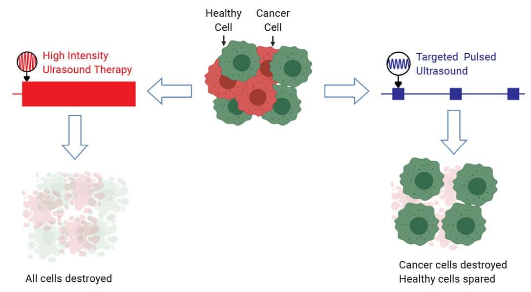
High-intensity focused ultrasound is sometimes used to destroy cancer cells deep within tissues, but the thermal energy transmitted can also damage healthy cells at the ultrasound focus. Low-intensity pulsed ultrasound (LIPUS), on the other hand, doesn’t heat up cells but is used to mechanically modulate cells, for stimulating bone repair, for example.
Computational modelling has previously shown that vibrations from LIPUS can selectively kill cancer cells. This led to the oncotripsy theory, which predicts that cells’ response to ultrasound waves depends upon cellular structure – analogous to how a building’s vulnerability to earthquake depends upon its structure. According to this theory, the altered internal structures of rapidly dividing cancer cells makes them vulnerable to certain frequencies of ultrasound. A recent collaboration between researchers at California Institute of Technology and the Beckman Research Institute supports this hypothesis (Appl. Phys. Lett. 10.1063/1.5128627).
“This project shows that ultrasound can be used to target cancer cells based on their mechanical properties,” says lead author David Mittelstein. “It’s an exciting proof-of-concept for a new kind of cancer therapy that doesn’t require the cancer to have unique molecular markers or to be located separately from healthy cells to be targeted.”
Selecting the magnitude
Mittelstein and colleagues tested LIPUS on suspensions of cancer cells (breast, colon or leukaemia) mixed together with healthy immune or red blood cells. They placed these suspensions in acoustically transparent plates, through which they applied short, 60 s, 0.5–0.67 MHz frequencies.
Targeted pulsed ultrasound exploits the unique mechanical properties of cancer cells to destroy them. (Courtesy: David Mittelstein)
The team tested varied ultrasound pulse patterns and found that 0.5 MHz with pulses 20 ms in duration were most effective. Cancer cells’ key structural protein, actin, was immediately disrupted after treatment, leading to almost complete destruction of the cancer cells.
“At certain ultrasound frequencies, low-intensity ultrasound caused the cellular skeleton of cancer cells to breakdown while nearby healthy cells were unscathed,” says Mittelstein.
Under pressure
The team found that acoustic standing waves, formed by a source wave interfering with its reflection, were present in their experiments. These types of waves have a static pattern, with areas of high and low pressure thought to be damaging within a cell. The scientists proved this by using a specially designed cuvette, which eliminated wave reflection and correspondingly cancer cell death.
Next, the researchers measured acoustic transmissions within cells and observed that standing waves rapidly amplified pressure in the cells, causing small cavity formation and collapse. This cavitation process occurred almost in sync with cell cytodisruption, and when cavitation was prevented by conducting experiments within an overpressure chamber, cancer cells survived. Although this proves that standing waves and cavitation were involved in cancer cell death, the scientists also detected cavitation in red blood cells surviving LIPUS.
“We found that standing waves and cavitation are necessary, but not sufficient, for the effect we observed,” Mittelstein explains. “So we suggest that this cell-mediated cavitation may act as a local amplifier of ultrasound. A given cell type’s response to the resulting mechanical stress depends on its biophysical properties.”
Targeted ultrasound in the clinic?
The researchers think that their ultrasound technique can be used to target blood cancer or circulating tumour cells. They also tested LIPUS targeting within solid gels, but this wasn’t as effective at killing the cancer cells. However, Mittelstein hopes that partial cancer cell death within a solid tumour could stimulate the immune system to respond and eliminate any remaining cancer.
“Our next step is to investigate the effect of combined therapy: using the benefits of this safe ultrasound technique to increase the effectiveness of chemotherapy, immunotherapy and other cancer treatments,” said Mittelstein.
About 80% of Chinese scientists who have gone abroad to study or work in the previous decade have returned home and are now publishing high-impact, collaborative research. That is according to an analysis by scientists in China, Europe and the US who found that the lure of attractive initiatives such as the Thousand Talent programmes is helping to foster China’s rise as a global scientific powerhouse.
As more researchers return home, the rise [of China] is going to continue
Caroline Wagner
The study used Elsevier’s journal database to search for authors who first published in China, then moved to another country before returning home. The team found that more than 4500 and 2300 Chinese researchers had left the US and Europe, respectively, to return to China in 2017 – roughly double the number seen in 2010 (Science and Public Policy scz056). The study also found that while more Chinese researchers moved to the US than Europe, a greater number went back to China from Europe than from the US. For every returnee from the US, about 1.4 overseas Chinese scientists stayed in the country – a ratio that was 1 to 0.9 for Europe.
Robust growth
A previous study found that between 1978 and 2007, more than 1.2 million Chinese went abroad to study or work but only about 25% returned (Asian Population Studies 4 331). In the following decade, however, according to the current study that return rate is 80%. The authors say this is partly due to the boost in research spending thanks to robust economic growth in China over the past couple of decades. “The pattern is expected, but the scope and scale of China’s rise is gigantic,” Caroline Wagner from Ohio State University who was involved with the study told Physics World. “As more researchers return home, the rise [of China] is going to continue.”
One of those who went to the US only to return is astrophysicist Lijun Gou from the National Astronomical Observatories in Beijing. Gou says he chose to return to China and start his own group via the Thousand Talent programme. “The years abroad gave me international perspectives and confidence, but I’m glad I made the choice so I can use my expertise [at home],” he says.
Fragments of fungal cells just 30 nm across have been found in the atmosphere at concentrations much higher than expected, according to experiments by researchers at the University of California, Irvine, US. These biological nanoparticles may play a role in cloud formation and could also trigger allergic reactions and asthma attacks.
Particles like soot and dust affect how clouds form by acting as “seeds” around which ice can nucleate or water can condense. Biological particles such as intact pollen and fungal spores can also make effective ice nuclei, but since these large particles are relatively uncommon in the atmosphere, scientists believe they are unlikely to play a significant role in ice-cloud formation under most conditions.
Now, however, Michael Lawler and colleagues, who are in James Smith’s group at UC Irvine, have detected smaller fungal particles being released during episodic bursts into the air over a rural area. The results, which were obtained at the Department of Energy’s Atmospheric Radiation Measurement (ARM) field site near Lamont, Oklahoma, showed that the concentrations of these fungal nanoparticles were orders of magnitude higher than large intact biological particles.
Size-selected nanoparticles
The team’s experiment involved drawing air into an instrument called a thermal desorption chemical ionization mass spectrometer. This instrument is size-selective, which meant the researchers could choose to sample only those nanoparticles measuring between 20 and 60 nm in diameter. They collected the particles onto a thin platinum filament, vaporized them by heating the filament and then detected the resulting gases.
Lawler and colleagues identified the fungal fragments by detecting chitin – the main component of fungal cell walls – and sugar alcohols such as mannitol, which the fungi use to store energy. Most of the particles they observed during their three-week measuring period were around 30 nm in size, and Lawler explains that some or all of them could make good cloud-condensation nuclei. However, he adds that more work is needed to test this hypothesis.
Allergies and asthma symptoms
During the detected fungal burst events, the concentrations of fungal nanoparticles increased from non-detectable levels (below perhaps 10 per cubic centimetre) to hundreds per cubic centimetre. Because particles smaller than 100 nm in diameter can be inhaled more deeply into the lungs, and are also more chemically active than larger particles, such bursts could have important health implications. “We know that fungal cells can cause allergies and asthma symptoms when they are released into the air,” Lawler says. “We have now essentially found fungal ‘shrapnel’ that likely contain the same irritants as larger particles, but which are more numerous.”
“We cannot unequivocally say from this study whether the fungal nanoparticles we detected could adversely affect health,” he continues, “but it seems possible that at least some of the health impacts associated with aerosol fungus could be caused by these smaller particles.”
Rainfall and the appearance of fungal nanoparticles
As for the causes of the “bursts”, the researchers speculate that they might be prompted by raindrops hitting the soil in which the fungi are growing. In their work, which they report in Science Advances, they found that particle counts increased within roughly a day of rainfall at the field site, but Lawler cautions that they know little about the exact processes involved. Unanswered questions include whether the cells burst in the air or on the surface of the soil, and whether rain or merely an increase in relative humidity controls the generation of the nanoparticles.
Future studies should shed more light on the mechanisms behind the bursts, and the researchers also hope that more measurements will help pin down the ice-nucleating potential of fungal nanoparticles. A third avenue for follow-up work would be to identify the specific fungal species involved. “This will help us define the likely health impacts of these nanoparticles, how they are generated and under which conditions,” Lawler tells Physics World.
Sweeping changes to US university-level teaching in physics and astronomy are required to address the long-running and systemic exclusion of African Americans. That is according to an American Institute of Physics (AIP) report, which finds that the percentage of bachelor’s degrees awarded to African American astronomy and physics students has fallen from 5% in the late 1990s to less than 4% now. That is despite the number of undergraduate degrees awarded in physics doubling over the past two decades.
The report is the result of a two-year investigation by the National Task Force to Elevate African American representation in Undergraduate Physics and Astronomy (TEAM-UP), which included interviews, surveys and visits to physics and astronomy departments. The report identifies five key factors affecting the success of African American students including academic and personal support as well as having a sense of belonging and identifying as a physicist or astronomer.
A systemic problem needs a systemic solution
Ed Bertschinger
The report provides recommendations to assist department, faculty, professional societies, funding agencies and donors in addressing and tackling these factors. These include fostering a greater sense of belonging for African American undergraduates, improving student services to better provide for them as well as providing recognition, rewards and other incentives to support minority scholars.
Financial strain
The task force found that many African American students are eschewing careers in astronomy and physics in favour of entering university programmes that are more supportive and financially rewarding. “A systemic problem needs a systemic solution,” says physicist Ed Bertschinger from the Massachusetts Institute of Technology, who is the task force’s co-chair. “The presence or absence of caring faculty members can make the difference between a student persisting or leaving.”
The AIP report recommends that departments more actively connect students with funding programmes and help them to find jobs that advance them academically during the time of their undergraduate study. The TEAM-UP task force aims to double the number of astronomy and physics bachelor’s degrees awarded to African Americans by 2030 and calls for physical science societies to raise $50m to support minority students experiencing financial burdens and enable implementation of the report’s recommendations.
“A lot could be done by alleviating the financial strain faced by African American students,” notes Jedidah Isler from Dartmouth College who is a TEAM-UP member. “I understand they are certainly not the only students who face financial strains, but there’s a long history of systemic discrimination against African Americans in this country that shows up in a tremendous wealth gap today.”
Artificial intelligence has been used to quickly and accurately model the 3D flow of light around arbitrarily shaped nanoparticles. Peter Wiecha and Otto Muskens at the University of Southampton in the UK demonstrated the modelling approach using a neural network that required just a single training procedure. Their technique could be used to design a wide range of optical devices that control the paths taken by light.
When light interacts with nanostructures that are smaller in size than the wavelength of the light, the result can be very different from how light interacts with larger structures and continuous media. The field of nanophotonics seeks to exploit this by designing nanoparticles with particular shapes and compositions with the aim of manipulating light in specific ways.
How light flows around such nanoparticles can be calculated using Maxwell’s equations of electromagnetism – at least in principle. In practice, however, the calculations can be very time consuming and it can take days to design and optimize complex structures.
Powerful tool
Artificial intelligence has recently emerged as a powerful tool for tackling optimization. Artificial neural networks can be taught to perform tasks through a knowledge of the basic rules underlying a system – and they have been used to approximate how spherical and H-shaped nanoparticles will interact with light. While successful, this technique can only be applied to simple, highly specific situations.
Wiecha and Muskens have taken a more generalized approach and have based their new technique on “convolutional” neural networks, which are commonly used for image analysis. The duo’s new system can quickly and accurately predict the 3D flow of light around nanoparticles with completely arbitrary shapes. A diverse variety of physical effects can be analysed with just a single training procedure – without the need to teach their neural network how to deal with numerous specific situations.
The researchers say that their approach could be applied to countless situations in nanophotonics. With further work, it could also be used for inverse design – whereby the required optical properties are input and the system designs the appropriate nanostrutures. Inverse design is currently extremely difficult to do and this capability could open a vast range of applications and areas of research that are unattainable today.
The duo’s neural network could soon enable researchers to monitor the performance of nanophotonic devices in real time, leading to more powerful physics experiments. Other applications could include computer chips with entirely optical components, nano-antennas which concentrate energy on molecular scales, and metasurfaces that can direct and control light.
Wiecha and Muskens are now aiming to improve the speed of their technique. They also hope to generalize the network even further to account for factors including multiple materials, arbitrary illumination, and larger geometries.