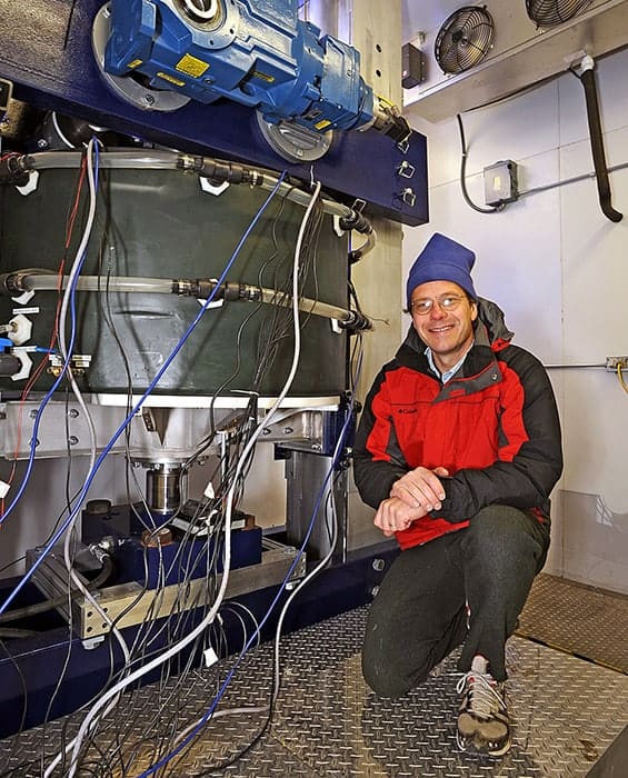
The submarine surface of Alaska’s LeConte glacier could be melting around 100 times faster than previously thought, direct observations have revealed. A US team led by David Sutherland at the University of Oregon came to this conclusion after monitoring the submerged portion of the glacier in a multibeam sonar survey. Their results highlight a pressing need to update existing models of glacial melting.
Commonly found on the coastlines of Greenland and Antarctica, tidewater glaciers form physical boundaries between oceans and continental ice sheets in high-latitude environments. They are known to be highly dynamic, with changes induced by submarine melting driving cycles of advance and retreat. The behaviour of these glaciers have a significant impact on factors including sea level, global ocean circulation, and the productivity of ecosystems.
Since the submarine interfaces of tidewater glaciers are so difficult to access, direct observations of their melting behaviours are sparse. This means that researchers predicting the impacts of climate change-driven ice losses on oceans and the atmosphere must rely on models with parameters based on limited observations.
To address this shortcoming, Sutherland’s team argues that models must be updated to include parameters such as melting rates along the vertical faces of tidewater glaciers. Such melting can form overhanging shelves of ice, which can eventually collapse under gravity to form icebergs; dramatically increasing ice loss.
Multibeam sonar surveys
In their study, Sutherland’s team carried out two week-long multibeam sonar surveys on the submerged face of the LeConte tidewater glacier in south-eastern Alaska, in August 2016 and May 2017. The 3D images they gathered revealed the complex shape of LeConte’s submerged faced to metre-scale resolutions.
By analysing time-varying changes in the glacier-ocean interface, Sutherland and colleagues discovered that significant melting occurred at the ocean’s surface in both spring and summer. However, they also observed a significant increase in melting of the deepest parts of the glacier during the summer, resulting in overhanging ice shelves at intermediate depths, which were vulnerable to collapse. They concluded that this pattern arose as sediment-laden meltwater penetrated through the glacier, emerging at its submarine face. This exiting runoff, which increased with depth, then created buoyant, turbulent plumes, which enhanced melting rates along the face of the glacier.
Although the team observed highly variable levels of melting, their results confirmed that overall, the submarine face of the LeConte glacier is melting up to two orders of magnitude faster than previous theories have predicted. With melting rates of tidewater glaciers universally predicted to accelerate due to climate change, the researchers’ insights into melting mechanisms could prove crucial in building reliable, parameterized models of its global impacts.
The “Best-in-Physics” electronic poster session at the AAPM Annual Meeting brings together the 15 studies that scored highest in the abstract review process. These studies – categorized this year as imaging, multi-disciplinary or therapy – are considered to reflect the highest scientific quality and innovation. As always, the session proved a popular attraction and drew in large crowds of delegates keen to find out more. Here is our selection from this year’s presentations.
Imaging the scatter could track tumour motion
Image guidance plays a central role in radiotherapy. With this in mind, Kevin Jones from Rush University Medical Center plans to use the photons scattered out of the megavoltage beam during treatment to visualize the irradiated anatomy. “Our hypothesis is that by collecting scattered photons with a pinhole collimator, we can identify the tumour position in real time during radiation delivery,” he explained.
Kevin Jones from Rush University Medical Center.
Jones and colleagues are developing a scatter-imaging system for tracking lung tumour motion during stereotactic body radiation therapy. He notes that one big benefit of this approach is that it doesn’t expose the patient to any additional radiation dose. It is also possible to place detectors at various positions around the patient to create a 3D image.
The prototype system comprises an off-the-shelf flat-panel X-ray detector and a pinhole collimator. The researchers optimized the collimator thickness using phantoms, and found that increasing the thickness from 7.6 to 35 mm increased the contrast-to-noise ratio by more than three times.
The team has now recorded the first scatter images collected from patients during lung SBRT. They placed the camera outside of the gantry rotation plane so it was not irradiated by the beam. Images recorded at two camera positions showed the location of the tumour. Comparison of the experimental and simulated scatter images revealed good agreement between the two at each camera position.
Jones noted that the detector – a 700 µm caesium iodide scintillator – did not collect as many photons as could a thicker detector. “The images are quite blurry, but we are optimistic that we can improve this is in the future by improving the detector and the collimator, and by moving the camera closer to the patient,” he told Physics World.
Ultimately, Jones hopes that the system will be used to track moving tumours, enabling the application of smaller treatment margins or verification of targeting accuracy. “We aim to develop an image-guided radiotherapy monitoring technique that does not require additional dose,” he said.
PEM plus ultrasound enhances biopsy targeting
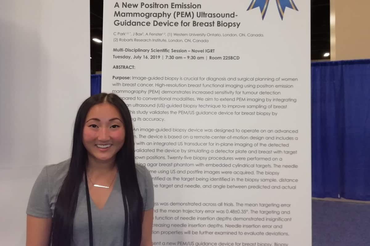
Image-guided biopsy is a vital stage in diagnosis and treatment planning of breast cancers, but conventional imaging modalities have limitations. Mammography, for example, cannot easily identify lesions in dense breast tissue. Claire Park from Western University Ontario hopes to address this shortfall by using positron emission mammography (PEM) to perform high-resolution functional breast imaging.
Claire Park from Western University Ontario.
PEM offers increased sensitivity and diagnostic accuracy for tumour detection, but as it is a functional imaging approach, it doesn’t provide an anatomical reference and biopsy workflow is not yet available. “So we are combining this functional imaging with a validated ultrasound-guided biopsy technique to improve needle guidance and tumour sampling,” she explained.
Park and colleagues are developing an ultrasound-guided biopsy device that will operate with an advanced PEM system. The idea is to align the ultrasound transducer between the PEM detector plates to enable real-time needle guidance. In phantom validation studies, the biopsy device demonstrated a mean targeting error of 0.35±0.20 mm and a mean trajectory error of 0.48±0.35°. “Under ideal conditions, it can target lesions that are less than 1 mm,” she told Physics World.
The next step will be to validate the complete robotic arm, including a 3D needle tracking system. The team also plans further software developments to visualize the PEM lesion locations and determine how to accurately guide the needle to those points.
Spectral CT sees multiple contrasts simultaneously
Spectral CT using multiple energy bins enables discrimination between different high-Z materials, such as the contrast agents used in medical imaging. By employing an advanced high-flux photon counting detector and selecting energy bins above and below the K-edges of each material, several contrasts can be imaged once, as well as structures such as bone – a feat that could not be achieved using conventional CT imaging.
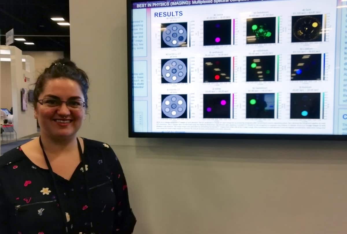
Chelsea Dunning from the University of Victoria.
Chelsea Dunning from the University of Victoria described the use of K-edge subtraction on a spectral CT system to simultaneously image a range of contrast agents in a phantom. In addition to commonly used contrasts such as iodine (I), gadolinium (Gd) and gold (Au), she decided to also examine novel elements with emerging clinical potential. These included lanthanum (La), holmium (Ho) and lutetium (Lu), which are close in K-edge energy and may be particularly difficult to distinguish.
Dunning and colleagues 3D printed phantoms containing seven vials, one filled with water plus two concentrations of each of: Gd, I and Au; Gd, La and Lu; and Gd, Ho and I. They used a spectral CT system with a cadmium zinc telluride detector to image each phantom, choosing detector energy bins to match the K-edge energies of each component.
K-edge subtraction images of each contrast agent clearly visualized all of the different materials with minimal artefacts. The detector could distinguish three contrast agents in the La-Gd-Lu phantom, each with a difference of seven in Z and close K-edge energies.
“Most remarkably, in the phantom with iodine, gadolinium and holmium, it can discriminate all of them really well; gadolinium and holmium only differ in atomic number by three,” Dunning explained. She noted that [to their knowledge], this is the first time that contrast agents with similar Z have been imaged simultaneously.
Auto-segmentation eases radiosurgery planning
Segmentation of brain metastases for stereotactic radiosurgery planning is a time-consuming process. A team at UT Southwestern Medical Centre aims to change that. “We are developing a platform for users to upload MR images and software that performs automatic segmentation,” explained Zi Yang.
Zi Yang (left) and senior author Xuejun Gu from UT Southwestern Medical Centre.
The platform imports high-resolution T1 contrast-enhanced images, and then uses a deep-learning based algorithm to carry out segmentation and labelling. At this stage, the auto-segmented brain metastases contours can be reviewed and modified by the user. The finalized contour sets are exported for use in treatment planning.
The researchers evaluated the algorithm with clinical data and found that it could perform segmentation and labelling in just 4–5 minutes, offering substantial clinical time savings and workflow improvement. Yang explained that the platform exhibits high accuracy for detection and contouring of lesions, although it currently detects some false positives. “We plan to improve the algorithm and add more functions,” she said. “Then we plan to implement the platform in the clinic.”
A new bioinspired adhesive made from a temperature-responsive hydrogel appears to speed up wound healing and so could find use in a wide range of application areas, including regenerative medicine and soft robotics. The material, which works like embryonic skin in that it draws wound edges together by contracting, has already been tested on the skin of mice with promising results.
Most treatments for skin wounds – be they cuts, blisters or burns – involve placing a barrier (such as cotton wool or gauze) over them to retain moisture and reduce exposure to infection by delivering, in some cases, antimicrobial agents.
Although researchers have developed more advanced dressings in recent years, containing, for example biologically active agents such as growth factors, these can be difficult to fabricate and are expensive. Growth factors can also have unwanted side effects.
Mimicking embryonic skin
A team led by David Mooney of the Wyss Institute for Biologically Inspired Engineering at Harvard University has now developed an active adhesive dressing (AAD) inspired by developing embryos, whose skin heals completely without producing scar tissue. It does this by producing cables made of the protein actin. These cables form at the edge of skin cells surrounding a wound and then contract, creating a mechanical force that draws the wound edges together like a purse string.
To mimic this mechanism, Mooney and colleagues have invented new wound dressings using a thermo-responsive hydrophobic polymer PNIPAm alginate hydrogel. Similar hydrogels developed recently are tough and have high adhesive energies of up to 1000 J/m2 on various tissues, including skin.
The hybrid hydrogel begins to contract at temperatures greater than 32°C. When placed on skin (which is between 35°C and 37°C), it transmits the force of contraction to the underlying tissue. The researchers also made it antimicrobial by adding silver nanoparticles (AgNPs) to it. AgNPs are already widely employed in wound care products, including commercially available alginate hydrogels and antimicrobial gauzes.
To test the antimicrobial function of their new alginate hydrogel, they analysed bacterial growth on agar plates in the presence of the material with and without AgNPs. They found that hydrogels containing AgNPs effectively inhibited bacterial growth. No AgNPs leaked from the hydrogel either, which proves that its antimicrobial function comes from the release of silver ions, not nanoparticles, from the gels.
Forming strong covalent bonds with skin
To form strong covalent bonds between skin tissue and the functional groups in the hydrogel matrix, the researchers primed the hydrogel surface with chitosan and carbodiimide coupling agents. The chitosan penetrates the skin and the hydrogel, while EDC [1-ethyl-3-(3- dimethylaminopropyl)carbodiimide] and NHS (N-hydroxysuccinimide) facilitate the formation of amide bonds between tissue proteins, chitosan, and alginate within the matrix, explains study co-first author Jianyu Li, who is currently assistant professor at McGill University in Canada.
The gel also closed wounds faster than other types of hydrogels as measured by the wound half-life – that is time required to reduce the wound area by half after application. The researchers found that the rate of wound closure with AAD compared well with that of photo-cross-linked chitosan hydrogels and microporous gel scaffolds. What is more, they say that the AAD appears to be safe for biological tissue since it does not cause inflammation or allergic reactions.
Finally, to simulate how the AAD mechanically interacts with wounded skin and to help optimize the closure process, the team also developed finite element models using the commercial software ABAQUS. “We are continuing this research with studies to learn more about how AAD performs across a range of different temperatures, as body temperature can vary at different locations,” explains team member Benjamin Freedman.
Chronic wounds could benefit
As well as skin wounds, the AAD could also be employed on chronic wounds, such as diabetic ulcers and pressure sores, and wounds in other epithelial tissues such as the intestine, lung and liver, adds Mooney. It may even be used in drug delivery and as a component of soft robotics-based therapies.
This work opens new avenues for developing wound dressings based on adhesive and stimuli-responsive hydrogels, he says. Unlike many other such materials, the AAD requires no additional reagents or sophisticated apparatus (such as UV light, for example) to work since it takes advantage of the natural temperature change when the dressing is placed on the body.
Reporting their work in Science Advances, the researchers told Physics World that they are now moving from the product development and preliminary in vivo study stages to preclinical animal trials.
Future experiments will also look at how the expression of genes known to be important in wound healing and collagen organization impact skin tissue healing over time. “It will be important to find out how the mechanical cues exerted by AAD affect the biological process of wound healing – and in particular how they affect the phenotype, migration, and activity of relevant cells such as fibroblasts,” they say.
A key aspect of Einstein’s general theory of relativity has passed its most rigorous test so far. An international team led by Tuan Do and Andrea Ghez at the University of California, Los Angeles confirmed the Einstein equivalence principle (EEP) by analysing the redshift of light from the star S0-2 at its closest approach to Sagittarius A* – the supermassive black hole at the centre of the Milky Way. The study combined over 20 years of existing spectroscopic and astrometric measurements of S0-2 with the team’s own observations.
Since Einstein first proposed his general theory of relativity in 1915, the idea has stood up to intense experimental scrutiny by explaining the behaviours of gravitational fields in the solar system, the dynamics of binary pulsars, and gravitational waves emitted by mergers of black holes.
In 2018, the GRAVITY collaboration carried out a particularly rigorous test – observing S0-2 at its closest approach to Sagittarius A* in its 16-year orbit.
As expected, the GRAVITY astronomers observed a characteristic relativistic redshift in light from S0-2. This redshift is a lengthening of the wavelength of the light and arises from both the motion of the star (the Doppler effect) and the EEP. The latter is a consequence of general relativity and predicts a redshift in light from a source that is in a gravitational field such as that of a supermassive black hole.
Further measurements
Now in a completely independent study, Do and Ghez’s team tested the EEP using three further spectroscopic instruments. These collected an additional three months of redshift data at similar times to the GRAVITY study. Their observations included S0-2’s closest approach to Sagittarius A* in May 2018, its maximum line-of-sight velocity in March, and its minimum line-of-sight velocity in September; spanning a range of 6000 km/s in radial velocity.
The researchers then combined their observations with S0-2 redshift data collected by others in 1995-2017 – which includes observations made by eight other instruments. This allowed them to test for biases between instruments, and to perform additional analysis of the systematic errors made in previous studies. Overall, their data included 45 astrometric position measurements spanning 24 years. This allowed the team to map S0-2’s orbit, and 115 redshift measurements spanning 18 years – 11 of which were the team’s own new measurements.
Just like the GRAVITY experiment, the measurements gathered by Do, Ghez and colleagues were consistent with the EEP, with statistical analysis revealing that Einstein’s theories were 43,000 times more likely to explain their observed redshifts than the Newtonian model of gravity. Therefore, while EEP violations are predicted by theories incorporating effects including quantum gravity and dark energy, general relativity still holds up for the time being. However, the researchers remain hopeful that with yet more rigorous analysis, Einstein’s theories may soon begin to break to provide a glimpse of new physics.
In this episode of the Physics World Weekly podcast we chat about new research that shows that global warming is not just another natural variation in Earth’s climate. We also meet a Russian planetary scientist who is eager for his country to return to the Moon and a physicist who co-founded a company that makes diamonds.
We also reveal whether our readers would sacrifice flying to a distant conference to reduce their carbon footprint and learn how to play quantum tic-tac-toe (or quantum noughts and crosses).
Researchers have found that electrons organize themselves into a new kind of ferromagnet in twisted bilayer graphene (TBG). In this system, which forms when two sheets of graphene are stacked on top of one another with a small twist angle between them, it is the orbital motion of electrons, rather than their spins, that aligns. Such behaviour could produce emergent topological states that might be exploited in applications such as low-power magnetic memory in the future.
Graphene is a flat crystal of carbon just one atom thick. When two sheets of the material are placed on top of each other and misaligned by rotating them relative to each other, they form a moiré pattern. Last year, researchers at the Massachusetts Institute of Technology (MIT) found that at a “magic” twist angle of 1.1°, the material becomes a superconductor (that is, it can carry currents with no losses) at 1.7 K. This effect, which occurs thanks to miniband flattening at this angle that strongly enhances interactions between electrons in the material, disappears at slightly larger or smaller angle twists.
A team of researchers led by David Goldhaber-Gordon of Stanford University has now found unambiguous evidence of ferromagnetism – as the giant anomalous Hall (AH) effect – in TBG when its flat conduction miniband is three-quarters filled.
Three-quarter filling with a difference
The crystal structure of a single layer of graphene can be described as a simple repetition of carbon atoms, which is known as its unit cell. A normal electronic band can accommodate two electrons (one of each spin) per unit cell. This miniband can accommodate four electrons (each of two spins, each of two orbital states) per moiré cell.
“Three-quarter filling would naively mean that each of the four nearly-degenerate bands (spin up, orbital 1; spin down, orbital 1; spin up, orbital 2; and spin down, orbital 2) would be three-quarters filled,” explains Goldhaber-Gordon. “But, what if the electrons organized themselves to completely fill three of these bands instead, leaving the other one empty?”
In this case, the electrons would be polarized in both spin and orbital states with the orbital polarization giving rise to the giant AH effect, he says. And this is exactly what the researchers have observed in measurements of the voltage drops in a Hall bar device made from the material. Indeed, they measured an AH effect as large as 10.4 kΩ.
“Incipient” Chern insulator
The presence of a giant AH effect in an apparent insulator is reminiscent of a ferromagnetic topological insulator approaching a Chern insulator state, says Goldhaber-Gordon. Such a state is one whose bands are all either filled or empty (as described above) and whose filled bands have a net total Berry curvature or Chern number. Such a system should have zero longitudinal conductance and quantized Hall conductance.
“We instead see a minimum in longitudinal conductance at three-quarter filling (but not zero longitudinal conductance) and a large Hall conductance (that is not fully quantized),” he tells Physics World. “This suggests the presence of a parallel conduction mechanism.”
And that is not all: the researchers also found that they can, surprisingly, reverse the magnetization of the sample by applying a small DC current. This could possibly have implications for low-power magnetic memory applications in the future given the orders-of-magnitude smaller critical current density required for flipping the magnetization compared to previous such devices.
Measurements on a Hall bar device
Goldhaber-Gordon and colleagues performed their measurements on a Hall bar device made from TBG with a target twist angle of 1.17°. They sandwiched the graphene between two hexagonal boron nitride (hBN) cladding layers to protect the graphene channel from disorder and to act as dielectrics for electrostatic gating. By then adding a silicon back gate and Ti/Au top gate, they were able to independently tune the charge density in the TBG and an electric field applied perpendicular to the graphene sheets.
They measured the longitudinal and Hall resistances using standard “lock-in” techniques with an AC bias current.
The team, reporting its work in Science, is now studying the properties of the magnetic states at three-quarter and other band fillings using a mix of techniques, including transport measurements and optical probes. Understanding the magnetic order and topological character of the correlated insulating states will be crucial to unravelling the rich phase diagram of TBG, says Goldhaber-Gordon.
Shining laser light through the skin can be used to form detailed images of tissues just below the surface, as a way to diagnose disease. High-power lasers can also be used therapeutically to disrupt diseased regions of tissue. A group at the Nello Carrara Institute of Applied Physics in Italy has developed and tested a new type of nanoparticle that could be introduced into tissue to react to the laser light more efficiently – and improve the effectiveness of these techniques (J. Biophotonics 10.1002/jbio.201900082).
One technique that the group hopes to use with the nanoparticles is photoacoustic imaging. This uses a short laser pulse to focus light into a precise region of tissue, up to a centimetre below the surface of the skin. The energy is absorbed and the region heats up a little, which causes it to vibrate and emit a small ultrasound pulse that can be picked up by a detector on the surface.
The magnitude of the emitted ultrasound is dependent upon the tissue properties, enabling different structures to be identified. Photoacoustic imaging can visualize much deeper tissues than imaging with laser light alone. This is because the light only has to travel one way through the tissue; the returning ultrasound waves are not scattered as much and can pass through the body far more easily. Nanoparticles that respond to the laser light might make photoacoustic imaging more effective.
Laser-stimulated gold nanoparticles have also previously been developed as a way to destroy cancerous cells. The nanoparticles can be coated with a compound that enables their absorption by specific cells. The particles respond to an applied laser light pulse and heat up their surroundings, killing the cells in a targeted way. Such nanoparticles have not been used clinically, however, either because of toxicity concerns or because they don’t respond well enough to wavelengths of light that penetrate deep into the body.
In their latest study, the researchers produced gold nanorods, around 75 nm long and 15 nm wide, and coated with polyethylene glycol and a peptide to reduce any negative biological effects and encourage their uptake by cells. They tuned the size of these nanorods to absorb infrared laser light at a wavelength of 1064 nm.
Previously, nanorods have only been able to absorb light of around 800 nm, which is less suited to medical applications, due to restrictions on maximum laser intensity at shorter wavelengths, and greater availability of longer wavelength lasers. These nanoparticles are also designed to respond to laser stimulation by producing bubbles, which break apart the cells, rather than destroying them through extreme heating.
The team tested their nanoparticles on isolated macrophages (white blood cells) and saw that the rate at which they were taken up was similar to previous types of nanoparticles. The nanoparticles did not appear to be toxic to the cells once absorbed, and no signs of apoptosis were observed. When the researchers applied laser pulses, the particles produced microbubbles within the cells that emitted detectable ultrasound waves. The bubbles were also able to cause cell death at laser intensity levels currently permitted under clinical guidelines.
“For clinical translation, I think that the use of nanoparticles to impart localized therapies is the most interesting, with the advantage of imaging support,” says lead author Lucia Cavigli. “Superficial cancers such as skin cancer, or cancers with easy access via an optical fibre (such as bowel cancer) would be particularly suitable for this approach.”
Further tests are needed to assess the safety of the nanorods, and how well they would function within the body. In particular, it’s important to see how long the particles remain in the body after they have been injected, to find out whether and how they are broken down and excreted.
Warming experienced since the middle of the 20th century has been truly global with a geographic consistency not seen during any other period in the Common Era, which began 2000 years ago. This is the conclusion of an international team of researchers, who used an extensive range of climate proxies to map the regional and temporal scale of temperature changes over the last two millennia. The team showed that no climate trend prior to the current warming period has affected the entire planet simultaneously, and that historical temperature anomalies once assumed to have been worldwide affected different regions at different times.
The Roman Warm Period, the Dark Ages Cold Period and the Little Ice Age are some of the better known excursions from global mean temperatures that have occurred in the last two millennia. First identified from tree-ring data and other climate proxies in the northern mid-to-high latitudes, researchers initially expected to find the anomalies reflected in paleoclimate records across the globe. When evidence did come in from further afield, the data were noisy enough, and the temporal boundaries blurred enough, for it all to be accommodated within the prevailing narrative of global-scale change.
“There’s a dominant paradigm in which [paleoclimate researchers] think, and it’s the climate epochs paradigm, in which there were globally coherent periods of cold and warm,” says Nathan Steiger of Columbia University in the US. Steiger, with Raphael Neukom of the University of Bern in Switzerland, and colleagues at Spain’s University of Murcia, MathWorks in the US and the Bjerknes Center for Climate Research Norway, tested whether this paradigm holds for the Common Era.
Using a community-sourced database of climate records spanning all of the Earth’s continents and oceans, the team reconstructed annual temperatures over a global five-degree grid for the period AD 1–2000. The lack of any strong, universal influence on climate in the pre-industrial period was immediately obvious: virtually every single year up to 1850 saw at least a tenth of the Earth’s area experiencing above-average temperatures, while at least the same fraction experienced below-average temperatures.
Strictly regional phenomena
Familiar climate epochs like the Little Ice Age only emerged from the data when multidecadal average temperatures were taken, but even then, no single trend encompassed the whole of the planet at the same time. Instead, when the researchers plotted the timing of peak warming or cooling periods for each grid cell, they found that the climate events previously thought of as global epochs were strictly regional phenomena, consistent with natural climate variability.
“That stands in stark contrast to the contemporary warm period,” says Steiger, “where it really is very much globally coherent in a way that’s totally different from the global variability that happened prior to that over the past 2000 years”.
Steiger and colleagues describe their study in Nature.
In agreement with Steiger’s assessment, a second team (the PAGES 2k Consortium — which includes Neukom) reports in Nature Geoscience the result of a parallel analysis of the same data set. This group studied the rate at which global average temperature changed over multidecadal timescales and tried to identify the factors that drove such changes.
Volcanic eruptions
Prior to industrialization, they found that global temperature changes were influenced mainly by major volcanic eruptions, with greenhouse-gas forcing contributing a relatively minor signal. Solar output variability was not detectable in the multidecadal record.
Again, for the post-industrial period the picture was very different, with rates of warming since the mid-20th century exceeding anything seen in the preceding two millennia. “We see from the instrumental data and also from our reconstruction that, in the recent past, the warming rate clearly exceeds the natural warming rates that we calculate,” says Neukom.
Unusually warm or cold periods in the climate record are often seized upon by those who seek to deny the existence or downplay the consequences of anthropogenic climate change. After all, if temperatures varied so much before industrialization, why think that recent trends are anything extraordinary?
Although neither study investigated the specific drivers of recent warming, the fact that the current period stands apart in terms of both spatial coherence and warming rate still says something about this argument. As Neukom points out, many attribution studies have been performed over the last few decades, and the evidence all suggests that there are anthropogenic causes. “We do not explicitly test this; we can only show that natural causes are not sufficient to actually cause the spatial pattern and the warming rate that we observed.”
Magnetic resonance imaging (MRI) is one of the most common techniques used to image internal body parts of animals and humans, in particular for cancer diagnosis. At a resolution of some few micrometres, conventional MRI instruments need relatively large samples to provide good images. In recent times, various research groups have sought to obtain resolutions down to atomic sizes. In the most recent breakthrough, a group of scientists from the US and Korea, led by Andreas Heinrich at the Ewha Womans University and the associated IBS Center for Quantum Nanoscience in Korea and Christopher Lutz at IBM Almaden in the US, has imaged single atoms on a surface. This boosts the potential of MRI for studying biomolecules with unprecedented resolution, as well as investigating the spin structure of atoms, molecules, solids, quantum systems and spin networks.
One of the quantum properties used to characterize protons, neutrons and electrons is their spin, which is related to the presence or absence of magnetic properties in a material. Conventional MRI is based on nuclear magnetic resonance, which measures the spin properties of atomic nuclei. To improve the resolution of the images Heinrich and Lutz and co-workers exploited scanning tunnelling microscopy (STM) – a technique that can resolve atoms by monitoring the tunnelling current between a tip and surface as it scans across a sample. STM resolution greatly exceeds what has been achieved by other alternative approaches to image spin properties but previous attempts to measure magnetic interactions with STM have been hampered by thermal motion. To image spin properties with atomic resolution the researchers use STM to detect changes in the electron spin resonance at cryogenic temperatures.
Combined techniques
They first adsorb single atoms onto a two-atom-thick layer of magnesium oxide and create a spin cluster on the STM tip by adding iron atoms. The conducting tip then provides the magnetic field gradient, electric read-out and the driving field required for the measurements. Applying a radio frequency (RF) voltage induces a transition between different spin states, so that at the atomic resonance frequency the tunnelling current peaks in the tip brought close to the surface. The magnetic field from the tip splits the spin states into additional energy levels so that the resonant frequency varies across the atom. Consequently scanning the atom scanned with the tip at a constant frequency maps out these spatially varying resonance patterns providing an atomic resolution spin image.
The scientists mapped out the three-dimensional (3D) magnetic interaction potential between spins of the magnetic tip and the surface atom. In addition, they show how various tip configurations and atomic species revealed details of the magnetic properties of both tip and surface atoms.
Throughout July the world has been celebrating 50 years since Apollo 11, when Neil Armstrong took those historic first steps on the Moon. In this episode of the Physics World Stories podcast, Andrew Glester looks to the future, at the prospects of returning humans to the Moon before setting our sights on Mars.
Glester reports from the Blue Dot festival at the Jodrell Bank Observatory in Cheshire, UK. There, he enjoyed a lively mix of contemporary music, scientific talks, and plenty of other creative performances. In the podcast, you will also hear from:
Monica Grady, professor of planetary and space science at the Open University
Michaela Musilova, an astrobiologist and director of the Hawaii Space Exploration Analog and Simulation (HI-SEAS).
Libby Jackson, director of human space flight at the UK Space Agency.
Kerry Sanz, operations director of MDA, a company offering LiDAR mapping technologies.
This podcast follows on from the June episode of Physics World Stories, which looked back at some of the lesser known stories from the Apollo era. For a comprehensive view of the Apollo legacy and future space travel, take a look at the July special issue of Physics World.