Particle telescope technology could help improve radiotherapy

KU team

A research team at the University of Kansas (KU) is developing a particle telescope to analyse charged particles emitted by the sun. The technology could also be used to measure doses delivered to patients during radiation therapy. So, what will be the key purpose of the telescope? And what work is underway to deepen understanding of its current and potential clinical applications?

Cosmic rays

The main purpose of the Advanced Energetic Ion Electron Telescope (also known as AGILE) is to identify particles in cosmic rays and to measure their energies.  As KU project leader Christophe Royon explains, this twin function is particularly useful because it is currently “very difficult to do both with a small solid-state detector”.

“This new telescope will be a breakthrough for NASA,” he says. “The idea is to use several layers of silicon sensors and to digitize the signal produced by the sensors following the passage of a particle.  This signal is then analysed to identify the particle type — electron, proton, oxygen, nitrogen and so on — and to measure its energy.”

Following the recent award of a $1.4 million grant from NASA, Royon and his team have embarked on a three-year project to design, produce and test a first prototype of the detector. The team will begin by simulating the response of the detector to different particles and different energies — before embarking on a second stage focused on measuring the response of the detector to a variety of beams of protons, electrons and heavy ions, using facilities at the Brookhaven National Lab or Los Alamos National Laboratory to tune the simulations.

“Usually, the signal emitted by a silicon sensor is quite low and it needs to be amplified.  At KU, we patented a new amplifier with striking performances that will be used in those applications,” Royon explains.

SAMPIC chip

The final step of the project will be to install the detector inside a CubeSat — a type of miniaturized satellite commonly used in space-based research — which will help to optimize power consumption, radiation hardness and the amount of data transmitted to Earth from orbit. Longer term applications involve the development of a network of hundreds of small satellites that are able to monitor the radiation levels around the earth. This is useful, among other things, to estimate the radiation that astronauts would be exposed to during their trips, for example during a flight to Mars.

Clinical applications

An interesting spin-off of the AGILE technology is the potential to use it in clinical settings — particularly for measuring the amount of radiation being absorbed by a patient undergoing radiotherapy. As Royon points out, the difficulty in measuring the exact dose of radiation accumulated inside patients with good resolution is currently a “big issue in cancer treatment”.

“We use a silicon detector to measure radiation in space and our idea is to use a similar technology to monitor in real time the radiation produced by medical particle accelerators,” he explains. “This project is just in its infancy, but all hospitals in the world are dreaming of getting high resolution measurements of the dose received by patients during cancer treatment.”

According to Royon, improving the spatial resolution of the detectors used to measure doses delivered during cancer treatments will allow clinicians to optimize the dose received by the patient and increase the effectiveness of radiation treatments. As a result of the completely new approach to these types of measurements being developed for the space-based project, Royon is also confident that clinical applications will be able to evolve quickly, generating innovative ways to improve radiation treatments.

Looking ahead, Royon reveals that he and his team are now very keen on working alongside the KU Medical Center in an effort to explore potential clinical applications in more detail — and to further understand the challenges faced in adapting the technology for use in radiotherapy.

“We are starting frequent meetings with the support of the vice-chancellor for KU medical research,” he says. “The challenge, on our side, is to understand in more detail the medical aspects and how radiation or low-energetic particles interact with the human body.  This is where the collaboration with medical physicists is essential in order to make progress.”

“I can also mention our collaboration with a hospital in Dublin, Ireland, where we used our detectors to characterize a beam to treat patients and we could understand in detail the beam structure,” he tells Physics World.

Wet weather sinks carbon into coastal temperate rainforests

Rainfall is the most important factor controlling how much carbon is stored in the soil of coastal temperate rainforests. That’s according to researchers who used machine-learning to characterize soils across British Columbia and Alaska. The results will inform models of how carbon stocks respond to climate change.

Temperate rainforests like those of Scandinavia and the Pacific Northwest contain the densest accumulation of above-ground carbon in the world, with up to 1500 tonnes per hectare. Storage beneath the forest floor is harder to estimate, but in places could exceed 500 tonnes per hectare in the top metre of soil alone. This would place coastal temperate rainforests above grassland, cropland and all other types of forest for its below-ground carbon concentration, and leave it second only to Arctic permafrost.

To pin down the figure, Gavin McNicol of University of Alaska Southeast, US, and colleagues compiled a database of more than a thousand soil profiles taken across the North Pacific coastal temperate rainforest. From each profile, the researchers determined the mass of soil organic carbon down to a depth of one metre. Then they associated each data point with geographical attributes such as annual precipitation, topography and underlying rock type, and used the combined data to train a machine-learning algorithm.

As the greatest concentrations of soil organic carbon occurred where the ground was wettest, annual precipitation was the most important attribute, followed by topographical factors like slope and elevation. Informed by these empirical relationships, the algorithm was set to work on a digital map of the study region, predicting soil organic carbon density based on the geographic characterization of each pixel.

The total mass of soil organic carbon calculated for North Pacific coastal temperate rainforest was 4.5 gigatonnes of carbon, which the researchers believe is probably an underestimate. The limited spatial resolution of the landscape data means that small, wet depressions were overlooked, yet these are places where soil organic carbon density tends to be highest. Another reason to suspect that the total figure is conservative is that carbon stores in many places probably extend much deeper than the one metre used in the study.

“By limiting our estimates to the top metre, we permitted our work to be readily compared to estimates for other systems globally,” says McNicol. “But while researching the literature for this project I came across some old work from the 1950s and 60s showing that the peatlands in southeast Alaska can be more than 5–6 m deep.”

Temporary reservoir?

For now, soil organic carbon is accumulating in the North Pacific coastal temperate rainforest. But that situation is not necessarily permanent. Soil organic carbon appears to decompose more quickly at higher temperatures, so some of this carbon could return to circulation as the climate warms.

“Some studies have estimated that 1 °C of warming could release over 50 GtC from the top 30 cm of soils globally by 2050,” says McNicol. For perspective, 50 GtC is about five times the amount of carbon released by human activity in 2018.

Conversely, if climate change brings more annual precipitation, and causes less of it to fall as snow, an increased area of water-saturated soil could bring even larger accumulations of soil organic carbon. Either way, by providing a robust estimate of how much carbon is in the soil, and an understanding of what influences its distribution, the research contributes to models of carbon-biosphere interactions.

Gavin McNicol and colleagues reported their findings in Environmental Research Letters (ERL).

Battle of the elements: three physics reasons why uranium should take the top-element crown

Uranium. It’s big. Clocking in (mostly) with 92 protons and 146 neutrons, it’s the heaviest “primordial” element. That makes it the most massive element on Earth to have been created before our planet existed. Not that you’ll ever find it as a pure element, only inside other compounds. When refined, it’s a silvery-white metal.

But uranium’s not just physically large. It’s also had a massive influence on our understanding of physics. More so, I’d argue, than any other element.

Radioactivity and isotopes

Remember Henri Becquerel? In 1896 the French physicist placed a uranium salt on top of a photographic plate with a copper sheet shaped in the form of a cross in between. Despite wrapping the plate in thick black paper and storing it in a drawer to keep sunlight at bay, the plate – when developed – revealed an image of the cross. Becquerel had discovered radioactivity.

So thanks to uranium, we know that atoms aren’t always unchanging, constant objects. Uranium’s radioactive decay therefore brought to physics the notion of intrinsically statistical events. You can’t precisely predict when any individual nucleus will decay – you can only analyse the probability based on the statistics of many nuclei. And as we know, statistics plays an essential part in understanding the weird quantum world.

Uranium’s second big physics claim to fame rests on work carried out in the early 1900s by Frederick Soddy and Ada Hitchins. Their experiments not only showed that uranium decays into radium, spitting out an alpha particle. It also revealed the existence of isotopes – the fact that one element can have more than one atomic mass. Each isotope of an element has the same number of protons, but different numbers of neutrons. So: think isotopes, thank uranium.

Splitting up

In uranium’s case, by far the most common isotope is uranium-238. But there’s also uranium-235, with 92 protons and 143 neutrons, making up 0.7% of all uranium. As Otto Hahn, Lise Meitner and Fritz Strassmann discovered in 1938, firing neutrons at uranium-235 splits it into barium and krypton nuclei, releasing further neutrons.

The discovery of nuclear fission was revolutionary, with Leó Szilárd later realizing it could be used to create a chain reaction. Nuclear bombs and nuclear power were only a step away.

We now know that uranium-235 is the the only naturally occurring “fissile” isotope, which means it can be split with low-energy, thermal neutrons. As opposed to “fissionable” nuclei, such as uranium-238, which can be split only with high-energy neutrons. Depleted uranium, which has only about 0.3% of uranium-235, meanwhile, is used as counterweights in aircraft, radiation shielding in medical radiation therapy, and containers for transporting radioactive materials.

Three of the best

So there we have it. Uranium is the top element in physics for three clear reasons: it led us to discover radioactivity, isotopes and nuclear fission. And while you might not like nuclear power or nuclear weapons, they have – for better or worse – revealed the huge influence that uranium has had on the fate and fortunes of the world.

What’s your favourite element? Contact us at pwld@ioppublishing.org with your pick – and the reason why – or via Twitter using the hashtag #battleofelements.

Optical physics collaboration targets early oesophageal cancer diagnosis

Oesophageal cancer is a bad news story in every sense. The disease occurs in the gullet – the long, hollow tube that helps to move food from the back of the throat to the stomach before digestion – and is currently associated with an extremely poor survival rate: just 10% of patients will still be alive five years after diagnosis in instances where oesophageal cancer is detected late. The number of cases is also on the rise. According to the World Cancer Research Fund International, there were more than 570,000 new cases of oesophageal cancer diagnosed in 2018 – a six-fold increase over three decades – making it both the seventh most common cancer and the sixth biggest cause of cancer deaths worldwide. To make matters worse, oesophageal cancer is one of the most expensive cancers to manage and treat in the oncology clinic, with average costs (over five years) of around €130,000 per patient.

This gloomy picture owes much to the fact that oesophageal cancer is notoriously difficult to detect in its early stages. At this point the disease is eminently treatable: the five-year survival rate jumps to 90% when cancers are caught early. The trouble is that conventional, white-light endoscopy – a visual inspection of the oesophageal tract using a long thin tube with a light source and camera on the end – is not very good at spotting or classifying the lesions that signify the disease in its early stages. Patches of suspect tissue can, of course, be sampled and analysed in the lab, but prescribing random biopsies of the gullet isn’t really a viable solution. Among other drawbacks, the process is time-consuming, covers less than 0.1% of the oesophageal surface, and is prone to sampling errors that miss suspect lesions.

Some good news

So far, so unsatisfactory. For the past two years, however, a cross-disciplinary team of scientists, engineers and clinicians from six European countries has been working to change the narrative for the better. The four-year, €4m ESOTRAC project – funded as part of the European Union’s Horizon 2020 research and innovation programme – is aiming for nothing less than a “paradigm shift” in the early diagnosis of oesophageal cancer. Front and centre to their efforts is a novel hybrid endoscope that combines two biophotonic technologies – optical coherence tomography (OCT) and multispectral optoacoustic tomography (MSOT) – into a single clinical instrument. The overall goal is to make 3D imaging of the oesophageal tract into a routine diagnostic procedure, one that can be carried out in under five minutes and without the need for patient sedation (typically required with current oesophageal endoscopic examinations).

Early detection: variations on the theme

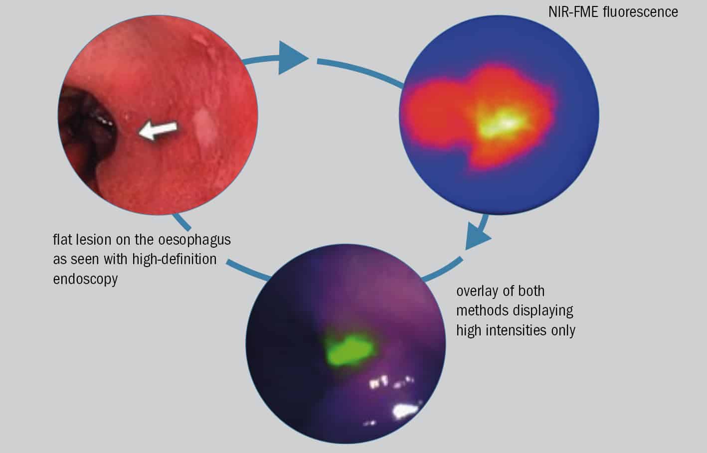
Innovation in biophotonics underpins another promising interrogation scheme for detecting early-stage oesophageal cancer and precancerous lesions. Near-infrared fluorescence molecular endoscopy (NIR-FME) relies on the administration of a fluorescence-labelled antibody (or tracer) that attaches selectively to suspect tissue on the lining of the oesophageal tract. When this tissue is then illuminated at NIR wavelengths, it becomes possible to visualize cancer-specific features both on the surface of the oesophageal tract and just below it. Conventional high-definition white-light endoscopy, in contrast, identifies surface-only morphology and tissue discolourations.

In a paper published in early 2019 (Gut 68 7), a Dutch–German collaboration led by Wouter Nagengast, a clinician at the University of Gröningen and Vasilis Ntziachristos, ESOTRAC project coordinator (see main article), reported on a small-scale clinical trial that highlights the potential of NIR-FME for flagging up oesophageal lesions early. In the study, a cohort of 14 patients, all of them scheduled to undergo a “resection” procedure to remove previously identified cancerous and precancerous lesions from the lining of the digestive tract, were simultaneously evaluated using NIR-FME and high-definition white-light endoscopy.

“Our in vivo results show that even flat and difficult to distinguish lesions were identified with NIR-FME,” the researchers write. What’s more, the subgroup of patients receiving a topical administration of the fluorescent tracer – effectively spraying the tracer onto the oesophageal lining immediately prior to NIR-FME inspection – saw a 33% improvement in early lesion detection compared with conventional white-light endoscopy.

“Fluorescence can guide better evaluation of suspicious lesions,” explains Ntziachristos, adding that the ESOTRAC consortium is investigating whether it might be possible to integrate fluorescence detection into its hybrid endoscope using a forward-looking camera.

If the collaboration hits that mark, the payback for patients (in terms of long-term survival prospects) and cash-strapped public healthcare systems (in terms of aggregate cost savings) will be substantial. “By detecting earlier states of oesophageal cancer, we can shift from high-risk and expensive treatments [surgery and chemotherapy] to simple lesion removal [via resection or ablation],” explains Vasilis Ntziachristos, the project co-ordinator for ESOTRAC and director of the Institute of Biological and Medical Imaging at HelmholtzZentrum München, Germany. “The situation is analogous to early diagnosis of skin cancer,” he adds, “where a simple procedure to remove a melanoma has curable effects, while late-stage disease comes with a very poor prognosis and low five-year survival rates.”

Biophotonic innovation

In January 2019 the ESOTRAC partners (see box above) gathered in Vienna, Austria, for a “half-time” review of their four-year programme. The meeting was essentially a progress report on ESOTRAC’s product development roadmap and, in particular, its stated goal of delivering a “market-ready hybrid endoscope with unparalleled capabilities to detect the early stages of oesophageal cancer”. During the first two years of the project, the partners designed a portable, laboratory-based instrument – essentially a proof-of-principle demonstration to combine MSOT and OCT into a single probe that can acquire complementary tissue information for pathophysiological mapping. In parallel, the teams have also been iterating and optimizing the prototype design for their hybrid endoscope in terms of size, biocompatible materials, optical and ultrasound components, as well as the integration of the core technologies into a practical device.

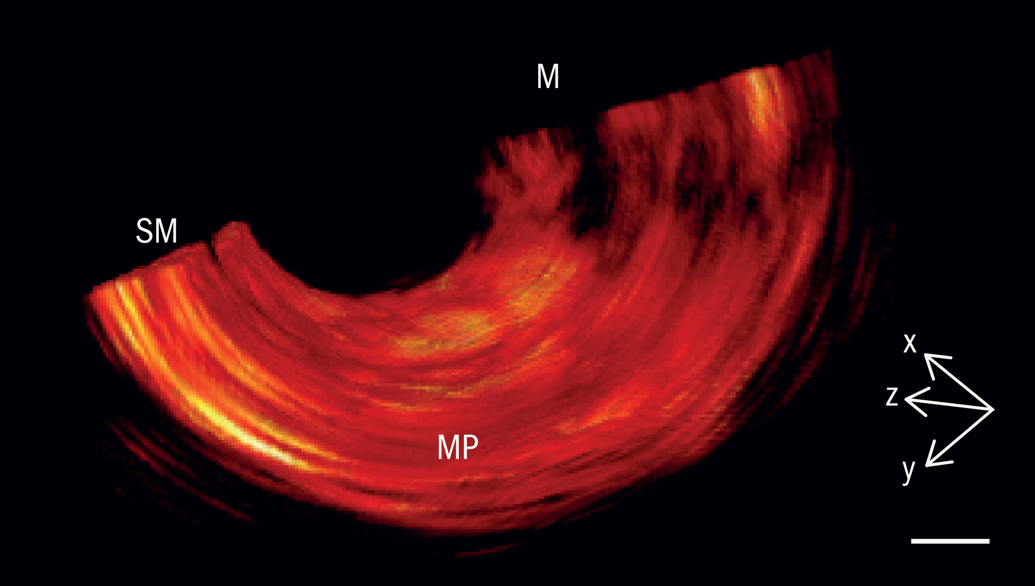
The two imaging modalities chosen for this next-generation endoscope are complementary in nature. MSOT delivers non-ionizing laser pulses into oesophageal tissue. Thanks to the photoacoustic effect, the tissue responds by emitting ultrasonically across a wide frequency band, producing a signal that can be used to identify abnormalities in metabolic activity – for example, changes in blood volume, tissue oxygenation, as well as bleeding and inflammation that are indicative of early-stage cancer. OCT, for its part, uses low-coherence near-infrared interferometry to map reflections from different depths within tissue. Those reflections yield micron-resolution cross-sectional images of subsurface lesions in the oesophagus wall and the growth of new microscopic blood capillaries that feed cancerous tissue.

ESOTRAC in brief

Project budget: €4m
Project duration: 2017–21
Academic partners: Institute of Biological and Medical Imaging, HelmholtzZentrum München, Germany (co-ordinator); University of Cambridge, UK; Technical University of Denmark; Medical University of Vienna, Austria
Industry partners: Sonaxis (France); Amplitude Systèmes (France); Statice (France); Ascenion (Germany); RayFos (UK)
Expertise: optoacoustics, OCT, oesophageal cancer (clinical research), ultrasound probes (manufacturing), ultrafast lasers (manufacturing), software development, biomaterials and biomedical devices (manufacturing), exploitation and intellectual property

As ESOTRAC moves into its third year, the partners’ immediate goal is to optimize the prototype of their hybrid endoscope and to analyse imaging data obtained during studies on “tissue phantoms” (specially designed test objects) and excised oesophageal tissue. Further iteration of the endoscope design will be informed by that image analysis. Ntziachristos calls the endoscope’s development “a photonics-intense and hardware-intense process,” and adds that the team is now finalizing the functional prototype and integrating and miniaturizing the various subcomponents that need to go into it, including optics, ultrasound and motorized control for volumetric scanning. The result, he says, will be “a world-first in terms of integration of optoacoustic and optical coherence tomography in a single endoscope”.

Translational science

From a biomedical perspective, the ESO-TRAC partners know that success will ultimately be measured by whether they can translate their R&D endeavours into a commercial product – ideally one that can be deployed for routine clinical application at scale. With this in mind, the consortium is working to secure patents for many of the project’s core innovations – spanning laser sources, ultrasound detectors, imaging software and the overall hybrid endoscope design. They also expect that, in the long run, the cost of the ESOTRAC-developed technology will be comparable to current commercial endoscopes, which are amortized after around 1300 examinations.

Even more compelling is the bigger picture associated with early detection of oesophageal cancer. A mere 1% increase in the number of people whose cancers are detected early, rather than late, would translate into vastly improved five-year survival rates for close to 6000 patients worldwide. It would also save a heap of money: the €130,000 five-year average cost of treating a patient whose oesophageal cancer was diagnosed late drops to around €12,000 when the disease is caught early, with the potential to yield annual savings in excess of €650m for national healthcare systems.

For the ESOTRAC partners, the end-game couldn’t be clearer. “With the increasing prevalence of oesophageal cancer worldwide, earlier detection has the potential to save tens of thousands of lives every year,” concludes Ntziachristos. “The consortium has big plans for the future, with a prototype that can be robustly deployed in clinical trials and one day translated into clinics everywhere.”

  • Enjoy the rest of the 2019 Physics World Focus on Optics & Photonics in our digital magazine or via the Physics World app for any iOS or Android smartphone or tablet.


Photons for a healthier world

The market for medical devices is highly regulated, with long product-development cycles and extensive procedures for obtaining regulatory approval. These characteristics make it challenging for companies and research groups to translate laboratory innovations into clinical practice. The National Healthcare Photonics Centre – part of CPI (Centre for Process Innovation), and located in Sedgefield, UK – aims to ease this translation process by providing practical support for developers of light-based tools for diagnosing and treating disease. On the centre’s opening day in March 2019, I spoke with CPI’s healthcare photonics lead, Tom Harvey, about its plans.

What are some key emerging applications of photonics in healthcare?

In terms of commercial importance, and applications that are in the clinic today, CT scanning comes first. People don’t always consider X-rays part of photonics, but for us, they are. CT scanning is a comparatively mature technique in terms of the hardware designs, but there’s still a lot of work being done on analysing the images it produces, and especially on the use of artificial intelligence (AI) in image analysis. Radiography is probably the most advanced clinical area where AI is making a difference.

The next technique I’d mention is optical coherence tomography (OCT). The importance of OCT is not to be underestimated. It’s used in ophthalmology to inspect the back and the front of the eye, and recent developments are lengthening its depth of field in a way that is analogous to earlier advances in the history of microscopy. We started with normal, 2D microscopy, and now everything is in 3D, going deeper into tissue using longer wavelengths of light. The combination of OCT and other types of imaging is also very powerful, especially for inside-the-body imaging. You can catheterize the OCT instrument, place it inside the body, and combine that information with fluorescence imaging to look at, for example, both the walls of the artery and the tissue type. Or you might inspect the bowel for tumours by giving the patient a fluorescent molecule to consume that sticks to those parts of the intestine wall that are not normal tissue, then go in and look at that.

There are also some interesting developments around using different lasers and laser techniques for healthcare. Medical lasers occupy a growing market sector in their own right. These laser types, such as the holmium:YAG laser, are designed to operate at wavelengths where absorption due to water in tissue is high, so that tissue absorbs the laser energy and is ablated. But they’re relatively large and expensive, so innovation is focused on making more compact or lower-cost laser sources or extending the range of laser sources available at different wavelengths. That’s where devices such as quantum cascade lasers (for mid-infrared wavelengths) and techniques that convert laser frequencies either up or down are coming into their own. We’re also seeing improvements in the performance of so-called “white-light” or “supercontinuum” lasers that give you both laser power and tuneability in the wavelength.

On a related note, fibre lasers are also very important in medical applications, as it is possible to get both high power and high beam quality at wavelengths between 1–3 µm. Also, improved methods for delivering the laser light to the patient are being developed. This is something that’s being done at Heriot-Watt University, for example, in partnership with researchers at the University of Leeds, using ultra-short-pulse lasers for tissue resection in surgery. One of the advantages of having a short laser pulse is that you avoid heating effects, so you get a very clean scalpel cut without any cauterization or damage to the surrounding tissue.

Another big area for healthcare photonics is Raman imaging. Larger, research-based Raman microscope systems have been around for a while, but until recently it wasn’t possible to do Raman spectral imaging through a fibre, because you would excite too much Raman signal from the fibre itself. Now, thanks to new developments in hollow-core fibres, you can point the fibre at something in the body and say, “Because I see this marker present, or this tissue is producing a different type of Raman spectrum, therefore it must be in a diseased or abnormal state.” The technique has advanced massively, but there is still room for improvement. Because the cross-section for Raman scattering is so small, you get very few photons scattered, so you need very sensitive systems to detect them – especially if you’re trying to do the imaging in real time, so that you can use the Raman signal for guidance as to what tissue to cut away during an operation. Real-time Raman imaging will require improvements in the electronics and better optical signal processing.

So we’re seeing some themes emerging around diagnostic imaging and therapy, but honestly, I never really know what clients are going to want when they walk through the door.

How will CPI help transform laboratory devices into commercial products?

We have a range of assistance that we can provide. First, there needs to be an entity that wants to collaborate with us – usually a spin-out company or some other commercial entity that wants to take an idea forward, sometimes an academic research group. We’re not looking to develop products ourselves. We’re not a business; we’re an independent, not-for-profit organization, so it’s not us that drives the process. Instead, clients come to us, and we ask them what they need to turn their idea into a product. It may be that their concept is fine and so is their design, but they don’t know where all the parts are going to come from for the real product, or they don’t know what the best method is for assembling it. The idea is that we provide that support.

We can also do more in-depth things, such as examining clients’ optical design or working with them on developing the electronics they need to control their system, if they haven’t done that already. We can give advice on whether their device is operating within a regulated, quality-control system, and if it’s not, we can show them how to build a technical file, which is what they’ll need if they’re going to get CE marking [European regulatory approval] for their device. We can guide them through that process and help them collect the appropriate information.

We’re also applying for a licence from the Human Tissue Authority, which would permit us to collect human tissue samples, bring them to our labs and use them to make measurements with the prototype devices that people bring to us. In some cases, products have failed because their makers only had access to a limited number of tissue samples, whereas patients are hugely diverse. It’s one thing making a device work one time in the lab. It’s completely another thing demonstrating that it will work across the range of different patients: men, women, young people, old people, people with different stages of a disease and so on. Getting that robustness is important, and that is what CPI is interested in doing. We want to do the legwork to help companies get to the stage where they’re ready to launch their products onto the market.

What would make product or service development in this field easier?

Smaller companies really appreciate the total package – they need help with funding, they need access to facilities and experienced people, and they need to be introduced to the regulatory environment. They may also need more than one part of CPI to help them. For example, we have groups devoted to biopharmaceuticals manufacturing, industrial biotechnology, printable electronics (including flexible and wearable electronics), and we have CPI’s capabilities for materials development. An example of such a project is one with some aspect of photonics in the product – a detector, for example, or a reader for an in vitro diagnostic device – but the company is also interested in is something else, such as the reagent they’re using or the antibody they’re developing that is specific to a particular condition or disease. Then we can bring in different parts of the organization to help them.

Larger companies, in contrast, usually have all the resources they need to do product development in photonics, but their people may be tied down with standard tasks, such that they don’t have the capacity do something a little bit different or disruptive. That’s the sort of thing that we can potentially work on with them. Somebody can come to our labs and carry out some measurements that are a bit unusual, on things that they wouldn’t normally do in the corporate lab.

Then there are companies in the middle. They might like to develop a new product line, but they don’t have the resource to do that, so they can pay our staff to work on it. For example, companies making spectrometers may not have good applications notes for the full range of possible medical applications. So if we develop those applications notes, that can really add value for them.

You’re a physicist by training. How do you see physicists contributing to the field of healthcare photonics? What do they bring that an engineer or a bioscientist wouldn’t?

I work with people from a lot of different disciplines, and I find that really stimulating. But photonics is definitely a physics thing for me. My PhD at Heriot-Watt was in the nonlinear optics of conducting polymer systems, so I’ve always been interested in the interaction of light with materials. I think as physicists, we bring an understanding of instrumentation and instrumentation systems design, but we also understand the basic science of what’s happening when we look at a sample. We understand things like refraction and light scattering and absorption and emission, and that understanding is helpful. It means that when I enter discussions with companies, I have a good grounding and understanding – most of the time! – in the basics of what they’re doing. I love engineering as well, and the crossover between physics and those other disciplines, but an understanding of the physical processes involved in photonics is key to the whole thing.

  • Enjoy the rest of the 2019 Physics World Focus on Optics & Photonics in our digital magazine or via the Physics World app for any iOS or Android smartphone or tablet.
Ultrasound guides particle in a midair display

A midair visual display that uses a single acoustically-levitated particle has been unveiled by researchers in Spain and the UK. Dubbed an “acoustophoretic display”, the image is created by using two ultrasound transducer arrays to levitate the particle and manipulate it to trace out the desired graphic at high speed.

In 2015, Asier Marzo at the Public University of Navarra and Bruce Drinkwater at the University of Bristol created a sonic tractor beam that used ultrasound to levitate, rotate and move objects. Using a single grid of 64 off-the-shelf loudspeakers controlled by a programmable array of transducers the device created 3D fields of sound – acoustic holograms – that could hold and manipulate a small polystyrene particle in mid-air.

Since then, the field has progressed and earlier this year Marzo and Drinkwater revealed an acoustic levitation device that used two grids of speakers to hold and individually manipulate up to 25 polystyrene balls at the same time. This opened up the possibility of new applications for sonic tractor beams, including visual displays created with multiple levitated particles.

According to Marzo, such acoustic generated images, which he calls acoustophoretic displays, would offer an advantage over current holograms as they would not suffer from clipping. “[With holograms] the image can only be viewed from determinate angles and the frame of the display occludes the image, it is like looking inside a window,” he explains. “With the acoustophoretic displays the images reside in the physical space and can be observed without clipping from 360°.”

Many-body problems

But when the team tried to create displays using many levitated particles they had some problems. “We were trying to levitate multiple particles and make them form the shape at any given time,” Marzo told Physics World. “However, this leads to coarse graphics given the limited number of particles and the minimum distance between them, which is governed by the wavelength – approximately 1 cm.”

Undeterred, Marzo, Drinkwater and colleagues came up with another idea: using a single illuminated particle travelling at high speeds to trace the path of the image. While using multiple levitated particles like pixels would be similar to the way liquid-crystal, plasma and organic LED displays work, Marzo points out that using a single particle to trace the image is reminiscent of the way cathode-ray tubes use electron beams to create displays.

The idea is that if you levitate a single illuminated particle and move it very fast and precisely you can create the illusion of an image. This relies on persistence of vision – the capacity of the eye to briefly maintain an image on the retina after it has disappeared, enabling successive images (that follow quickly enough) to be perceived as one image.

University logo

At the recent 177th Meeting of the Acoustical Society of America the team revealed its new system , which comprises two grids of 30 ultrasonic speakers and an illuminated, 0.7 mm polystyrene particle to create a 12×12 mm image of the University of Bristol logo (see figure).

Creating the display involved the development of a novel calibration method that allowed them to improve the accuracy with which they could position the particle. They claim that they reduced the positional error of the levitation device down to 0.11 mm and 0.03 mm in the horizontal and vertical axes, respectively. These developments allowed the particle to move at a speed of 60 cm/s, while maintaining an accurate enough trajectory to create the image.

“We needed to increase the communication speed so that we can rapidly reconfigure the sound field emitted by the device,” explains team member Tatsuki Fushimi. “The biggest challenge has been understanding the particle dynamics and the operation of our electronics at these high speeds. For the next iteration we are going to need even faster hardware and also more powerful acoustic emitters.”

Bristol’s Tom Hill told Physics World that while the current graphics are quite simple he believes the technique is promising. He adds that the team are developing algorithms to compensate for the inertia of the particle to create more accurate traces. They are also working to increase the speed of the particle. This would allow longer traces and therefore larger images. Soon the researchers hope to move to speeds of 400 cm per second, Hill explains, which would allow them to draw a 10 cm square or a 3 cm cube – graphics that require the particle to travel 40 cm ten times a second.

Looking to the furure, Marzo says the team would like to have multiple particles tracing the graphics as “that would allow for more complex figures to be created, but at the moment we are focused on the dynamics of a single particle”.

  • In related research, Marzo has joined forces with scientists at Bristol and the University of Glasgow to create an acoustic levitator that is controlled by hand gestures. The system levitates multiple polystyrene spheres that are 3 mm in diameter. A user gestures towards the sphere to be manipulated, which can then be directed by the user’s hand. The device provides haptic feedback to the user and the system could find use in the manipulation of fragile objects such as biological samples.

Growing better single crystals with the help of machine learning

Machine learning could help grow high-quality single crystals by predicting whether a particular crystal can be successfully grown under given experimental conditions. The approach could be an inexpensive and rapid alternative to growing crystals in the laboratory.

Machine learning (ML) is the process by which a system learns from a training set so that it can autonomously deliver an appropriate response when supplied with new data. Although pioneered in the middle of the 20thcentury, increasingly powerful computers and faster calculations speeds mean that ML is now being applied to solve problems in areas as diverse as finance, healthcare, transport and, of course, science.

Single crystals are crucial for many research fields, including condensed matter physics, surface science, lasers and nonlinear optics. Fundamental studies, such as those on the quantum Hall effect and Wyle semi-metals, to name just two examples, all require high-quality single crystals.

It is difficult to grow single crystals in the laboratory, however, because so many interrelated factors, such as temperature and material composition ratios, need to be considered. Growing ternary compounds is particularly problematic because of the lack of ternary phase diagrams. Single crystal growth is thus labour-intensive and time consuming.

ML could come into its own here, say researchers at the Chinese Academy of Sciences in Beijing, and Renmin University, also in Beijing, who have focused on the single-crystal growth of ternary compounds by the so-called flux method, which is one of the most widely used methods in laboratories to grow crystals.

Data on crystal growth used to train ML

In their study, the researchers collected data on crystal growth from two different groups in their laboratory. This data includes growth temperature curves, raw elemental compositions and ratios, and growth conditions. Both the quantity and quality of the data is important for generating successful models, they say. Issues like the presence of noise and outliers, missing, inconsistent or duplicate data, and data that are biased or unrepresentative of the phenomena at hand, thus need to be taken into account.

The team trained four kinds of ML (support vector machine, or SVM, decision tree, random forest and gradient boosting decision tree) on the experimental data. The SVM method works well and can predict with an accuracy of 81% which crystals will grow under specific, artificially selected, growth conditions. To compare, this figure is just 36% for crystal growth experiments in the laboratory. The decision tree model, for its part, reveals which features are critical in a particular growth process.

“We use the model obtained by the decision tree on the training data to analyse the important factors of single crystal growth,” explains team member Yu-Jie Sun. “Based on the information obtained, we then use the SVM to predict whether the growth conditions selected can successfully guide crystal growth in the laboratory.”

Accuracy will continue to improve

“The accuracy of the technique will continue to improve as we increase the amount of laboratory data we use to train the ML,” he adds. “We adjust the weight of different new data when adding this to the training model.

“Our work shows that such a learning model can predict and instruct crystal growth in the laboratory,” he tells Physics World. “The approach could help grow high-quality crystals more efficiently, further automate the process and so promote the development of the disciplines that rely on single crystals.”

The researchers, reporting their work in Chinese Physics Letters 10.1088/0256-307X/36/6/068101, are now trying to broaden the scope of their technique. “At present, our research is quite limited, so we are going to try new learning algorithms, grow more kinds of single crystals and look into other growth methods and larger databases,” says Sun. “We hope that our method will be able to not only predict growth conditions, but also the appropriate growth method for any type of single crystal.”

PET scanner images free-moving, interacting rats

PET images of rat brains

Anaesthesia is widely used in preclinical imaging to keep animals stationary within the scanner’s field-of-view and avoid motion artefacts. However, anaesthesia also creates one of the biggest limitations in imaging studies as it alters the animal’s normal physiological state. A collaborative study between McGill University and the Molecular Imaging Center Antwerp (MICA) aims to remove this constraint.

The researchers have developed a new PET imaging platform that’s capable of simultaneously scanning awake and interacting rats. The proposed method involves performing PET scans of free-running rats using a high-resolution, large field-of-view human brain PET scanner (NeuroImage 10.1016/j.neuroimage.2019.02.064).

During scanning, the rats are housed inside an acrylic imaging cage and are free to move in a 19.4 x 23 cm area. A plastic grid platform in the cage positions them in the centre of the scanner’s field-of-view. Four small lightweight positron-emitting point sources are attached to each animal’s head, enabling head motion to be tracked using an algorithm developed by MICA researchers. After image acquisition, motion-corrected images are reconstructed from the PET tracking measurements.

The authors note that previous methods developed to avoid anaesthesia required external tracking or surgical implantation of devices to scan the brain of a moving animal. The approach developed through this collaboration offers a less invasive means of conducting imaging studies and allows animals to interact in a “natural” manner during scans.

“We think our breakthrough will open a new era of small-animal PET imaging research and unprecedented experimental designs that many researchers have been anxious to test for a long time,” says McGill’s Pedro Rosa-Neto.

The researchers first validated the system performance using a motion resolution phantom. The point source-based motion tracking approach exhibited a precision of 0.359 mm. A minor loss of spatial resolution was seen in motion-corrected reconstructions of the phantom compared with motion-free reconstructions.

The team then performed 20-min long FDG-PET scans of three awake single rats, as well as an FDG-PET scan of two interacting rats. During the awake scans, the rats moved extensively around the platform, with an average head motion of 1.69 cm/s. After each awake scan, the researchers anesthetized the rat and performed a 20 min motion-free scan for comparison.

During the single rat scans, the animals exhibited exploratory behaviour, with recurrent changes in posture and sniffing behaviour, as well as long periods of motionless. Rats 1, 2 and 3 travelled 21, 15 and 14.9 m, respectively, during the 20 min scan. The tracking success rate was 81%, 54% and 90%, respectively, for these rats (the low success rate of rat 2 was due to low activity of one of the point sources).

In the simultaneous scan, both rats remained calm, exploring the cage for brief periods of time and at times with their heads overlapping. These rats travelled 12.7 and 13.0 m, respectively, during the 20 min scan, with tracking success rates of 84% and 90%, respectively.

The researchers reconstructed motion-corrected and motion-free images of the rats’ brains and calculated the regional FDG uptake for both reconstructions. They observed high FDG uptake in brain regions such as the cerebellum, cortex and hippocampus — with strong correlation between the relative regional brain uptake in motion-corrected and motion-free images.

“From the start of this project, our goal was to develop a practical approach to imaging awake animals,” says corresponding author Jeroen Verhaeghe from MICA. “After more than three years of development, we delivered an approach that can be easily implemented so scientists can focus on new exciting biology questions that can be answered rather than on technical issues.”

Using this innovative platform, the researchers will continue their collaboration and hope to answer questions that have long eluded scientists, for example, the extent to which brain cells use glucose as the main energy source. The new scanning method could also help understand the neurochemical basis of sympathy, fear, learning and memory in real time in awake animals, questions that could not previously be answered because of the use of anaesthesia.

Greener labs, sounding out sharks, and Einstein in Oxford: the June 2019 issue of Physics World is now out

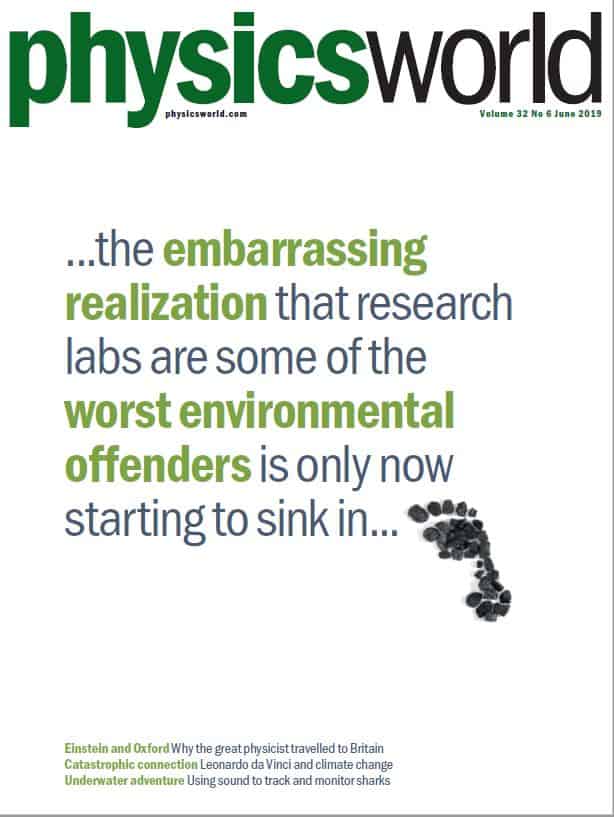
The cover of the June 2019 issue of Physics World could not be more direct: the embarrassing realization that research labs are some of the worst environmental offenders is only now starting to sink in.

The cover of the June 2019 issue of Physics World

It’s the simple message from the lead feature in the new issue of Physics World, which examines how many scientific laboratories are terrible at wasting energy and resources. Fortunately, there are initiatives to improve the situation – and to see tips to make your lab greener, check out the feature by Benjamin Skuse.

Elsewhere in the issue, you can find out why the world’s greatest physicist Albert Einstein came to the University of Oxford three times in the 1930s, discover what Leonardo da Vinci has to do with climate change, and see why physicists must do a much better job at using videos to teach students.

You can enjoy the June 2019 issue of Physics World magazine via our digital apps for iOSAndroid and Web browsers (membership of the Institute of Physics required). Let us know what you think about the issue on TwitterFacebook or by e-mailing us at pwld@iop.org.

For the record, here’s a run-down of what’s in the issue.

• NASA contractor hit with $47m fine – Falsified tests by a US metals manufacturer were behind the failure of two high-profile climate missions, as Peter Gwynne reports

• Transformative thinking – Best known for his work on invisibility cloaks and “transformative optics”, Ulf Leonhardt from the Weizmann Institute of Science in Israel talks to Joe McEntee about science in Israel, his career – and his new novel

• The master of disaster – Leonardo da Vinci died 500 years ago, but his work contains clues for how to think about the modern problem of global warming, finds Robert P Crease

• Patently obvious – James McKenzie explains why physicists who want to succeed in business must nail down their intellectual property rights

• Rutherford’s legacy – With the International Year of the Periodic Table of Elements in full swing, John Campbell celebrates the immense contribution of Ernest Rutherford, who first split the atom 100 years ago

• Leading by example: going green in the lab – With so much research focused on creating technologies that reduce our collective carbon footprint, the embarrassing realization that research labs themselves are some of the worst environmental offenders is only now starting to sink in. Benjamin Skuse examines the problem and what is being done to solve it

• Einstein in Oxford – Amid growing political unrest in Germany, Albert Einstein paid three short visits to Oxford in the early 1930s. Andrew Robinson reveals why this  celebrated physicist travelled to Britain and how Einstein seduced – and then shocked – his audiences with his new thinking about how science works

• Listening for sharks – Pelagic thresher sharks are a sight to behold, but their numbers are dwindling as a result of illegal hunting and fishing. Now, however, scientists are using acoustics to learn more about these animals’ behaviour and how they can be better protected. Louise Murray looks under the surface

• Black holes and revelations– Andrew Robinson reviews Gravity’s Century: From Einstein’s Eclipse to Images of Black Holes by Ron Cowen

• Solve for X –Ian Randall reviews All of Physics (Almost) in 15 Equations by Bruno Mansoulié

• A versatile technology – Julia Zimmermann, co-founder of the German start-up firm Terraplasma, speaks to Julianna Photopoulos about how a desire to develop  applications for cold atmospheric plasmas led her to co-found a family of companies

• Once a physicist: Ben Cowie – meet the man who opened a cycling specialist shop and cafe in Ontario, Canada, in 2017.

• Video versus the blackboard – Manuela Ramos Marques da Silva and Pedro Sidónio Pereira da Silva explain why we need to do a much better job creating high-quality physics videos for students as that’s the way many people now learn.

Making physics greener: why research labs must do more to save energy and resources

When I was a PhD student at the Cavendish Laboratory, Cambridge, in the 1990s, I was once standing in the queue for the lab’s storeroom when the then head of physics, Archie Howie, wandered by. Overhearing a physicist ask the storeroom supervisor for two ballpoint pens. Howie demanded to know why the physicist needed two Biros when you can only write with one at a time. Although I can’t remember if I slyly pruned back my own wish list for glass vials and bottles of solvent, Howie’s comment struck me as a small but perfect example of the old approach in physics of getting by with less.

Physics at Cambridge (and elsewhere for that matter) was not supposed to be about burning cash on expensive and unnecessary equipment, but about using money, materials – and probably even pens – ingeniously, prudently and wisely. It was the “string and sealing wax” way of doing physics developed in the early 20th century, which stipulated that rapid progress could be made (and indeed was more likely) with rudimentary resources. Some physicists even frowned on buying equipment from instrument manufacturers: far better (and quicker) to build it yourself.

Such attitudes might seem quaint in today’s world, where there’s pressure to get ever-bigger grants, plus countless hi-tech manufacturers that offer products far superior to anything that researchers could hope to knock up for themselves (just check out some of the companies that have placed adverts on this website.) Still, that old-fashioned, prudent approach to physics, exemplified by Howie, has one big modern-day advantage: it’s much better for the environment too.

As the cover feature of the June 2019 issue of Physics World makes clear, being green doesn’t only involve using fewer disposable rubber gloves, though wasting less is vital. It’s also about expending less energy. The trouble is, many physics experiments are appallingly energy-hungry. With its countless radiofrequency cavities and liquid-helium-cooled superconducting magnets, CERN’s Large Hadron Collider, for example, uses 1.3TW of electricity a year, which the lab says is enough to power 300,000 UK homes.

So far, most initiatives and efforts to reduce researchers’ carbon footprint have come in the biological and life sciences, for example, through the My Green Lab in the US and the Laboratory Efficiency Assessment Framework in the UK. It’s now time for physicists to do their bit too – especially given that many physicists love to boast how they’re at the forefront of developing technologies for dealing with climate change.

At a simple level, that means turning off kit when not needed, closing fume cupboards and recycling or donating old electrical items (check out a list of top energy-saving tips in this feature. But it also means making energy efficiency an inherent part of all physics-research equipment – finding innovative ways to make them use as few resources as possible. Funding agencies may even have to insist on projects – especially big-science facilities – proving their green credentials before they get financial support.

It’s a cultural shift that’ll take a generation, but with the growing impact of climate change, there’s little time to lose.

Copyright © 2026 by IOP Publishing Ltd and individual contributors