
Anaesthesia is widely used in preclinical imaging to keep animals stationary within the scanner’s field-of-view and avoid motion artefacts. However, anaesthesia also creates one of the biggest limitations in imaging studies as it alters the animal’s normal physiological state. A collaborative study between McGill University and the Molecular Imaging Center Antwerp (MICA) aims to remove this constraint.
The researchers have developed a new PET imaging platform that’s capable of simultaneously scanning awake and interacting rats. The proposed method involves performing PET scans of free-running rats using a high-resolution, large field-of-view human brain PET scanner (NeuroImage 10.1016/j.neuroimage.2019.02.064).
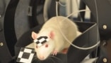
During scanning, the rats are housed inside an acrylic imaging cage and are free to move in a 19.4 x 23 cm area. A plastic grid platform in the cage positions them in the centre of the scanner’s field-of-view. Four small lightweight positron-emitting point sources are attached to each animal’s head, enabling head motion to be tracked using an algorithm developed by MICA researchers. After image acquisition, motion-corrected images are reconstructed from the PET tracking measurements.
The authors note that previous methods developed to avoid anaesthesia required external tracking or surgical implantation of devices to scan the brain of a moving animal. The approach developed through this collaboration offers a less invasive means of conducting imaging studies and allows animals to interact in a “natural” manner during scans.
“We think our breakthrough will open a new era of small-animal PET imaging research and unprecedented experimental designs that many researchers have been anxious to test for a long time,” says McGill’s Pedro Rosa-Neto.
The researchers first validated the system performance using a motion resolution phantom. The point source-based motion tracking approach exhibited a precision of 0.359 mm. A minor loss of spatial resolution was seen in motion-corrected reconstructions of the phantom compared with motion-free reconstructions.
The team then performed 20-min long FDG-PET scans of three awake single rats, as well as an FDG-PET scan of two interacting rats. During the awake scans, the rats moved extensively around the platform, with an average head motion of 1.69 cm/s. After each awake scan, the researchers anesthetized the rat and performed a 20 min motion-free scan for comparison.
During the single rat scans, the animals exhibited exploratory behaviour, with recurrent changes in posture and sniffing behaviour, as well as long periods of motionless. Rats 1, 2 and 3 travelled 21, 15 and 14.9 m, respectively, during the 20 min scan. The tracking success rate was 81%, 54% and 90%, respectively, for these rats (the low success rate of rat 2 was due to low activity of one of the point sources).
In the simultaneous scan, both rats remained calm, exploring the cage for brief periods of time and at times with their heads overlapping. These rats travelled 12.7 and 13.0 m, respectively, during the 20 min scan, with tracking success rates of 84% and 90%, respectively.
The researchers reconstructed motion-corrected and motion-free images of the rats’ brains and calculated the regional FDG uptake for both reconstructions. They observed high FDG uptake in brain regions such as the cerebellum, cortex and hippocampus — with strong correlation between the relative regional brain uptake in motion-corrected and motion-free images.
Open-field PET enables brain scans of rats in motion
“From the start of this project, our goal was to develop a practical approach to imaging awake animals,” says corresponding author Jeroen Verhaeghe from MICA. “After more than three years of development, we delivered an approach that can be easily implemented so scientists can focus on new exciting biology questions that can be answered rather than on technical issues.”
Using this innovative platform, the researchers will continue their collaboration and hope to answer questions that have long eluded scientists, for example, the extent to which brain cells use glucose as the main energy source. The new scanning method could also help understand the neurochemical basis of sympathy, fear, learning and memory in real time in awake animals, questions that could not previously be answered because of the use of anaesthesia.


