The first-ever seismic signal to be measured on Mars has been reported by scientists monitoring NASA’s InSight mission. Researchers believe that the tiny “Marsquake” originated from within the planet rather than being the result of wind or other surface phenomena. Studying the seismology of Mars should provide important information about the interior of the planet and how it was formed.
The quake was detected on 6 April by a seismometer called SEIS, which InSight had placed on the Martian surface in December 2018. A weaker signal had been detected about three weeks earlier, but its origin is unclear. Signals with ambiguous origins were also measured on 10 and 11 April.
It’s so exciting to finally have proof that Mars is still seismically active
Philippe Lognonné
“We’ve been waiting months for a signal like this,” says Philippe Lognonné of the Institut de Physique du Globe de Paris in France – who leads the team that built SEIS. “It’s so exciting to finally have proof that Mars is still seismically active.”
Quieter than Earth
Mars is not the first place that scientists have detected extraterrestrial seismic activity. Five seismometers operated on the Moon between 1969 and 1977 and measured thousands of “moonquakes”. Like the Moon, Mars does not have tectonic plates and therefore is expected to be much quieter than Earth when it comes to seismic activity.
Scientists believe that seismic activity on Mars is related to the ongoing cooling of the interior of the planet, which is causing the planet to contract. This results in the build-up of stress, which is released by a break in the crust that generates a quake. A similar process is expected to occur on the Moon.
NASA’s director of planetary science Lori Glaze says that the 6 April signal is “exciting because its size and longer duration fit the profile of moonquakes detected on the lunar surface during the Apollo missions”.
Lognonné and colleagues are now analysing the four signals and expect to detect more in the future. “We’re looking forward to sharing detailed results once we’ve had a chance to analyse them,” he said.
Gaining insights into the interior of Mars is a primary goal of the InSight mission, which landed on the planet in November 2018 and is expected to operate for at least two years.
The above video from NASA is an audio and video illustration of the signal, which has been sped-up by a factor of 60 and amplified greatly in order to be audible.
In this episode of Physics World Weekly, we’re focusing on measurements and precision in physics. Physics World’s Hamish Johnston visits researchers at the National Institute for Standards and Technology (NIST) in Colorado, US. He learns of an innovation to make atomic clocks even more impressive timekeepers, using aluminium ions. NIST researchers also speak about the facility’s project to integrate its calibration services onto computer chips – which have even been launched into space.
As always, we bring you a round up of some of the other research highlights making the headlines on our website this week. That includes the story that some Roman theatres and amphitheatres appear to be protected by “invisibility cloaks” made from acoustic metamaterials. Whether the Romans based their designs on an understanding of the fundamental physics remains a mystery.
If you enjoy what you hear, then you can also subscribe to Physics World Stories via Apple podcasts or your chosen podcast host.
Piezoelectric materials are widely employed as transducers and sensors in a variety of applications, including ultrasound medical imaging systems. Researchers have now nearly doubled the performance of a class of piezoelectrics, known as relaxor ferroelectrics, which were discovered 20 years ago, by adding trace amounts of the element samarium (Sm) to them. The rare-earth doping technique, which appears to increase local structural heterogeneity in the crystals, could be a general strategy for enhancing the piezoelectricity of these materials.
Lead author of the story, Fei Li. Courtesy: Fei Li
The discovery of relaxor ferroelectric single crystals, such as Pb(Mg1/3Nb2/3)O3-PbTiO3 (PMN-PT) and Pb(Zn1/3Nb2/3)O3-PbTiO3 (PZN-PT), was a milestone achievement in ferroelectric research, explain the researchers of Xi’an Jiaotong University in China, Pennsylvania State University in the US and the University of Wollongong in Australia. These materials, which have been optimized over time, have very high piezoelectric coefficients (the amount of charge generated for each unit of force) of 1200 to 2500 picocoulombs per newton.
Although these materials far outperform mainstay piezoelectrics, such as lead zirconate titanate ceramics, researchers have had a hard time improving the properties of PMN-PT and PZN-PT further because it has proven difficult to grow single crystals of the materials with uniform properties.
The Xi’an Jiaotong-Penn State-Wollongong team has now grown Sm-doped PMN-PT singe crystals that boast piezoelectric coefficients ranging from 3400 to 4100 picocoulombs per newton.
Enhanced local structural heterogeneity
The researchers, reporting their work in Science, imaged small volumes of the PMN-PT structure (about 20 x 20 unit cells, or around 40,000 atoms). Thanks to these and first-principles calculations, they showed that the giant piezoelectric properties come from the fact that the Sm3+ dopant enhances local structural heterogeneity within the crystals. “What is more, the trace dopant magically offsets the phase variation induced by Ti from the B-site component of the crystals, so making them more uniform,” they explain.
There are only around 50 Sm atoms present in the volume of sample imaged, which corresponds to a doping of one Sm atom per 1000 atoms of the PMN-PT crystal structure. This is enough, however, to double the material’s piezoelectric coefficient.
The effect might be mediated by nanoscale strain fluctuations in the material, writes Jiří Hlinka of the Institute of Physics of the Czech Academy of Sciences in a related Perspectivearticle. “These intriguing findings not only represent good news for the designers of electromechanical transducers and low-field actuators but also raise questions about the origin and limits of the distinctive piezoelectric performance of the PMN-PT family of crystals,” he says.
The crystals could find use in various room-temperature piezoelectric applications, say the researchers, and especially in high-frequency medical imaging transducers and low field-driven actuators. Since the new doping technique works on as-grown PMN-PT crystals, it should help decrease the cost of the final devices and reduce material waste.
Around three-quarters of US female physics undergraduates have experienced sexual harassment during their studies. That is according to an analysis of surveys that were carried out in 2017 at conferences for undergraduate women in physics. The authors of the study, which is published in Physical Review Physics Education Research, say that their results show the pervasiveness of sexual harassment in physics and its impact on female physicists.
More than 450 female undergraduates answered the surveys, which asked them about their experiences of sexual harassment within the previous two years that was associated with physics – defined as being in research labs, classrooms, instructional settings or student organised events. Given that 1349 physics bachelor degrees in the US were earned by women in 2015, the researchers say that their study covers a large proportion of female physics undergraduate students.
Recognising and condemning gender harassment will both address the harm of gender harassment and prevent other types of sexually harassing behaviour
Lauren Aycock
The authors split sexual harassment into three categories. This includes “sexist gender harassment” in which women receive sexist comments or are treated differently, ignored or put down because of their gender. Another category — sexual gender harassment — involves women being subjected to inappropriate sexual remarks, jokes or stories or having sexual comments made about them. The final category — unwanted sexual attention — includes being repeatedly asked out after they had declined or being touched without their permission in a way that made them feel uncomfortable.
Overall, 74% of respondents had experienced at least one form of sexual harassment with 68% having experienced sexist gender harassment, while 51% had been subjected to sexual gender harassment and 24% had dealt with unwanted sexual attention. Indeed, many students had experienced more than one form of sexual harassment with one-fifth stating that they had experienced all three categories of harassment and a quarter saying that they had been subjected to both sexist and sexual gender harassment.
Addressing the problem
The study’s lead author, physicist Lauren Aycock, who is a science and technology policy fellow at the US Department of Energy, told Physics World that she undertook the study as she had “concerns” that the culture of physics is hostile to women and wanted to quantify the scope of sexual harassment in physics to enable “productive discussions that extends beyond personal anecdotes”.
Aycock says that the way to tackle sexual harassment in physics is to address gender harassment. “[This type] is the most common form and a type of sexual harassment that conveys derogatory attitudes about women that is often dismissed as not real harassment,” she says. “However, it has been shown to have substantial negative consequences on individuals”.
Indeed, the students were asked questions designed to assess the impact of sexual harassment with the study finding that it eroded students’ sense of belonging in physics and made them doubt their accomplishments as well as become fearful of being exposed as a fraud — also known as imposter syndrome. “Both a sense of belonging and imposter phenomenon have been shown to impact persistence in the field,” says Aycock.
Aycock adds that the study highlights the scale of sexual harassment in physics without the need for women to tell their stories. “Recognising and condemning gender harassment will both address the harm of gender harassment and prevent other types of sexually harassing behaviour,” she says. “I hope our findings will be used as a good starting place for physics departments to address gender harassment.”
Meanwhile, a joint report by the International Society for Optics and Photonics (SPIE) and The Optical Society (OSA), released last week, found that 24% of women reported being harassed while attending meetings organised by SPIE and the OSA compared to 4% of men. Harassment was most likely to occur to those aged between 40 to 49.
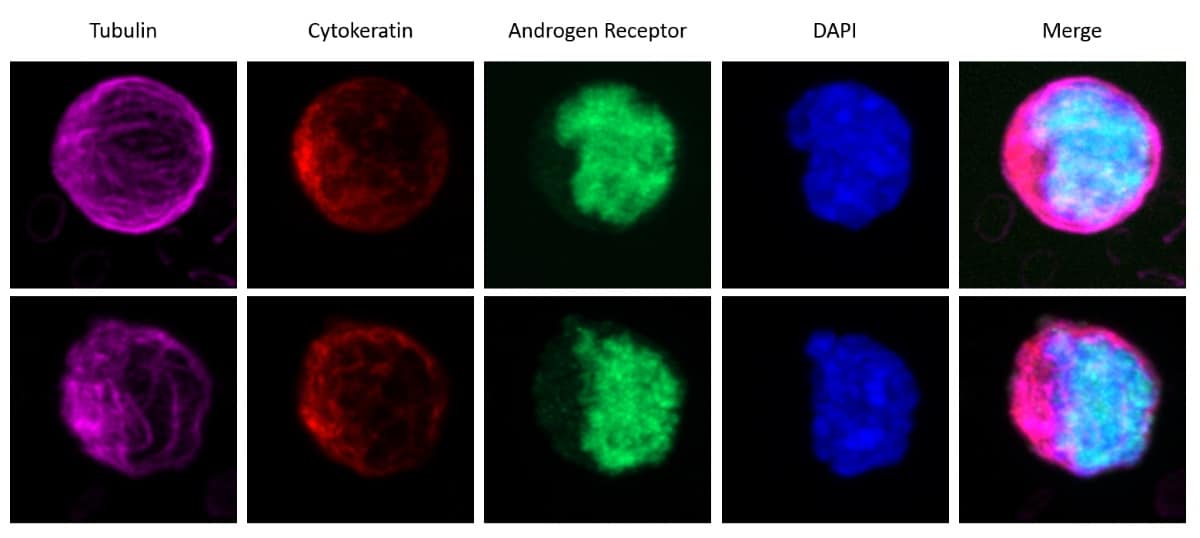
Circulating tumour cells from metastatic castration-resistant prostate cancer patient before taxane chemotherapy. (Courtesy: Daniel Worroll)
A sampling and image-analysis method that spots and characterizes cancer cells in the blood has been developed by researchers in the US. The high-throughput technique quantifies the presence of androgen receptor (AR) proteins in cell nuclei — a prognostic indicator and measure of treatment efficacy for metastatic prostate cancer. Less invasive than a conventional biopsy, monitoring circulating tumour cells (CTCs) also provides a more representative picture of tumour properties in real time (Phys. Biol. 10.1088/1478-3975/ab073a).
Growth of metastatic prostate cancers is associated with the build-up of androgen receptors in cell nuclei. Usually restricted to the cell’s cytoplasm, these proteins are only transported into the nucleus when bound to the hormone androgen and a polymeric cellular component called a microtubule. In the nucleus, ARs bind to the DNA, activating a set of genes that promote tumour growth.
Most chemotherapy strategies for prostate cancer aim to prevent the activation of these genes by excluding ARs from the nucleus, which they achieve by inhibiting the function of microtubules. Measuring the presence of nucleic AR in cancer cells, then, offers a diagnostic tool and a way to monitor a treatment’s effectiveness.
The usual way to obtain cancer cells for such purposes would be to conduct a biopsy at the site of the tumour itself. For prostate cancer, this is an invasive procedure with a real risk of complications, and the information that it provides reflects just a single point in time and space. Seeking to improve on the convenience and diagnostic power of this technique, Daniel Worroll and colleagues at Weill Cornell Medicine developed a method to process and analyse liquid biopsies instead — blood samples that can be obtained easily and as frequently as treatment requires.
The researchers took blood samples from 15 patients being treated for metastatic castration-resistant prostate cancer (mCRPC), as well as four healthy donors whose blood was then “spiked” with a prostate-cancer cell line. First, they applied a cocktail of cell-specific antibodies to remove white and red blood cells from the samples, leaving a residue enriched in CTCs. The conventional way to isolate CTCs is to capture them directly using a cancer-specific cell-surface marker, but this method misses potentially important cancer cells in which the marker is absent.
“The biological relevance of CTCs that do not express traditional epithelial markers is something that requires more research,” says Worroll. “Based on other groups’ results, the hypothesis is that those tumour cells that lose epithelial markers may have a higher metastatic potential and be more resistant to chemotherapy.”
Next, the researchers stained the samples with a range of fluorescent antibodies that highlighted certain cellular components. This allowed the first step of the image-analysis process, in which an algorithm classified CTCs based on cell shape, size and fluorescence profile. Using a higher-resolution image of the same sample, the algorithm then identified the nuclei of individual CTCs, and determined the ratio of AR-related fluorescence in and outside of the nucleus.
Taking this ratio as a measure of AR nuclear localization, the researchers compared the results with the patients’ therapy outcomes. Patients who responded the least to chemotherapy tended to be those whose CTCs showed a greater quantity of nuclear AR. The explanation for this, Worroll and colleagues suspect, could imbue the technique with additional clinical value.
Although ARs rely on the presence of microtubules to access the cell nucleus — making them susceptible to treatments that target this vector — an AR variant exists that is not so dependent. Lacking the molecular structure that interacts with microtubules, variant AR-V7 gains entry to the nucleus by another (currently unknown) route, and is associated with a chemotherapy-resistant form of prostate cancer. The researchers measured the expression of AR-V7 messenger RNA in CTCs and found that the variant was more likely to be present in those cells with more nuclear AR.
“With multiple studies showing a zero-response rate of AR-V7-positive patients to AR-targeted therapies, detection of AR-V7 mRNA status prior to initiation of these therapies would have an obvious clinical impact,” says Worroll. “Furthermore, due to the non-invasiveness of liquid biopsies, longitudinal monitoring while on therapy for the emergence of AR-V7 would offer therapeutic benefit.”
Machine learning and automation technologies are gearing up to transform the radiation-therapy workflow while freeing specialist clinical and technical staff to dedicate more time to patient care. That message will be front and centre for visitors to the booth of RaySearch Laboratories, the Stockholm-based oncology-software company, at the annual congress of the European Society for Radiotherapy and Oncology (ESTRO 38), which gets under way in Milan, Italy, on 26 April.
The ESTRO conference represents the official European unveiling of RayStation 8B, the latest release of RaySearch’s treatment-planning software. Among a raft of new features, perhaps the most anticipated are the machine-learning innovations that underpin RayStation’s capabilities in automated organ segmentation – in other words, quantitative 3D visualization – and automated treatment planning.
The goal is twofold: to deliver workflow efficiencies versus manual treatment planning, with plans delivered in minutes rather than hours; and to generate personalized treatment plans tailored to the unique needs of each patient. (See also “Machine learning: a game-changer for radiation oncology”.)
“Our machine-learning and deep-learning applications will help improve efficiency and consistency in the radiation-oncology clinic, reducing the dependence on an individual planner’s knowledge,” explains Emil Ekström, chief functionality owner for RayStation. What’s more, adds Ekström, “the machine-learning framework in RayStation 8B will facilitate knowledge-sharing, with radiation oncologists and medical physicists able to learn from each other through the machine-learning models.”
Significantly, deployment of the machine-learning models is independent from the version of the treatment-planning software, with RaySearch adding models on a rolling basis so that customers will be able to access them without waiting for a new software release.
Clinics will also be able to train their own models for both segmentation and planning as well as share models with other facilities. “The nature of machine learning makes it possible to share models without the inclusion of personal data and thus creates unique opportunities for collaboration between cancer centres,” says Ekström.
User-centric innovation
Ekström, for his part, is at the sharp-end of RayStation product development, working alongside a 70-strong cross-functional team comprising computer scientists, UI/UX specialists, physicists, mathematicians and testers – all of them co-located at RaySearch’s Stockholm headquarters. Collectively their remit is to translate RayStation product strategy – which is owned and articulated at executive board level – into continuous improvement and product innovation for clinical customers.
“We put a lot of focus on the user experience, making RayStation look good and feel good to the user,” explains Emil Ekström, chief functionality owner for RayStation. (Courtesy: RaySearch Laboratories)
“We put a lot of focus on the user experience, making RayStation look good and feel good to the user,” explains Ekström. “That means smooth workflows, not too many clicks, and access to the tools you need when you need them. The mindset is to make the software as intuitive and friction-free as possible, plus with every release we look to enhance the speed and efficiency of our algorithms.”
A case in point is RayStation’s new Monte Carlo dose engine, which delivers fast and accurate dose computation. RaySearch claims that the Monte Carlo dose for a dual-arc volumetric modulated-arc treatment (VMAT) plan can be computed in less than 60 seconds – “at least one order of magnitude faster than any other system on the market”.
“Users can expect improvements in the dose accuracy compared to the analytical dose engines in cases with large inhomogeneities, small fields and for dose out of field,” says Ekström. While the Monte Carlo dose engine for photons will ultimately be made available to all RayStation users, Ekström advises that it will be limited to selected clinical sites for the latest release.
Another prominent theme in RayStation 8B is robustness – specifically regarding the software’s “toolbox” for evaluating and comparing treatment plans and plan approval. The creation of robust treatment plans and the evaluation of robustness prior to delivery is one of the main challenges in radiation therapy.
“The quality of a radiation treatment plan depends strongly on the ability to deal with uncertainties that may originate from patient set-up, errors that may be associated with planning CT images, as well as from changes in patient anatomy over the course of treatment,” explains Ekström.
With this in mind, RayStation’s robust evaluation module provides a set of tools to compute and assess multiple error scenarios with different uncertainty settings. “That all translates into rapid decision support through the display of different robustness metrics versus the clinical goals of the treatment,” Ekström adds. (See also the webinar “Robustness in radiation therapy”.)
Next-generation therapies
Meanwhile, RaySearch continues to strengthen and diversify its support for a range of particle-therapy techniques and beamlines. Alongside new features for proton and carbon-ion planning, RayStation 8B’s planning capabilities have been extended in scope to cover boron neutron-capture therapy (BNCT). This emerging modality has shown significant promise in clinical trials to treat malignant brain tumours and recurrent head-and-neck tumours, both of which are difficult to combat with conventional radiotherapy techniques.
According to Ekström, the BNCT planning application – the result of a collaboration between RaySearch and the BNCT equipment makers Sumitomo Heavy Industries and Neutron Therapeutics – represents a significant milestone in the wider roll-out of BNCT technology.
“By adding support for BNCT planning in RayStation,” he explains, “we are creating the world’s first CE-marked and regulatory-approved treatment planning system for BNCT. This is crucial for the deployment of BNCT not only within clinical trials but also as an approved treatment from which more patients could benefit.”
In terms of market introduction, RaySearch will showcase RayStation 8B at the main spring and summer conferences in radiation oncology, starting with ESTRO 38 in Milan and followed by, among others, ICCR-MCMA 2019 (Montreal), PTCOG 58 (Manchester) and AAPM 2019 (San Antonio, Texas).
“Our strategy is to offer all customers, both existing and new ones, the latest version of RayStation,” Ekström concludes. “In some markets, though, it takes longer to get the necessary market approvals, so there is always a gradual roll-out of any new version.”
RaySearch Laboratories will be exhibiting on booth 1100 at the ESTRO 38 annual congress in Milan from 26-29 April.
Innovation and collaboration: Judith Driscoll. (Courtesy: Judith Driscoll)
You have collaborated with different companies during your career. How did those relationships develop?
The relationships have developed around my knowledge and expertise in thin-film electronic oxide materials. The longest-standing collaboration arose from my time as a postdoctoral fellow at Stanford University/IBM Almaden in the US. One of my research assistants, Connie Wang, ended up working at Applied Materials, which is a big semiconductor firm, after she got her PhD. I was still vaguely in contact with her when the company started getting interested in superconductors. It identified a market within the electrical grid for superconducting fault current limiters, which are very useful as more renewable energy sources become connected to the grid. It needed some technical help, and since I’d already been working on superconductors for many years and my former student knew that, she contacted me. Applied Materials funded a postdoctoral fellowship for three years and we did some really interesting new science together.
I’ve also worked with several smaller companies and start-ups. For example, there’s a company in Oxford called Tokamak Energy that is trying to make fusion magnets out of high-temperature superconductors, and I have a PhD student who is helping it understand how good its conductors are and how to benchmark them. I’ve also worked with a small company in Wales called Deregallera, which was started by a mechanical engineer with an interest in electric vehicles. Deregallera is building prototypes and is interested in energy-storage materials. It funded a postdoc for a couple of years, and we are now trying to translate a superior new dielectric material that we demonstrated in thin-film form in my lab into a large-volume material in Deregallera’s lab. I also have a long-term collaboration with a Cambridge firm, PragmatIC, that makes flexible electronics for Internet of Things devices. I’m working with PragmatIC to develop p-type oxides that are suitable for CMOS technology in flexible electronics. And I have a more informal connection with Murata, a Japanese corporation that makes electronic components. It is trialling some experiments on a new ferroelectric material in its labs, trying to mimic what we’ve done in my research group, but using a slightly different, more scalable process.
Finally, I work with a South Korean company called SuNAM, which makes superconducting wires based on a process that I demonstrated in 1995. It’s doing really well, and it sponsors a couple of students in my group. I see SuNAM’s researchers three times a year and we have a good relationship – they’re fantastic scientists, very knowledgeable, and they’ve developed all the technology themselves, in-house. We very much appreciate each other’s complementary skills.
Can you say a bit more about the process you developed and how SuNAM adapted it?
Sure. If you make superconducting wires via a thin-film process, it’s very slow. You make a vapour and deposit it inside a vacuum chamber to make a solid film, but you’ve got to get the material perfect and that doesn’t happen quickly. What I showed was that if you put a liquid in the film as well, and you start at a much lower oxygen pressure than you normally would, when you change the oxygen pressure the superconductor will crystallize rapidly. This process is much faster, and during my postdoc at Stanford University/IBM Almaden I demonstrated that in a very simple form, using powders on a silver tape.
After my postdoc, I came back to the UK to take up a faculty position at Imperial College London. However, in parallel with my work, an academic at Stanford, Bob Hammond, started work on the liquid process for thin films, using electron beam evaporation. We kept in close contact over the next 20 years. When I moved to Cambridge, my group used pulsed laser deposition to make the films with liquid in them so as to demonstrate the concept. SuNAM is using the electron beam evaporation process today to make long lengths of commercial, high-temperature superconducting wire.
Most of your work is with oxides, which have applications in many industries. What is it about oxides that makes them so useful?
They have a huge range of electrical properties, all the way from insulating through to superconducting. And they’re stable, which is important; there are lots of interesting, exotic materials out there that show wonderful performance, but they are often not stable over time. The biggest challenge with oxides is their high melting points. For many electronic applications, you need the thin-film form of the material to be perfect, and it’s very hard to make an oxide perfect at a reasonable growth temperature. Typically, thin films are grown at about 700 °C, but that’s way below the melting point of oxides, which can be more than 2000 °C. That means there will be more defects, and the properties of the oxides are sensitive to these defects because the oxides often contain transition metals, which have variable valence states. If you don’t get the oxygen right, you can end up changing the average valence state of the transition-metal ion, which gives the material a whole different performance.
Because of these challenges, the processing must be spot on. This is one reason it took 10 years to develop hafnium dioxide for use in gate dielectrics in transistors.
What are some future applications of more complicated oxides?
Because of their huge variety of properties, they could go almost anywhere, from quantum computing and medical devices to computer memory and data storage. That’s both good and bad. The possibilities are so wide open that it is hard to know which application to focus on. Also, it takes a long time to develop a new material or method before it gets to the point where a company takes it up. This can make patenting things challenging. The university really wants to get an industry partner involved within one or two years of a patent application, and in complex advanced materials that’s always too soon.
What are some other benefits you’ve gained from working with industrial partners?
I think it’s helped to point me in the right direction. One of the things we miss out on in the UK is that we don’t have large industrial research labs that operate between basic science and applications. My first research experience, aged 19, was as a summer student at GEC Hirst Research labs, which was just down the road from my house. I was really impressed by the industrial know-how of the different groups and I loved working there. When you’re an academic working in applied science, but you don’t have labs to connect to that take your research to the next level, you may not know the answers to quite basic questions surrounding the technology being targeted. In some cases, you are working in the dark.
The main focus of university groups is to publish original research, which is as it should be. But it’s much easier to publish nice papers than it is to get something into industry, and if we had some “in-between” research labs, they could test whether the academic results are viable at the next level. It is extremely satisfying to translate science into technology, and I believe this is the ultimate driving force of the research of most applied scientists – or at least, in my opinion, it should be. Having contacts with people in industry research labs helps with that. They know things that you don’t.
Can you give me an example?
Suppose you’re developing a new material for a next-generation memory device. You may have discovered the best material, but you may not know, for example, (a) whether the process you’re using is going to be applicable in industry, (b) what specific performance you need to achieve, or (c) what the timescale will be for refining your process before the next generation of devices comes along. That information isn’t in research papers; your typical academic researchers will put in a nice data plot showing “good performance” without giving details about, say, what temperature the real-world device will be operating at; how large the effect needs to be; or what endurance it needs to have. Or you may be working on a material where the process for making it is so complicated that it’s never going to be practical. In that case, you may have done interesting science and shown a nice effect, but if you keep developing it, no-one is ever going to use it. No-one will care if you keep making marginal improvements. After you’ve done the discovery part and published a few papers, you need to know when it is time to move on to something else. Industry will let you know!
Enjoy the rest of the 2019 Physics World Focus on Nanotechnology & Nanomaterials in our digital magazine or via the Physics World app for any iOS or Android smartphone or tablet.
Pelagic thresher sharks visit the Monad Shoal seamount in the Philippines so that cleaner wrasse eat the parasites that infest their skin and gills. In turn, the sharks attract scuba divers from around the world. The local economy near Malupascua Island receives 80% of its income from dive tourism and local people have taken measures to protect the sharks – the Bantay Dagat community–based organisation guards the area against illegal fishing day and night.
Simon Oliver, diving on a rebreather, watches as a freediver tags a thresher shark with an acoustic tag behind the dorsal fin. Neither a freediver or a scuba rebreather emits bubbles, allowing a closer approach to the shark for tagging. (Courtesy: Thresher Shark Research Project)
When they venture further afield, these sharks, which are found in the Indian and Pacific Oceans, are at risk from fishing lines set for smaller fish, and from capture for their meat, fins, skin, and liver oil. Combined with the animals’ slow reproduction, these pressures mean that numbers of pelagic thresher sharks are declining. The species is listed as vulnerable by the International Union for the Conservation of Nature.
Until recently, however, scientists had little idea how far individuals that visit the seamount roam. With that in mind, Simon Oliver of the University of Chester, UK, and his colleagues tagged pelagic thresher sharks and tracked their whereabouts acoustically.
“We decided that we needed to understand how these animals use this part of the Indo-Pacific Ocean in order to better protect them,” said Oliver, who has studied threshers for 15 years and was the first to explain why these sharks frequent the seamount.
A free diver attached acoustic tags behind the dorsal fins of 14 sharks. Free divers can get closer than scuba divers to the sharks, which are sometimes skittish. Each tag transmitted pulses of sound – or “pings” – at a unique transmission frequency, enabling the researchers to identify individual animals.
Oliver’s team moored four submersible data loggers at known cleaning stations at the seamount. These picked up pings from the tags and showed that individual sharks preferred particular cleaning sites, in the early morning. The researchers reported these findings in Biological Conservation.
Oliver and colleagues followed the tagged sharks as they arrived at and left the seamount using acoustic telemetry from a boat. Most swam along a specific corridor to leave the cleaning areas to forage for food; the researchers dubbed this the “shark highway”. The threshers’ average speed was nearly 4 km/hour.
The tracking data also revealed that each day the sharks move through up to five jurisdictions. Here their degree of protection varies. In the Leyte jurisdiction, the sharks are at risk of becoming bycatch in Indian sardine fisheries or of being targeted because they foul fishing gear. In Cebu they are protected because they generate tourist revenue but may be at risk from fisheries in areas with fewer tourists. In other jurisdictions the pelagic thresher sharks are targeted for their meat and fins. Effective protection of the sharks would need at least five territorial governments in the Philippines to coordinate.
The study could also enable scientists to estimate shark numbers from dive records, by providing a guide for linking the two. Now Oliver’s Thresher Shark Research and Conservation Project aims to use satellite tags to monitor these animals over a larger scale and to better inform conservation measures throughout the Philippine archipelago.
Neurological conditions or injuries that result in the inability to communicate can be devastating. Patients with such speech loss often rely on alternative communication devices that use brain–computer interfaces (BCIs) or nonverbal head or eye movements to control a cursor to spell out words. While these systems can enhance quality-of-life, they can only produce around 5–10 words per minute, far slower than the natural rate of human speech.
Researchers from the University of California San Francisco today published details of a neural decoder that can transform brain activity into intelligible synthesized speech at the rate of a fluent speaker (Nature 10.1038/s41586-019-1119-1).
“It has been a longstanding goal of our lab to create technology to restore communication for patients with severe speech disabilities,” explains neurosurgeon Edward Chang. “We want to create technologies that can generate synthesized speech directly from human brain activity. This study provides a proof-of-principle that this is possible.”
Chang and colleagues Gopala Anumanchipalli and Josh Chartier developed a method to synthesize speech using brain signals related to the movements of a patient’s jaw, larynx, lips and tongue. To achieve this, they recorded high-density electrocorticography signals from five participants undergoing intracranial monitoring for epilepsy treatment. They tracked the activity of areas of the brain that control speech and articulator movement as the volunteers spoke several hundred sentences.
To reconstruct speech, rather than transforming brain signals directly into audio signals, the researchers used a two-stage approach. First, they designed a recurrent neural network that decoded the neural signals into movements of the vocal tract. Next, these movements were used to synthesize speech.
Electrodes placed on a participant’s brain, from which activity patterns recorded during speech (coloured dots) were translated into a computer simulation of their vocal tract (right), which could then be synthesized to reconstruct the spoken sentence. (Courtesy: Chang lab/UCSF Department of Neurosurgery)
“We showed that using brain activity to control a computer simulated version of the participant’s vocal tract allowed us to generate more accurate, natural sounding synthetic speech than attempting to directly extract speech sounds from the brain,” says Chang.
Clearly spoken
To assess the intelligibility of the synthesized speech, the researchers conducted listening tasks based on single-word identification and sentence-level transcription. In the first task, which evaluated 325 words, they found that listeners were better at identifying words as syllable length increased and the number of word choices (10, 25 or 50) decreased, consistent with natural speech perception.
For the sentence-level tests, the listeners heard synthesized sentences and transcribed what they heard by selecting words from a defined pool (of either 25 or 50 words) including target and random words. In trials of 101 sentences, at least one listener was able to provide a perfect transcription for 82 sentences with a 25-word pool and 60 sentences with a 50-word pool. The transcribed sentences had a median word error rate of 31% with a 25-word pool size and 53% with a 50-word pool.
“This level of intelligibility for neurally synthesized speech would already be immediately meaningful and practical for real world application,” the authors write.
Restoring communication
While the above tests were conducted in subjects with normal speech, the team’s main goal is to create a device for people with communication disabilities. To simulate a setting where the subject cannot vocalize, the researchers tested their decoder on silently mimed speech.
For this, participants were asked to speak sentences and then mime them, making the same articulatory movements but without sound. “Afterwards, we ran our speech decoder to decode these neural recordings, and we were able to generate speech,” explains Chartier. “It was really remarkable that we could still generate audio signals from an act that did not create audio at all.”
So how can person who cannot speak be trained to use the device? “If someone can’t speak, then we don’t have a speech synthesizer for that person,” says Anumanchipalli. “We have used a speech synthesizer trained on one subject and driven that by the neural activity of another subject. We have shown that this may be possible.”
“The second stage could be trained on a healthy speaker, but the question remains: how do we train decoder 1?” adds Chartier. “We’re envisioning that someone could learn by attempting to move their mouth to speak — although they cannot — and then via a feedback approach learn to speak using our device.”
The team now has two aims. “First, we want to make the technology better, make it more natural, more intelligible,” says Chang. “There’s a lot of engineering going on in our group to figure out how to improve it.” The other challenge is to determine whether the same algorithms used for people with normal speech will work in a population that cannot speak — a question that may require a clinical trial to answer.
An exotic and extremely rare nuclear decay that involves the simultaneous capture of two atomic electrons by a xenon-124 nucleus has been observed in a dark-matter detector. Physicists in the XENON Collaboration have measured the half-life of the two-neutrino double electron capture process to be about 1022 years, which is about one trillion times the age of the universe. Studying this rare decay could shed light on a related process called neutrinoless double electron capture, which if observed, would reveal important information about the nature of the neutrino that goes beyond Standard Model of particle physics.
Electron capture is a common mode of nuclear decay that occurs when an atomic electron interacts with a proton in the nucleus to create a neutron and an electron neutrino. Two-neutrino double electron capture occurs when two electrons are captured at once and is a much rarer process. As well as providing important information about the structure of the nucleus, observing two-neutrino double electron capture could help physicists devise experiments to observe neutrinoless double electron capture. This hypothetical process can only occur if the neutrino is its own antiparticle, which is not predicted by the Standard Model. As well as establishing the neutrino as the first elementary particle to be a Majorana fermion, detecting neutrinoless double electron capture would provide important information about the absolute mass of the neutrino.
Buried deep under Gran Sasso mountain in Italy, the XENON1T dark matter detector contains about 3 tonne of ultrapure liquid xenon. The experiment ran for one year in 2017-18 looking for hypothetical dark-matter particles called WIMPS, which are expected to collide occasionally with xenon nuclei. Collisions would produce light and electrons, which can both be detected by the apparatus.
Auger electrons
While XENON1T has seen no evidence for WIMPS, physicists were also on the look-out for X-rays and Auger electrons emitted from tellurium-124 atoms – which are produced when xenon-124 undergoes two-neutrino double electron capture. The XENON team spotted 126 such events in the detector over the course of a year. Although xenon-124 makes up only about 0.1% of the xenon in the detector, there is still a huge number of the nuclei present. Knowing the number of xenon-124 nuclei in the detector and the number that decayed in a year allowed the team to calculate the half-life of two-neutrino double electron capture to be 1.8x10²² years, which is the longest half-life ever measured directly.
XENON1T is now being upgraded to create XENONnT, which will comprise 8 tonne of xenon and have even lower background radiation levels. The XENON team say that their discovery “sets the stage” for searches for neutrinoless double electron capture and other exotic decay processes using XENONnT as well as two other upcoming detectors – LZ to be built in the US and PandaX-4T in China.