Biopsy needle makes brain surgery safer

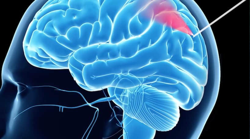
Researchers in Australia have made a new type of imaging needle that can be used during brain biopsies to detect cerebral blood vessels. The device, which is the first of its kind, works thanks to an optical coherence tomography camera attached to the needle and could reduce the risk of fatal haemorrhaging during neurosurgery. It has already been successfully tested out in vivo on 11 human patients.

“We start off with an optical fibre around 125 microns thick and build a tiny lens on the end of it,” explains Robert McLaughlin of the University of Adelaide, who led this research study. “We then angle-polish the end of the lens so that light will come out at its side. We then glue this fibre-optic probe into a commercial brain biopsy needle.

“Standard biopsy needles have a hole at the side, near the tip, and this is where they suck the tissue that is to be removed from the brain for subsequent testing and diagnosis. Our probe ‘looks out’ through this hole so that it can warn the neurosurgeon if they are about to cut a blood vessel.”

Roughly 80,000 people are diagnosed with primary brain tumours each year in the US alone and 14,000 of these patients undergo brain biopsies. Although the procedure is minimally invasive, it still carries a risk because there is a chance that the needle will damage an important vessel. Indeed, about 1% of patients die and 2-3% are left disabled following this damage.

Optical coherence tomography to detect blood vessels

McLaughlin and colleagues’ needle makes use of optical coherence tomography (OCT) to detect blood vessels by differentiating this tissue from blood that is flowing. OCT is similar to ultrasound but uses light waves instead of sound waves.
needle makes use of optical coherence tomography

The technique is routinely employed in ophthalmology and cardiology and does not require any fluorescent or other labels. It instead measures the light scattered from tissue when it is illuminated using near-infrared light. Researchers reconstruct the backscattered optical signal into a structural image of the tissue, and spatial resolutions of 5 to 20 microns are possible.

“Flowing blood has a particular characteristic appearance in these images,” says McLaughlin. “We developed an intelligent image processing algorithm to distinguish between this blood and stationary brain tissue. During neurosurgery the surgeon can see the scan in real time and our software marks it in red when the needle is next to a blood vessel.”

Two types of studies

“One of the major challenges in this work was to determine how accurately the needle could detect blood vessels that can’t be seen with other, standard imaging methods such as magnetic resonance imaging (MRI) because they are too small,” he explains. “We thus performed two types of studies.

“We tested the needle out on 11 patients, all of whom were undergoing craniotomies as part of other types of surgery,” he explains. “This is where the surgeon removes a part of the skull to reveal the brain, so we could see the blood vessels on its surface. For each patient, we chose several blood vessels and moved the needle over the vessel to see if the needle could detect it. We then moved the device over areas without blood vessels to make sure nothing was being detected. This is how we were able to calculate that the needle has a sensitivity of 91.2% and a specificity of 97.7% in blood vessels measuring more than 500 microns across.”

For three of the patients (with grade IV astrocytoma), McLaughlin’s team also deeply inserted the needle into brain tissue – as is done in a normal biopsy. “Before this particular surgery, we identified a blood vessel that was visible on an MRI scan of the patient’s head. We then drilled a small hole in their skull and inserted the needle so that it was adjacent to the blood vessel. In all three cases, the needle was able to detect that it was indeed next to the blood vessel.” This is how the researchers were able to calculate that the needle can detect blood vessels at depths of 21.9 mm, 25.3 mm and 27.2 mm.

“The patients were at Sir Charles Gairdner Hospital in Western Australia and of course gave their consent to be part of this study and weren’t undergoing biopsies,” McLaughlin tells Physics World. “They were actually having other types of brain surgery and in all cases were going to have a diseased part of their brain removed. It was very important for us to come up with a way to test this new device without putting the patient at any risk at all. So, for each patient, we only did experiments on parts of the brain that were going to be removed.”

More advanced needle could detect cancerous tissue too

“Ours is the first reported use of such a probe in the human brain during live surgery and is the first step in the long process required to bring new tools like this into clinical practice,” he adds. “There are many types of operations where a needle is inserted into tissue and there is risk of a bleed, but brain biopsies are the most important because the impact of bleeds in the brain can be so catastrophic.”

The researchers, reporting their work in Science Advances 10.1126/sciadv.aav4992, say they are now working on a version of their needle that can detect cancerous tissue as well as blood vessels. “In current practice, a neurosurgeon may take eight to ten tissue samples during a brain biopsy using a standard needle. If we can develop a needle that can detect when it is in cancerous tissue, they may then only need to take one sample, which would be much safer.

“Developing the next generation of such needles will take several more years of research and development because we need to be very careful when developing tools that will be used on patients.”

Deep-learning algorithm estimates gestational age from smartphone images

Prematurity is a significant cause of mortality in neonates. Knowledge of an infant’s gestational age is critical in post-delivery treatment plans to reduce neonatal deaths. In high-income countries, prenatal ultrasound scans – the ground truth measure – are the gold-standard method to track gestational aging, but in lower-income countries, access to ultrasound technology and medical experts is limited.

“If we could accurately estimate gestational age for newborns using simple, portable technology, we would be able to administer a proper post-treatment plan to reduce the risk of mortality in many under-serviced regions,” says Arjun Desai from Duke University, first-author on a new study into an automatic system for gestational aging.

The cross-disciplinary team, led by Sina Farsiu, has developed a system based on the previously reported inverse correlation between blood vessel density in the anterior lens capsule region, and gestational age. Located behind the pupil, the anterior lens capsule vasculature (ALCV) can be assessed by an expert using an ophthalmoscope.

In this new study, an ophthalmoscope has been attached to a handheld, smartphone-based device to take videos of the ALCV of 124 premature neonates in their first 48 hours of life. The team has now reported on their fully-automatic, deep-learning algorithm that estimates gestational age (Biomed. Opt. Express 10.1364/BOE.9.006038).

Configuring machine-learning

Recording good quality videos during the clinical trial wasn’t easy, and the doctors involved often had to use cotton-buds (or Q-tips) to gently keep the newborn’s eyes open for filming.

“Most of the information acquired in the videos was pretty irrelevant. We wanted to focus on the eye, but each video frame mostly contained the externals of the infants’ face, the room etc. Our algorithm first removed all extraneous information,” explains Desai, who helped develop the deep-learning algorithm that extracted the local eye-region-of-interest from each frame of the videos.

Blind image analysers were used to select the clearest, single frame of each neonate’s eye region, which were passed to a pre-trained neural network to extract representative features. “Deep-learning is still a pseudo black box and one of the challenges is optimizing it to look for important features, while not really knowing ahead of time what these features are and the best methods for extracting them,” says Desai.

The selected features were then clustered using support vector machines to produce a binary classification. The team trained their algorithm on the ground-truth ultrasonic gestational age results to produce a binary yes (1) or no (0) response to multiple thresholds. Each threshold posed the question of whether features from an image were of a specified gestational week or lower. Six thresholds were used, from 33 to 38 gestational weeks.

Manual versus automatic

The researchers tested their automatic results against several manual methods, which used manually-extracted features from ALCV in premature neonates to estimate gestational age. Analysis was a time-consuming task, with the manual selection of the clearest frame from videos of each infant, and then annotation of vasculature to estimate features, such as ALCV density, branch length, and tortuosity (“bendy-ness”). The best performing manual method fit a linear regression between ALCV density and gestational age.

“The automatic algorithm performed as well or better than the manual methods at all gestational ages, except 33 weeks,” said Desai. This new automatic method is far less time consuming than manual segmentations of images and doesn’t require medical expertise to operate.

Data driven

“There may still be some work required to fine tune this algorithm, but as we collect more data we’ll be better able to do that,” says Desai.

It’s not clear how ALCVs correlation with gestational age is affected by nutritional status or racial profile, and Desai points out that the study’s focus on neonates within the United States is a “good start” but may not be “representative enough”. In collaboration with Jennifer Griffin, a research epidemiologist from RTI International in Los Angeles, the algorithm will be tested and fine-tuned in a large-scale clinical trial, funded by the Bill and Melinda Gates Foundation, in sub-Saharan Africa and South Asia.

The automated software is open-source so that communities in low-income countries can freely access, what the team hopes will become, an influential tool for remote neonatal care.

Tiny Casimir torque is measured at long last

A very subtle macroscopic effect caused by quantum fluctuations has been observed more than four decades after it was predicted. The “Casimir torque”, which causes two optically anisotropic objects to rotate relative to one another, has been seen for the first time by physicists in the US, who say that the demonstration could lead to improvements in tiny electromechanical devices and liquid crystals.

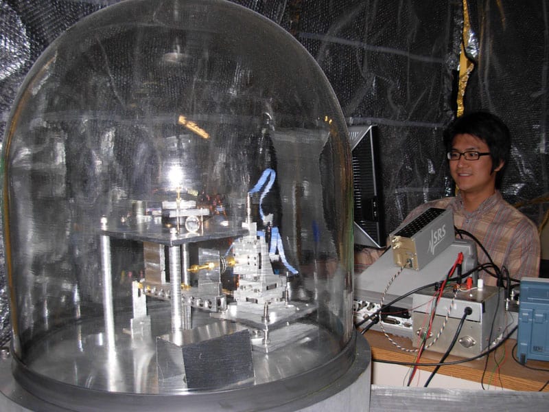
Physicists know that quantum fluctuations in the vacuum can give rise to what is known as the Casimir force. Predicted by Dutch physicist Hendrik Casimir in 1948, this arises between two uncharged parallel metal plates placed within a micron of each other such that they create an optical cavity that restricts the number of different electromagnetic waves that can exist between them. The higher density of waves, and hence energy, outside the plates leads to a tiny net force that pushes the plates inwards.

But in 1972 American physicists Adrian Parsegian and George Weiss predicted that replacing the metal plates with optically anisotropic materials could also lead to a torque between the two objects. That is because the internal asymmetry of each plate would change the boundary conditions of the cavity when one is rotated relative to the other. The torque would arise spontaneously, causing a rotation that would stop when the cavity reaches its lowest energy state.

Realigned gratings

One very simple optically anistropic material is a grating. If two gratings are aligned parallel to one another they trap a limited number of electromagnetic waves – those with the right wavelengths and polarization to bounce back and forth between the gratings’ strips. But if positioned at right angles to one another the gratings do not trap any waves. This means that all waves can exist between the gratings, just as they do outside of them. And because this leads to a higher energy density between the two objects the Casimir torque will cause them to realign.

This effect has now been experimentally confirmed by Jeremy Munday and colleagues at the University of Maryland, College Park, thanks to a novel combination of anisotropic materials – one a solid and the other a liquid crystal. As Munday explains, it is hard to bring two relatively large flat objects close to one another without them touching, and as such most experiments measure the Casimir force between a plate and a sphere. However, he says, fashioning a sphere from an optically anisotropic material – such as a birefringent crystal – is very difficult to do.

This means that most attempts to measure the Casimir torque have reverted to the two-plate system. These experiments typically rely on a pair of solid birefrigent crystals, materials whose refractive index depends on the polarization and direction of an incoming light beam. However, in addition to the original problem of alignment, says Munday, these systems face a second challenge – how to rotate one crystal relative to the other.

Liquid solution

The new experiment overcomes this problem by replacing one of the solid crystals with a nematic liquid crystal. The idea is that the liquid crystal serves as both an isotropic medium and as a torque sensor. The rod-shaped molecules at one end of the liquid crystal are fixed in place while those at the other end are rotated by the Casimir torque, given the presence of the birefringent solid some fixed (small) distance away.

This set-up twists the liquid crystal, which means that a polarized light beam fired through the solid and then liquid crystals has it axis of polarization rotated. The researchers work out this rotation by measuring the intensity of the light after it passes through a second polarizer, and then use the known elastic properties of the liquid crystal to calculate the Casimir torque.

The team measured the torque using four different brirefringent crystals – calcite, lithium niobite, rutile and yttrium vanadate – while varying the orientation of the crystal’s optical axis and the distance from the liquid crystal. The variation in torque almost exactly matched that predicted by theory. “We didn’t know the optical properties for the materials we used over all frequencies,” says Munday. “But for the data we did have the experimental and theoretical results agree very well with each other.”

Macroscopic quantum effect

Munday points out that the Casimir torque is “one of the few macroscopic manifestations of a quantum effect,” and reckons its discovery could have practical applications – such as reducing unwanted rotations within micromechanical devices and the creation of very sensitive rotation sensors.

The research is described in Nature. In an accompanying commentary piece, Slobodan Žumer of the University of Ljubljana and Jožef Stefan Institute in Slovenia agrees that the demonstration “paves the way for the development of complex micrometre- and nanometre-scale mechanical devices.” But he adds that it might be possible to find liquid crystals that experience greater torque and are more easily twisted.

Munday explains that he and his colleagues used a very common liquid crystal called 5CB to minimize unknown effects. The team is now investigating more obscure but potentially more anisotropic materials. He adds that they are also extending the inter-crystal distance beyond their current maximum of 40 nm, in part to establish whether – as predicted theoretically – an increase in separation can flip the direction of rotation for certain pairs of birefringent materials.

Physics-related festive jumpers and Mumbai’s plastic ban

In this final Physics World Weekly of 2018, you can hear the story behind Physics World’s festive jumper competition (or Christmas sweaters if you prefer). Features editor Sarah Tesh is joined by our reviews and careers editor Tushna Commissariat to describe their geeky designs and provide inspiration for you to create your own.

Later in the podcast, multimedia editor James Dacey is in conversation with general physics editor Hamish Johnston about a new Physics World film examining the single-use plastics ban in Mumbai, India. Part of our Sustainable Futures collection, Mumbai’s Plastic Ban explores the motivations behind the ban – which came into effect in June – and the challenges of implementing the change.

Closing the episode, Hamish Johnston speaks about the accelerating growth of the quantum technologies sector. The quantum realm is explored in Physics World’s book of the year 2018 – Philip Ball’s Beyond Weird: Why Everything You Thought You Knew About Quantum Physics is Different. That book is discussed along with loads of other festive reading suggestions in the December episode of our other podcast, Physics World Stories.

If you enjoy what you hear, you can subscribe to Physics World Weekly and Physics World Stories via the Apple podcast app or your chosen podcast host.

Join us for more podcasts in 2019!

Journeys in intellectual property

Scientists, by and large, are more than happy to turn money, whether from government funders or industrial sponsors, into research output. Flip that around, though, and it’s clear that many researchers, and not just those working in academia, are way outside their comfort zone when it comes to transforming scientific breakthroughs into commercial innovation and cold, hard cash.

One of the blockers is the uneasy fault-line between science and commerce, not least the often byzantine world of intellectual property (IP) law. Patents are a case in point. The lifeblood of research impact and commercialization, patents give scientists and their employers legal protection of their invention – effectively the right to take legal action against would-be competitors who make, use or sell an invention without permission over the lifetime of a patent (typically 20 years).

Trouble is, the patenting process and research commercialization rarely follow a linear path from A to B. Here Physics World talks to an academic research manager, an IP professional and a technology entrepreneur about what works well – and what doesn’t – when taking science out of the lab and into the marketplace.

The research collaboration

Anne Moore is project manager of Proteus, a UK-based interdisciplinary research collaboration (IRC) that’s pioneering photonics-based technologies to diagnose and manage lung diseases in the intensive-care environment.

Anne Moore

Proteus is backed by the UK’s Engineering and Physical Sciences Research Council (EPSRC), a government funding body, to the tune of £9.5m. Three consortium universities –  Heriot Watt, Bath and Edinburgh – contribute a further £2m in support.

For Moore, who’s based at the University of Edinburgh, the strength of the IRC model lies in its fusion of research capabilities and domain knowledge across a range of disciplines. “It’s all about team science,” she explains. “Integration is everything – there’s nothing like sharing the same space.”

In the case of Proteus, “team science” means 16 postdoctoral researchers and 24 PhD students spanning chemistry, fibre optics, imaging, signal processing, machine learning and front-line clinical care – all with a shared objective to develop, translate and ultimately commercialize technologies for the rapid, accurate diagnosis of bacterial infection in the lungs.

This emphasis on team science, and specifically broader collaboration and colocation of experts across science, legal and business disciplines, is what really marks Proteus out. “It’s not them and us,” explains Moore. “Proteus is all about putting the scientists, engineers and business development people together.”

As a result, claims Moore, translation and commercialization of research are hard-wired into the IRC’s working model. “There are financial benefits for the IRC universities and the researchers in making sure technologies reach the marketplace,” she says. “Increasingly translation of the opportunities is expected by sponsors and government, while innovation and entrepreneurship form one of the key pillars of our strategic plan here at the University of Edinburgh.”

In fact, Proteus is setting itself up to be a case study in how to patent research breakthroughs in a university setting. “It’s important to dream big: assume your IP is the next big thing,” says Moore. “Across the collaboration we have already filed 12 patent applications, and more are in the pipeline.”

With this in mind, disclosure and assessment of Proteus inventions are handled in-house, and for the benefit of the entire consortium, by Edinburgh Innovations (the innovation management service of the University of Edinburgh), while filing and prosecuting of patent applications are outsourced to Marks & Clerk, a specialist IP legal services firm.

As the Proteus project manager, Moore’s “integration is everything” mindset is crucial in managing the sensitive trade-offs around best-practice IP strategies in an academic research context – for example, the balancing act around research collaboration versus management control; the tensions between academic publication versus patenting; and the relative emphasis on blue-skies research versus translational research.

The bottom line: “It’s never too early to start thinking about IP,” advises Moore. “IP can appear in unexpected places – you can’t take back a disclosure.”

The established manufacturer

Dominic Ashmole is an innovation and IP analyst at Canon Medical Research Europe, the Edinburgh-based subsidiary of Canon Medical Systems, an international manufacturer of diagnostic medical imaging systems spanning CT, MR, X-ray and ultrasound technologies.

Dominic Ashmole

Canon Medical Research Europe traces its origins to the mid-1990s and a University of Edinburgh imaging software spin-out called Voxar, which was subsequently acquired by Barco (2004) and Toshiba (2008) before ending up as part of the Canon group earlier this year.

Ashmole, who’s been an ever-present since the Voxar days, says the 120-strong staff team at the company – comprising software engineers, research scientists, clinical specialists and verification engineers – boasts an impressive track-record of innovation in medical visualization software, evidenced by a portfolio of 70+ granted patents in the past 20 years.

“Working on patents together with our scientists and engineers over the years, we’ve been surprised by many things,” he explains. “For starters, the vast majority of our patents are from a mere handful of inventors. Once they succeed, however, these inventors persist in succeeding. Unfortunately, the bug does not seem to be terribly infectious.”

There’s also what Ashmole calls the “strategy shmatergy” conundrum. “There’s always grand talk about patent strategies [in IP circles], but our reality has seemed more messy than that. Basically, the task is to keep trying things to motivate and enable employees to propose ideas for patents. Then keep navigating the internal and external hoops to get those ideas filed and granted.”

He continues: “To be honest, I’ve never heard of one of our patents being infringed or generating licence revenue. But sometimes large companies don’t sweat the details, instead focusing on building up an overall ‘war chest’ of hundreds or thousands of patents that they can they can cross-licence in bulk with their competitors.”

As a more general advisory, says Ashmole, it’s also worth noting that many, if not most, patents are granted for incremental improvements. “We can’t all invent the lightbulb or the CT or MRI scanner. Consider the maturity of your technical field and don’t self-censor your patent potential.”

Conversely, by the time they are granted, many patents have become so narrow that their only value for big manufacturers is as part of a broader “smokescreen” – essentially a distraction or false trail for competitor companies to follow. “More than half of our patented ideas have not made it into a product,” Ashmole explains, “while many of our product ideas are not patented.”

Ultimately, says Ashmole, patents are sufficiently time-consuming and difficult to read that they are often misunderstood and over-estimated. “Beware the ‘empty shell’, when meagre claims protect little of the grandiose description. It’s all in the claims, and the description and figures may entirely mislead the unwary and hasty.”

The early-stage start-up

Dave Hughes is co-founder and chief technical officer at novosound, a Scottish start-up that’s developed a fully additive thin-film fabrication technique for the volume manufacture of printable ultrasound sensors. Those sensors are being lined up for diverse non-destructive-testing applications spanning oil and gas, industrial process control and medical imaging – target markets that collectively run to billions of pounds each year.

Dave Hughes

Novosound is the first spin-out to emerge from the University of the West of Scotland and back in April closed a seed investment round with £1.5m in funding.

That investment, claims Hughes, is a bet on novosound’s differentiation, along a number of coordinates, versus traditional ultrasound sensor technologies – for example, multipoint devices that work above 250 °C (current ceramic sensors cease to be piezoelectric above around 160 °C); thin-film sensors that offer consistent sub-mm resolution; and printed sensor materials compatible with the complex geometries required in many industrial end-use cases.

Technical innovation notwithstanding, Hughes and colleagues are also majoring on a robust IP strategy. This year, for example, novosound secured a UK patent for its ultrasound sensors in various imaging and measurement contexts, while formal patent protections are in progress for core material science and manufacturing, as well as a fully printed, flexible and high-resolution imaging array for ultrasound applications.

Hughes says the focus on IP is mandatory for would-be entrepreneurs: “The best bit of advice that I got when thinking about commercializing was to ‘stop publishing everything’ and to really assess what was protectable – taking into account the application and market as well as the novelty of the technology and science. Once the IP is protected, there is plenty of time to publish academic findings in a safe manner [versus the commercial potential].”

The university technology-transfer office (TTO) is an invaluable resource in this regard, providing information about previous IP that has (or has not) been protected from the university as well as links to commercial contacts and funding opportunities.

For Hughes, the TTO was instrumental in supporting his start-up funding application to Scottish Enterprise, enabling him to recruit a “commercial champion” – a technology entrepreneur called Richard Cooper, who subsequently became the co-founder and CEO of novosound.

Now that the company is up and running, Hughes and Cooper must keep a constant eye on IP issues to remain on top of any developments inside novosound and further afield. “To achieve this, we have a culture of IP protectionism, whereby every member of the team is on high alert for new IP so that it can be documented and protected – formally if needed. We also use a third-party consultant with a long history of expertise in managing an IP strategy for exploitation.”

Elsewhere, says Hughes, there’s plenty of value to be had from the trial-and-error approaches that typify many start-up operations. “Negative IP affords an opportunity to protect your failures as well as your successes. There is immense value in the knowledge a team builds from things that don’t work.”

  • This article is based on presentations given at the Institute of Physics (IOP) workshop “Journeys in Intellectual Property: Perspectives from Physics and Technology” in Edinburgh, UK. More information on IOP events in 2019 can be found here.

TOF PET/MRI reduces dose for breast cancer detection

© AuntMinnieEurope.com

Clinicians can use as little as 10% of the conventional FDG dose and still produce diagnostic-quality images to detect breast cancer using a time-of-flight (TOF) imaging technique with PET/MRI, according to a study published in the December issue of the Journal of Nuclear Medicine.

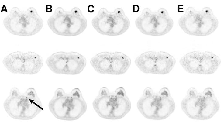
Researchers from Switzerland, the US and Canada used five image reconstruction scenarios with varying amounts of FDG and found that TOF PET images with only 10% of the standard dose of the tracer were sufficiently adequate in terms of image quality, sharpness, noise and lesion detectability.

Concurrently, the reduced dose would bring the radiation burden for women with breast cancer to less than what they would receive from a single digital mammogram.

“Having the possibility of reducing the amount of injected tracer for specific indications will significantly reduce the radiation burden of patients, potentially resulting in a paradigm shift in PET imaging,” wrote lead author Bert-Ram Sah from University Hospital of Zurich and colleagues. “A reduction of injected dose as presented in our results will render partial-body examinations with PET/MRI justifiable.”

PET images

TOF PET potential

PET has long been a primary modality in detecting many cancers, but it also has the downside of radiation exposure for patients. More recently, newer PET imaging techniques, such as TOF imaging, have lessened the need to increase the tracer dose to improve image quality. In addition, the advent of silicon photomultipliers (SiPMs) on PET scanners has improved sensitivity compared with conventional PET/CT and PET/MRI scanners, so users can better balance dose reduction with image acquisition time, the authors noted.

The question is: What is the minimum amount of tracer — in this case, FDG — that will provide clinically useful images? To that end, Sah and colleagues analysed 26 consecutive women (median age, 51 years; range, 34-83 years) with confirmed cases of breast cancer.

Most of the patients were diagnosed with invasive breast cancer (19 cases, 73%). There were 36 primary breast lesions (51%); 26 lesions (37%) of the axillary, hilar or internal mammary chain lymph nodes; and nine lesions (13%) of the bone, lung or abdomen (J. Nucl. Med. 10.2967/jnumed.118.209007).

The patients fasted for four hours before intravenous injection of 3 to 3.5 MBq of FDG per kilogram based on body weight. Clinical TOF PET/CT (Discovery 690, GE Healthcare) was performed approximately one hour after tracer administration. No additional FDG was given for the PET/MRI scan. The mean uptake time for the 20-minute PET image acquisition was 34 min (±6 min).

TOF PET/CT scans followed clinical oncologic imaging protocols before imaging on a simultaneous TOF PET/MRI scanner (Signa PET/MR, GE) with an eight-channel breast coil. There was a mean time of 38 min (±3.5 min) between the start of image acquisitions. The PET/MRI protocol included Dixon-based MRI acquisition for attenuation correction and a dedicated breast MR imaging protocol for diagnostic purposes.

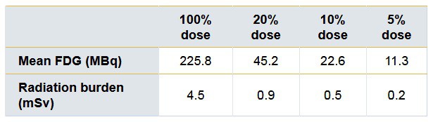
To evaluate TOF PET images at different doses, the researchers reconstructed the 20-minute PET acquisition time for each patient into five different image sets. They began by reconstructing the first two minutes of the scan with three iterations and 28 subsets to use as a standard of reference and for comparisons. They then followed with image reconstruction simulations with FDG dose amounts of 100%, 20%, 10% and 5% for the clinical PET scans in the 20-minute acquisition time.

“In this way, the total scan time remained 20 minutes and, as a result, the data still included normal effects such as decay, biodistribution and eventual patient motion,” the authors wrote.

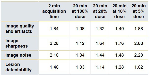
The process created a total of 130 reconstructed PET datasets, with five different reconstructions for each of the 26 patients. Two nuclear medicine physicians/radiologists with experience in PET and MRI rated the image sets based on image quality and artefacts, image sharpness, image noise and lesion detectability using a four-point scale (1 = excellent, no artefacts, 2 = good, 3 = average, and 4 = inadequate, marked artefacts).

Seeing is believing

The 20-minute reconstruction with 100% dose rated the best in all four categories of image quality, sharpness, noise and lesion detectability. The 20-minute reconstructions with doses of 20% (p < 0.001) and 10% (p = 0.001) significantly outperformed the two-minute reconstructions in three categories: image quality and artefacts, sharpness and noise. Image reconstruction with only 5% FDG dose fared rather poorly when compared with the other higher-dose parameters and proved significantly inferior to the two-minute scan for image sharpness.

Mean ratings

Interestingly, lesion detectability rated as good to excellent in all four dose scenarios, with 100% dose reconstruction significantly better than the two-minute reconstruction for primary lesions (p = 0.38) and lymph nodes (p = 0.001). There also was a statistically significant difference between the 20% dose (p = 0.001) and 10% dose (p = 0.032) reconstructions and the two-minute reconstruction for lymph nodes, but no significant difference in primary lesion detection. Reconstruction with 5% dose was inferior to the two-minute reconstruction, but the results were not statistically significant for primary lesions or lymph nodes.

Radiation burden

So how would exceptionally low-dose PET/MRI scans for breast imaging affect patients’ radiation exposure? In this study, the mean full 100% dose of FDG was 225.8 MBq (±5.5 MBq), which means patients would be exposed to approximately 4.5 mSv (±1.1 mSv) of radiation. In comparison, the average effective dose for a digital mammogram is in the range of 0.44–0.56 mSv. Naturally, less dose means proportionally less radiation burden.

Mean radiation burden

Sah and colleagues concluded that a reduction in dose of as much as 90% can produce viable images with less radiation exposure for breast cancer patients.

“To date, PET imaging in oncologic examinations is restricted mainly to whole-body examinations in patients with different cancer indications,” the authors wrote. “This restriction was somewhat justified by the radiation burden in the current clinical setting (5–15 mSv for a PET/CT scan). With the results of this study, this restriction might be reconsidered.”

  • This article was originally published on AuntMinnieEurope.com © 2018 by AuntMinnieEurope.com. Any copying, republication or redistribution of AuntMinnieEurope.com content is expressly prohibited without the prior written consent of AuntMinnieEurope.com.

Can ‘living well’ stop climate change?

Humanity has just 10 years to turn things around. The most recent IPCC report says that we need to reduce anthropogenic carbon dioxide emissions by 45% by 2030 or else be locked into a warming scenario that none of us wants to see. So, what to do? The answer, according to a new study, requires a fundamental change in society. It lies in satisfying human needs like happiness and health instead of focusing on economic growth.

“We need to start thinking, ‘is the carbon footprint that comes from different economic activities actually worth it in terms of societal outcomes?’,” says Gibran Vita from the Norwegian University of Science and Technology. “There is potential to live fulfilling lives with much less environmental impact.”

The challenge is to build societies where our activities are more holistic, enabling us to balance achievement, relaxation, work, play, romance, responsibility and freedom

Gibran Vita

Quality of life is correlated with economic growth. As a result, economic growth is often viewed as the best way of improving quality of life. But there comes a point where more possessions don’t necessarily make us much happier.

To investigate, Vita and his colleagues looked at the carbon footprints that result from meeting people’s fundamental needs. They considered the nine needs in the system developed by Chilean economist Manfred Max-Neef: affection, identity, protection, participation, subsistence, understanding, creation, freedom and leisure.

The researchers calculated the carbon footprint associated with each need by looking at the goods and services that people used to meet it. The team employed EXIOBASE 3, an open-access database containing information on economic activity and associated greenhouse gas emissions and resources for 200 goods in 44 countries. Finally, quality of life measures assessed how effective these carbon “investments” were.

Budget needs

Not all needs are equally polluting, the results showed. Meeting subsistence and protection needs blew nearly half the global carbon budget. Leisure, identity, creation and freedom took most of the other half. But understanding and participation were relatively “cheap”, responsible for less than 4% of the carbon emissions.

Comparing the carbon spend between different countries, Vita and his colleagues found that some needs quickly reach a saturation point in terms of carbon expenditure, whilst others are a bottomless pit.

“No matter how wealthy you are, you can only spend so much money on store-bought food or in heating your house,” explains Vita, who published the study in Environmental Research Letters (ERL). “But there is virtually no limits to the money you can spend on holidays, entertainment, leisure, restaurants, education and so on, even if you are not getting more quality of life bang for your buck.”

Using indicators of quality of life such as those produced by the World Bank and the Human Development Report from the United Nations, Vita and colleagues assessed how well people in each country they investigated felt their needs were satisfied. The team compared this with how much carbon people “spent” on each need.

On average, the results showed that meeting all a person’s physical needs — affordable housing, good health, clean water and so on — required carbon emissions of between 1 and 3 tonnes of carbon per person per year. However, countries like the US and Australia blow around 6–8 tonnes per person per year to meet these same needs, whilst low income nations averaged closer to 1 tonne per person.

For many of the objective measures, such as electricity access or child survival, it was clear that there was a threshold above which more consumption didn’t bring greater satisfaction. It is these areas where Vita and his colleagues believe there are most gains to be made; where carbon emissions can be cut without negatively affecting people’s health and well-being.

People-centred

Countries such as Denmark already lead the way on this front – Danish people have two-thirds the leisure footprint of citizens of the Czech Republic, but similar levels of satisfaction.

“Denmark has people-centred city design, people-centred public spaces, subsidies for sport, good food, accessible cultural events, shorter working days and decent holidays,” says Vita. “None of this requires more carbon. Similarly, countries including Bhutan and some Latin American nations focus on ‘living well’, where helping people to flourish is the driving concern, rather than economic growth.”

To achieve this change to a low-carbon society, the human-needs centred view needs to permeate institutions, businesses, households and individuals, Vita and his colleagues conclude.

“The challenge is to build societies where our activities are more holistic, enabling us to balance achievement, relaxation, work, play, romance, responsibility and freedom, without each of these pursuits being mutually exclusive,” says Vita.

The researchers believe there are interesting options to explore for this transition, including supporting sustainability-oriented grassroots initiatives, deploying universal basic income, reducing the work-week, designing cities for people and editing out polluting choices such as fossil fuel energy, driving, throw-away electronics, plastics and eating meat and processed food.

For wealthy countries this change is easier because they have already invested in housing and infrastructure to meet many basic needs. But there is an opportunity for emerging countries to learn from wealthy nations’ mistakes.

“Emerging countries have a golden opportunity to leapfrog directly to a more sustainable version of development,” says Vita. The question is, can the world transition to a ‘human needs’ economy in time?

Twisted linkage geometries hint at new organic chemistry

Linkages are fundamental components to machines with moving parts. The term describes any assembly of rigid bodies connected by joints. From this vast catalogue of structures, the linkages that most often interest engineers are those with just one internal degree of freedom, meaning they can only change shape in one way, which allows greater mobility control.

Until recently only a few linkages met this requirement – scissorlike elements, the Sarrus linkage, the Bennett linkage, and the Bricard linkage. Now researchers at Okinawa Institute of Science and Technology in Japan have unearthed a whole new class of ring linkages with just one internal degree of freedom – Möbius Kaleidocycles. Reporting in the Proceedings of the National Academy of Sciences, they suggest that technological applications of their discovery may include robotics and “the design of new organic ring molecules with peculiar electronic properties”, as well as raising fundamental questions about geometry, topology, and the limitations of mobility for closed loop linkages.

A degree of freedom

The researchers – Johannes Schönke and Eliot Fried – began by examining the classic sixfold conventional kaleidocycle described by six tetrahedra joined by orthogonal hinges at opposing edges to form a closed ring. The only internal degree of freedom of these structures is an everting motion where different faces of the tetrahedra are exposed as the structure is continuously turned inside out. In contrast for example an eightfold kaleidocycle has two internal degrees of freedom, pressing Schönke and Fried to enquire “How many degrees of freedom does a ring of N tetrahedra linked by N revolute hinges generally have?”

To tackle this the researchers devise a definition for the degrees of freedom of the corners of the kaleidocycle tetrahedra as constrained by the tetrahedron edges. However after subtracting the external six degrees of freedom corresponding to translations and rotations of a rigid body in space, their definition leads to a sixfold kaleidocycle with no internal degrees of freedom.

“It transpires that a K6 [sixfold kaleidocycle] is a nontrivial example of an overconstrained mechanism,” explain Schönke and Fried in their report. “Its high symmetry allows for a hidden degree of freedom.”

A twist in the tale

Next the researchers expanded their investigation to kaleidocycles of conjoined tetrahedra with a twist so that the formally orthogonal hinged edges are no longer at right angles. They found that for kaleidocycles of seven or more tetrahedra there is a critical angle below which it is no longer possible to form a closed loop.

In addition, at the critical angle, all internal degrees of freedom collapse leaving just one as in the case of the classical sixfold kaleidocycle. Since the rings formed in this way share the topology of a Möbius band – recently familiar as the recycle icon – the researchers describe them as Möbius kaleidocycles.

Analogous annulene chemistry

Robotics is one of the fields most likely to benefit from this new class of kaleidocycles with their incorporation in robotic arms or self-propelling swimming rings. However, Schönke and Fried also highlight possibilities to explore in organic chemistry.

They highlight previous work on Möbius annulenes, monocyclic hydrocarbon compounds like benzene but with the twisted Möbius topology. In the 1920s Edgar Heilbronner showed that Möbius annulenes should have novel electronic properties. In 2014 Gaston Schaller at the University of Kiel in Germany and colleagues synthesised an annulene with a threefold Möbius band topology that was energetically favourable over its former onefold counterpart. Drawing an analogy between the “twist into writhe” strategy Schaller and colleagues use to produce an annulene with a lower strain, and the minimum critical twist angle of the topologically equivalent Möbius Kaleidocycles, Schönke and Fried conclude, “We hypothesize that an annulene made from molecular building blocks (equivalent to our tetrahedra) with a twist angle close to the critical angle should exhibit minimal strain.”

Future topics for further fundamental investigations include the possibility of other ring linkages with more than seven elements and only one degree of freedom, an explanation for the everting motion that the ring geometries can undergo, and other possible topologies and geometries.

Full details are reported in the Proceedings of the National Academy of Science.

Doughnut waveguide preserves twisted light

A new waveguide that can transmit twisted light within a photonic chip has been unveiled in China. It was created by Xian-Min Jin at Shanghai Jiao Tong University and colleagues and allows twisted light to propagate without significant degradation to its helicity. The waveguide could lead to the development of new chips for optical and quantum computing.

Characterized by its corkscrew-shaped wave fronts, twisted light carries orbital angular momentum (OAM) in a series of independent states. Each of these OAM states can be used to encode information, and this has already been used to boost the amount of data that can be transmitted using light. The quantum-mechanical nature of these states could also make twisted light useful for creating quantum computers.

An important challenge for those creating practical devices is that the effective index of refraction of a conventional solid wave guide is too low to allow the transmission of individual OAM states. Instead, states with similar OAM values will blur into each other.

Notoriously difficult

The solution has been known for some time: create a “doughnut” waveguide with an inner core and concentric rings made of materials with the appropriate indices of refraction. However, this complex structure has proven notoriously difficult to fabricate.

Jin and colleagues achieved the first practical implementation of a doughnut waveguide by using a laser-writing technique. This involves firing precisely-focused, femtosecond pulses of green light at a borosilicate glass wafer. Absorption of the pulses causes highly-localized transformations of the refractive index of the glass, resulting in a 10 µm-diameter, 20 mm-long doughnut waveguide.

Intensity profiles

The team tested their on-chip waveguide using twisted light with OAM values of +1, 0, and -1, as well as each combination of superpositions of the three states.  The researchers measured the intensity profiles of twisted light entering and exiting their waveguide, while also testing the helicity of the emerging light by interfering it with a separate twisted beam fired through free space alongside the waveguide.

They found that about 40% of the initial intensity of the twisted light was lost inside the waveguide. Even greater losses were measured for states with greater OAM. They also observed that the waveguide also filtered-out photons with poorly-defined OAM. As a result, the helicity of the light remained remarkably well-preserved as it passed through the waveguide.

Jin and colleagues believe their doughnut waveguide could be used in commercially-viable, high-capacity optical communications in the near future. The physicists will now work towards increasing the transmission rates of twisted light with higher values of OAM in their waveguide, potentially creating further opportunities in the rapidly-growing fields of quantum and optical computing.

The waveguide is described in Physical Review Letters.

Pristine graphene produces giant photoresponse

Graphene is a sheet of carbon just one atom thick and has many unique properties, such as extraordinarily high electron mobility and high thermal conductivity. It can also, in principle, absorb light over all frequencies of the electromagnetic spectrum, which makes it ideal for use in photodetectors, biosensing and bioimaging applications, as well as in night vision devices. Now a team of researchers at the University of California at Riverside and the Massachusetts Institute of Technology (MIT) has discovered that pristine graphene can produce large amounts of photocurrent when illuminated with light if it is made into special, constricted, shapes. The unexpected thing about this photocurrent is that it occurs at graphene’s charge neutrality point – at which current flow is not expected at all. The new finding could be important for developing more efficient and ultrafast photodetectors and even improved light-harvesting devices like solar panels.

In 2011, a team of researchers led by Pablo Jarillo-Herrero discovered that, when exposed to light, graphene produces “hot electrons” that then generate a photocurrent. This so-called hot-carrier regime is very unusual and is normally only seen at extremely low temperatures or in very non-linear processes, but in graphene it occurs at all temperatures from very low up to room temperature – and in the linear regime – when the material is excited with a laser.

Graphene can also produce a photocurrent when it is subjected to electric fields. Despite the fact that the processes responsible for producing these two types of photocurrent (thermoelectric and photovoltaic) are very different, they do share a common feature in that they are prominent at high charge densities but suppressed at the charge neutrality point. This is the point at which the valence and conduction bands meet and is also called the Dirac point. This behaviour is in complete contrast to what the researchers have now observed in pristine graphene – that is, graphene free of excess electronic charges such as those induced by doping or defects.

Unusual configurations

The team observed the Dirac-point photocurrent in samples cut and shaped into unusual configurations, like ladder-like linear arrays, narrowly constructed rectangles and samples with tapered and tailored edges. The largest photocurrent emerges in constricted areas, such as in the regions where a narrow ribbon connects wide regions, when they are illuminated with light.

Jarillo-Herrero and colleagues obtained their results by exciting their samples with a focused 850-nm continuous-wave laser and measuring this photocurrent through source and drain electrode contacts.

“Until now, converting light into electricity in graphene relied on the photothermoelectric effect,” explains Jarillo-Herrero. “For this effect to work, we need a junction formed between electron- and hole-doped graphene. This is the same as what we need in most other solar energy harvesting or photodetection devices. Here, the electrical current is generated in the junction region and moves through the distinct areas between the differently doped parts.

“What is different in graphene, however, is that the Fermi level shifts off the Dirac point and into the conduction or valence band once the material is doped with charge carriers. And this unfortunately blocks the material’s ability to absorb low-frequency photons.”

No special junctions required

“In our new work, we show that pristine graphene can convert light into electricity very well without the need for doping and without the need for a p-n junction structure. This means it continues to absorb light at any low frequency.”

“When we shine a laser on a perfectly neutral device, electrons are pushed away from the light beam, driving a local current, adds team member Nathaniel Gabor. “When you shine the laser on a corner or constriction, however, the electrons are forced to travel in only certain directions (they have to be along the same momentum line). And since the electrons travel long distances at the Dirac point in graphene, they eventually produce a strong photocurrent.”

Strongly interacting electrons

The phenomenon comes from the fact that electrons in pristine graphene interact very strongly with each other since they are confined to a very thin, atom-sized membrane, he adds. They are thus expected to behave as a liquid, transferring their energy collectively as they move.

“Combining the hot electrons that exist in graphene with the fact that we don’t require any sort of junction in it, means that we can engineer and control the behaviour of light-harvesting devices made of this material by simply exploiting the shape and size of a sample. What is more, because the sample is only one atom thick, we could use it to make devices that are semi-transparent and shaped to generate large populations of hot electrons. These could be embedded in structures such as windows panes, for example, or combined with other more conventional photosensitive materials to harvest excess energy from sunlight that is not usually absorbed.

The researchers, who detail their work in Nature Nanotechnology 10.1038/s41565-018-0323-8, say that they now plan to further study graphene’s intrinsic photocurrent effect over a broad range of infrared and THz frequencies and measure its response speed. “Our current work was carried out on mechanically exfoliated samples of the carbon sheet and we now hope to use material grown by chemical vapour deposition (CVD) as the next step for scale up,” says Jarillo-Herrero.

Copyright © 2026 by IOP Publishing Ltd and individual contributors