Investing in the climate

Hurricanes Harvey, Irma, Jose and Maria, which swept across the Caribbean last year, caused an estimated $200bn of damage. Recent studies indicate the frequency of Harvey-like downpours over Texas may have already increased by up to six-fold since the late 20th century. Since a hotter atmosphere has a more energetic water cycle, and warmer air can hold more moisture, future climate change is likely to increase the intensity and perhaps the frequency of hurricanes still further.

The study of extreme weather events such as hurricanes is one example of how science can raise thought-provoking and important questions regarding the appropriate actions of both investors and companies. In the wake of the devastation of the 2017 Atlantic hurricane season, the question is whether some companies should be held at least partially liable for their activities, with possible implications for investment. Could carbon-intensive industries be held liable for some of this damage? And how would you even quantify their responsibility?

Risk and responsibility

The economic impact of extreme-weather damage is already beginning to be incorporated into risk assessments, with some fund managers considering climate issues in decision-making. Yet the financial liability of carbon-intensive industries for such damage may not be reflected in companies’ market valuations.

Apportioning responsibility for such damage is, in principle, possible. We know that cumulative carbon-dioxide emissions are the primary cause of changes in the global climate, which means we can start to quantify contributions from individual nations and companies, including extreme weather-event frequency increases. In 2015, for example, the fossil-fuel sector accounted for 91% of global industrial greenhouse-gas emissions. From 1988 to 2015, some 25 companies and state producers generated 51% of global industrial emissions. Seven of these were publicly owned companies, collectively accounting for 9.5% of “scope 1” and “scope 3” emissions and with a combined market capitalization of around $1220bn.

If such firms contributed 9.5% of the 2017 hurricane damage ($19bn), this would decrease their share price by 1.6% – a not insignificant sum, particularly if contributions are requested for other past and future extreme-weather events. If global warming increases hurricane losses, under a hypothetical climate-liability regime, damage contributions approximating 1–2% of companies’ market capitalizations might become more usual with each annual hurricane season. This ignores other climate impacts, such as sea-level rise, which could readily run to much larger sums.

Despite the science, however, no legal precedent yet exists for extreme weather-event climate-damage liability. The 2015 Paris Agreement explicitly rules out damages associated with climate change as a basis for liability. It is hard to say how investors might react to the possibility of companies having to contribute for damages associated with climate change caused by their past emissions. Barriers to successful climate-damages compensation cases remain substantial, but as insight develops, the possibility remains. For major insurance companies or governments footing the bill, the prospect of multi-billion-dollar pay-outs may focus attention on whether legal barriers could be overcome, potentially allowing them to pass on costs.

Climate change highlights the challenges faced in making research accessible and relevant to the broader community. Members of the public, industrialists and financiers rarely read scientific journals, so society’s response to such research can therefore take a long time. The need for scientists to be open to broader uses for their research is now more crucial than ever. Regular engagement outside academia would help to ensure that wider society has a more robust scientific understanding, as well as a clear demarcation of where the current knowledge boundaries lie.

Based on scientific insights that highlight and communicate possible investment risks, environmentally aware investors can actively nudge companies away from destructive behaviours towards a more constructive role. Selective investment in firms facilitating the transition to a net-zero-carbon economy supports them, while refusal to buy shares in those companies failing to do so can make it harder for them to raise capital. Even small investors’ accumulated views matter, just as individuals should believe that recycling their plastic bottle or casting their democratic vote makes a difference.

The ever-developing world of “sustainable investing” is a valuable way for climate scientists to have a real influence on financial markets. Environmentally focused investors are integrating climate risks into financial decision-making in many different areas.

Ethical investment

Apart from reaching out to broader society, scientists have another route to express their insights. As individuals, many invest savings in funds and pensions schemes. By actively seeking out sustainable, environmentally focused investments, they too can support companies that share their values while avoiding those that do not contribute to climate solutions.

To influence social and corporate behaviours, scientists need to engage with the wider world through social media and other routes not normally used by academics. Physicists with cross-disciplinary skills are also contributing to this effort by making their research accessible and relevant to finance and business.
Experts agree on advantages of direct current power in buildings

Experts broadly agree that a widespread adoption of direct current (DC) power systems in commercial and residential buildings could offer significant advantages over alternating current (AC) systems, according to a new study. The finding could help change perceptions of DC among industry professionals, potentially resulting in safer, more reliable, more energy-efficient buildings.

Brock Glasgo at Carnegie Mellon University, US, and colleagues consulted 17 experts in a variety of industrial and academic fields to identify the advantages of a more widespread adoption of DC power systems while acknowledging their potential challenges.

Our society’s use of electricity is changing rapidly. While developments in renewable energy generation have seen sources for the power grid becoming more dispersed, increases in modern electrical components and devices in buildings have caused power consumption to grow steadily. In light of these changes, many scientists have encouraged increased adoption of DC power distribution in commercial and residential buildings.

According to Glasgo, the case for DC power has three key aspects. “Firstly, we now have semiconductor-based power electronics that function as DC-DC transformers and are nearly as efficient as modern AC-DC and DC-AC transformers,” he says. “Secondly, we’re seeing consistent growth in the installation of solar PV [photovoltaic] and other distributed generation sources that generate DC. And thirdly, a growing fraction of the electricity consumed in modern buildings is either consumed as DC or passes through a transient DC state on its way to being consumed.”

To some, these advantages prove that power systems that incorporate both AC and DC are becoming overly-complex and outdated. “Eliminating unnecessary DC-AC and AC-DC conversions by distributing DC power would not only simplify our building-level power supply but would also save energy,” says Glasgo.

Yet despite its technical and economic advantages, the widespread adoption of DC faces major barriers in other areas. Through their interviews, Glasgo’s team identified the two biggest obstacles as an unfamiliarity with DC among industry professionals, and the under-representation of DC devices and components in the market.

“The AC grid has been in place for over 120 years, and all of the physical components, the design, maintenance, construction, operation, and end users’ interactions with the electric transmission and distribution system are based on a long history and the physics of AC,” Glasgo says. “A transition to DC-powered buildings will depend on far more than the technical feasibility of the systems themselves.”

However, the experts generally remained hopeful that these challenges could be overcome; identifying areas which they believed should be prioritised to make a better case for widespread adoption.

“Our experts proposed training engineers and electricians on DC systems and identifying niche use cases where DC power distribution holds a clear advantage over AC and building pilot projects to help build the market for DC devices and components,” says Glasgo. “The professionals responsible for DC power systems and the markets needed to support them will need to undergo a major transformation before they [DC power systems] can be employed to more efficiently, safely, and reliably meet the demands of future buildings.”

Glasgo and colleagues reported the findings in Environmental Research Letters (ERL).

Lithium-oxygen batteries broach 100% coulombic efficiency

If you’re reading this with a rechargeable battery powered appliance, the chances are it’s a lithium-ion battery based on intercalation chemistry. But with increasing demands for higher energy density power banks the search is on for alternatives.

“Intercalation of a cation into a structure (along with the accompanying stored electron) doesn’t change the framework very much.  Charging and discharging is like driving a car in and out of a parking garage, where the framework remains intact,” explains Linda Nazar, a professor in the Department of Chemistry at the University of Waterloo in Canada. “But if you try to drive too many cars in you get irreversible changes to the structure.” In addition to this fundamental limitation to the energy storage capacity this poses, lithium ion batteries use metals such as cobalt, whose cost is increasing and where sustainable mining is problematic.

The push towards alternatives to the intercalation chemistry of lithium-ion batteries has led to increased interest in lithium-oxygen batteries, which charge and discharge by converting lithium and oxygen into a metal oxide and back again. However parasitic side reactions have plagued efforts to maximize the efficiency and reversibility of this reaction for several years.

After years of working on lithium-oxygen batteries, Nazar’s group came to the conclusion that the organic solvents used for the battery electrolyte were simply not workable. Using selected inorganic electrolyte counterparts that operate at temperatures of 150 °C, and a bifunctional electrocatalyst, her team was able to demonstrate high-energy-density reversible lithium-oxygen battery charging and discharging with close to 100% coulombic efficiency that operates with a four electron redox reaction.

Chemistry coming together

Previous designs of lithium-oxygen batteries have largely used a carbon electrode and an organic electrolyte and form lithium peroxide (Li2O2) on discharge. Instead Nazar and her team use a noncarbonaceous composite cathode composed of nickel nanoparticles for the cathode and a lithium nitrate/potassium nitrate (LiNO3/KNO3) eutectic molten salt as a liquid electrolyte.

The molten salt electrolyte has a number of key properties. Reduction of oxygen forms reactive superoxide that is primarily responsible for the parasitic side reactions that impinge on the reversibility and efficiency of the cell. However in Nazar group’s design the nickel nanoparticles at the cathode become coated with LixNiO2 in situ, and this LixNiO2 catalyses the four electron conversion of oxygen into lithium oxide (Li2O) a process that is reversed on charge with the aid of the electrocatalyst. Reduction-oxidation to Li2O also doubles the electron storage, increasing the energy density of the cell by 50% compared with the peroxide.

In addition, the electrolyte provides just the right solubility for the ions, sufficient for Li2O crystals to nucleate at the electrode but not so great that they accumulate all over the cell and lose contact with the electrode.

“There are numerous facets of this chemistry that have to come together to make it work,” says Nazar. “While I wouldn’t say I was surprised that it worked I was pleased – there has been a lot of frustration in the field, so it was nice to see that oxygen chemistry can work well.”

Molten inspiration

Nazar points towards a wealth of literature on the properties required for a good oxygen evolution and reduction catalyst, including several papers by Yong Shao Horn at MIT. However she attributes the success of their battery design largely to inspiration from previous work published on molten salt oxygen electrochemistry by Liox Power and colleagues at Caltech and Berkeley that used a carbon cathode, and the tenacity of her postdoc Chun Xia for attempting to build on the work. Finding the right bifunctional catalyst/metal cathode was key.

While the elevated operating temperatures of the battery may limit applications, there are examples of other types of battery and fuel cells with higher operating temperatures that are already commercialized. However Nazar tells Physics World, “For us the main impact is demonstrating that lithium-oxygen chemistry is reversible.” Future work will focus on improving the electrochemistry.

Full details are available in Science.

Dispersible nano-electrode sensors could detect early-stage cancer

Dispersible electrodes based on gold-coated magnetic nanoparticles modified with DNA can detect microRNA in unprocessed blood samples at extremely low concentrations and over a broad range – a first for sensors of this kind. The devices, which have been tested on mice, can produce results in just 30 minutes and might be used to make a finger-prick test for early-stage cancer diagnosis.

“There are many microRNAs (short ribose nucleic acid sequences between 19 and 25 bases long) that are post-transcriptional gene expression regulators – that is, they can turn genes on and off,” explains John Justin Gooding of the University of New South Wales in Australia, who led this research effort. “The levels of these miRNAs are indicative of a range of pathologies, including cancers. If we could detect these RNAs in blood, where they circulate, we could make a finger-prick test as an early cancer diagnostic.”

 Detection levels as low as 10 attomoles

“The problem is that the RNAs are found at very low concentrations of 10 femtomoles (fM) to 1 picomole (pM), so this is no easy task. Our new technique can detect levels as low as 10 attomoles (aM) and above 1 nanomoles (nM), so covering this entire range. What is more, it produces a result in just 30 minutes.”

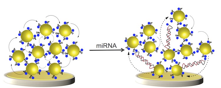
Gooding and colleagues developed gold-coated magnetic nanoparticles (Au@MNPs) modified with DNA that is complementary to the miRNA they want to detect. “We call these magnetic nanoparticles ‘dispersible’ electrodes because they diffuse throughout the sample to capture the miRNA,” says Gooding. “When we then apply a magnetic field, these tiny electrodes reassemble to form a bigger electrode.

Collecting Au@MNPs

“When miRNA is bound to the DNA of the nanoparticles, the electrochemical current through the macro-electrode changes. The electrode measures this change and produces a signal,” he explains. “Since the nano-electrodes disperse throughout a sample, they capture nearly all the miRNA in it and the more they capture, the bigger signal. This is why our device is so sensitive.”

Fast response time

The sensor also has a fast response time since it makes use of an applied magnetic field to “bring back” all the captured miRNAs to the macro-electrode, he adds. “It can be likened to a hunter-gatherer that is on a motorcycle rather on foot. When sent out to find ‘food’, it covers more territory, so collects more food and brings it back faster.”

The technique is better than the current gold standard to profile miRNA, the real-time polymerase chain reaction (qRT-PCR), which does not work on samples of whole blood (it requires isolated and purified RNA). Although highly reliable, qRT-PCR is also labour-intensive and time consuming.”

“Our sensor is the first to be able to detect concentrations of miRNA from 10 aM to 1 nM in unprocessed blood samples,” Gooding tells Physics World. “We found that it can also distinguish small variations in miRNA concentrations in blood samples taken from mice with growing tumours.

“We believe that our work is an important advance for developing liquid biopsies for early cancer detection, that is before symptoms of the disease actually appear, and to monitor how well, or not, a treatment is working,” he adds.

The researchers, reporting their work in Nature Nanotechnology 10.1038/s41565-018-0232-x, will now be trying to multiplex the technology so that they can simultaneously measure different types of miRNAs.

Augmented reality device aids interventional oncology

© AuntMinnieEurope.com

An Italian-led research team has developed a technique that can use augmented reality instead of conventional intraprocedural imaging to guide needle insertion for interventional procedures, according to research published in European Radiology Experimental.

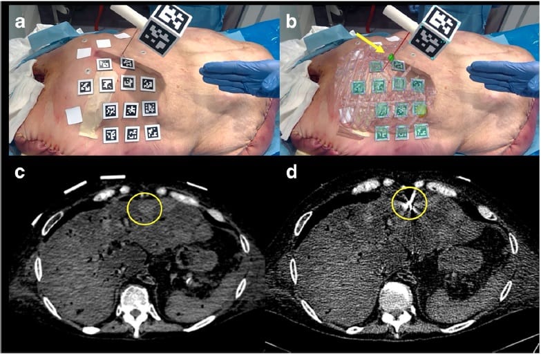
The group, led by first author Marco Solbiati of advanced visualization firm R.A.W., created augmented reality software capable of using a tablet to project 3D models of CT scans directly onto a target. They found that applying this technology to needle insertion — a common first step for the interventional oncology procedure thermal ablation — allowed clinicians to simulate the task well within a 5-mm accuracy threshold (Eur. Radiol. Exp. 10.1186/s41747-018-0054-5).

Augmented reality

This proof-of-concept study for the device was the first time that clinicians were able to target focal liver lesions using augmented reality only, wrote Solbiati and colleagues from various institutions in Italy, Israel and the US.

“Indeed, our investigation demonstrates that an augmented reality system can provide accurate guidance for interventional oncology procedures … without the need for further real-time intra-procedure imaging (such as ultrasound or CT), thereby avoiding possible exposure to ionizing radiation and the need for an additional co-registered modality,” they wrote.

Three-step experiment

Highly accurate image guidance is critical for the success of minimally invasive procedures performed in interventional oncology, a growing offshoot of interventional radiology, the authors noted. During surgery, clinicians in this field most commonly refer to a display of MRI or CT scans to guide intricate procedures such as thermal ablation. But this requires them to frequently look back and forth between the patient and the screen as well as mentally reconstruct the 2D scans into 3D images while performing the operation.

“Efforts have recently been made not only to improve the efficacy of ablative devices, but also to increase the accuracy of image-guiding systems,” the authors noted. “Several navigation systems have been developed using augmented reality techniques … [that] allow the operator to see 3D virtual objects superimposed upon the real world and not on a different screen.”

Seeking to improve the precision and efficiency of image guidance for interventional procedures, Solbiati and colleagues developed a new technique that relies on augmented reality instead of conventional radiographic imaging for visualization.

They created their augmented reality software (Endosight, R.A.W.) in a computer program (Unity 2017.1.1, Unity) and designed the software to superimpose 3D images directly onto a target. The augmented reality device includes a customized needle handle and a tablet (Microsoft Surface Pro 4, Microsoft) attached to a tripod that can display 3D images through its camera.

To test the accuracy of the technology, the researchers simulated needle insertion for thermal ablation using the augmented reality device on three different models: a custom-made silicone phantom, a pig and a cadaver with liver metastases.

First, they placed radiopaque skin markers on each of the models, acquired and processed CT scans of the models, and converted the scans into 3D images for the augmented reality software. Then the investigators used the augmented reality device to project these 3D images directly onto the corresponding model. This allowed the clinicians to visualize the targeted area for insertion in relation to relevant internal structures as they manoeuvred the needle.

High targeting accuracy

Overall, the tests for augmented reality guided needle insertion demonstrated extremely high accuracy — within the threshold of 5 mm from the targeted site. The average distance between the geometric centre of the model and the augmented reality images was 2 mm for the phantom, 3.9 mm for the pig model and 2.5 mm for the cadaver. The mean distance was 2.8 mm for the two liver tumours in the cadaver.

For the pig model, the mean amount of time it took to set up the device was 5.3 minutes, and the average time to perform needle insertion was 7.2 minutes. For the cadaver, the average set-up time was 5.8 minutes and the average procedural time was 9.4 minutes.

With the aid of the augmented reality device, clinicians may be able to visualize 3D models of organs and thermal ablation targets superimposed on real patients rather than on a different screen — improving the treatment result, the authors noted.

They are currently working on additional improvements for the prototype augmented reality device, including reducing the number of skin markers required for alignment and using fibre optics to detect needle bending during insertion in real-time.

“The application of augmented reality to interventional procedures might have a relevant impact in further improving the precision of guidance during ablation and holds the potential for a large diffusion in the near future,” they concluded.

  • This article was originally published on AuntMinnieEurope.com © 2018 by AuntMinnieEurope.com. Any copying, republication or redistribution of AuntMinnieEurope.com content is expressly prohibited without the prior written consent of AuntMinnieEurope.com.

Barnacles are no match for mushroom-shaped microstructures

It could soon be much more difficult for barnacles to cling onto the hulls of ships, thanks to a coating of mushroom-shaped microstructures that has been developed by researchers in Germany. The sea creatures – which are a serious problem in marine environments – are unable to completely wet the coating with their glue and form a strong, reliable bond, the researchers say. They add that their coating could help reduce the use of toxic anti-fouling coatings.

Biofouling is the unwanted accumulation of microorganisms, plants, algae and animals on surfaces. Aquatic biofouling affects the aerodynamics of ships and boats, increasing drag and therefore energy use. It can also corrode the surfaces of watercraft and structures such as bridges and wind turbines – and the build-up of organisms makes visual inspections and structural checks difficult.

Biofouling is also an important vector for invasive species. “Imagine your container ship is somewhere in Asia, in the harbour, and marine organisms from that ecosystem attach to the ship hull and then the ship travels to Europe or the US and the organisms detach, or release offspring,” explains Lars Heepe, a materials scientist and biophysicist at Kiel University in Germany.

Permanent fixtures

Of particular concern are so-called hardfoulers, like barnacles and mussels. These sessile marine organisms use adhesive substances to fix themselves permanently to surfaces. Most current strategies to tackle them use anti-fouling paints that stop larvae settling. But, these toxic coatings release harmful chemicals that contribute to marine pollution.

A barnacle’s cement works like most glues, explains Heepe. It starts as a liquid, so that it can flow across surfaces and into any defects before setting hard. As barnacles rely on thoroughly wetting the surface before hardening, to achieve a strong, permanent bond, Heepe and his colleagues wondered if it would be possible to tackle biofouling using purely physical means. They hypothesized that a non-wetting surface topography that the cement cannot completely coat would reduce the adhesive strength of barnacles.

Mushroom microstructures

To test this, they used a silicone-based material covered with mushroom-shaped microstructures – essentially pillars that bulge at the top. The bulges create an upward force on water droplets that prevents them collapsing into the space between the pillars, keeping the cavities dry. By contrast, on a similar surface with micropillars – without the mushroom-like structures at the top – water flows into the cavities between the pillars.

The researchers placed surfaces covered with mushroom-shaped microstructures and micropillars in the Baltic Sea for 17 weeks. For controls, they also submerged flat surfaces covered in the same silicone-based material and acrylic glass.

For the first three weeks barnacles only attached to the control surfaces. From week four to seven they steadily increased on all four surfaces, reaching an average density of 0.7 per 10 cm2 on the mushroom-shaped microstructure and micropillar surfaces, compared with a density of more than 2 per 10 cm2 on the control surfaces.

Zero coverage

Between week seven and 13 the number of barnacles on the mushroom-shaped microstructure surfaces dropped to zero, while the density on the micropillar-covered surfaces remained stable. The density on the silicone control surfaces also dropped to 0.7 per 10 cm2 by week 17, while the barnacles on the acrylic surfaces increased to 4 per 10 cm2.

Dennis Petersen, who is also based at Kiel University, told Physics World that it was already known that barnacle larvae settle at lower rates on surfaces with pillar microstructures – they appear to sense that they may not be good surfaces to attach to. Over time, however, they do slowly build up on these surfaces. The problem is that at some point the larvae have to attach somewhere. “It becomes a matter of life and death,” explains Petersen.

Because of this, Petersen says that the team were not interested in a surface that would deter settling – although the surface with the mushroom-shaped microstructures does – but one to which barnacles could not remain attached.

Knocked off

Petersen and Heepe say that their results suggest that the right surface topography can prevent the permanent adhesion of barnacles. Specifically, a surface that resists wetting reduces the contact area between the barnacle and the surface, resulting in a weak bond. They are then knocked off by forces such as natural currents and water flows created as the boat moves.

Marine-science expert Tony Clare of Newcastle University says the results “are interesting and show promise”. He encourages the Kiel team to “explore the utility of the approach beyond barnacles”.

The research is described in the Journal of the Royal Society Interface

Brownies get a new space badge, taking NASA selfies, Hey LIGO helps with debugging

Brownies in the UK have a new space badge thanks to a partnership between the Royal Astronomical Society, the UK Space Agency  and Girlguiding UK. For girls age 7-10, the badge “aims to spark girls’ curiosity to explore the universe around them by providing opportunities to develop the skills and confidence to engage in astronomy, planetary and space science,” according to a statement from the organizations. “Badge activities include stargazing with the challenge of identifying constellations on a clear night, creating a sunspot viewer and plotting a sunspot map, and designing an astronaut training programme.”

Cat in space

Once you have bagged your space badge, you could celebrate by taking a selfie that is out of this world using the NASA Selfies app. This puts you in a spacesuit with a selection of back drop images acquired by NASA’s Spitzer Space Telescope. Also new from NASA is the Exoplanet Excursions virtual reality app, which will send you on a tour of the Trappist-1 system of seven exoplanets. Both apps have been released to celebrate the 15th anniversary of the launch of Spitzer.

Moving from infrared astronomy to the detection of gravitational waves, Nikhil Mukund of India’s Inter-University Centre for Astronomy and Astrophysics has created an app called Hey LIGO. Inspired by digital assistants such as Apple’s Siri, Hey LIGO combs through LIGO logbook entries to see if solutions have already been found for specific problems with the huge detectors – something that could save operators a great deal of debugging time.

“If you ask some questions about the interferometer, [Hey LIGO] could intelligently come up with some answers that could help someone in debugging an issue or in knowing more about the detector,” explains Mukund. You can read more about it in Symmetry.

Growing risk of extreme heat and humidity

By the close of the century, the two-fisted assault of extreme heat and humidity could make the North China plain a deadly zone.

As water vapour rises from irrigated farmland, in heat extremes which are likely if humans go on burning ever-greater quantities of fossil fuels, then air temperatures and moisture conditions could become such that outdoor workers could no longer cool by perspiration.

In such circumstances no normal healthy person could survive more than six hours. And since 400 million people already live on the North China plain, by 2070 the consequences of ever-greater temperatures could be devastating, according to new research in the journal Nature Communications.

Simultaneously, a second study in a separate journal confirms that by 2080 excess deaths from extremes of heat will have risen in the tropics, subtropics and even the temperate zones.

In three of Australia’s great cities, deaths from heat waves will have risen by more than 470%.

The warning for China – which already emits more greenhouse gases than any other nation – is based on what meteorologists call “wet bulb” temperature, the combination of heat and humidity. When this climbs towards the natural body temperatures of humans and other mammals, conditions become dangerous. The North China plain covers 400,000 square kilometres of fertile floodplain irrigated by three great rivers.

The alarm is sounded by Elfatih Eltahir and a colleague at Massachusetts Institute of Technology in the US. Eltahir first identified the additional hazard of humidity in extremes of heat with a simulation of close-of-the-century temperatures that pinpointed the Gulf region, between Iran and the Arabian peninsula, as the zone where temperatures could become lethal. But the worst extremes would be over water.

A second examination of likely conditions under what climate scientists call the “business-as-usual” scenario, in which nations go on burning fossil fuels and emitting greenhouse gases in ever-increasing quantities, pinpointed Asia as the continent most at risk of lethal heat extremes for the greatest numbers of people.

The latest study is a refinement of the projections, and is based on evidence from the most recent three decades. Warming in the North China region has been double the global average – 0.24 °C per decade compared to 0.13 °C for the rest of the world. In 2013 there were extremes of heat that lasted for up to 50 days, and maximum temperatures topped 38 °C (around the accepted limit for humans).

Irrigation key

And the potential lethal factor for the region is likely to be irrigation: rainfall in the north is low, and evaporation from the soil moisture adds around another 0.5 °C to local temperatures. Water vapour is itself a greenhouse gas.

“This spot is just going to be the hottest spot for deadly heat waves in the future, especially under climate change,” said Eltahir.

That extremes of heat combined with higher hazards from humidity are already on the increase, and will continue with ever-greater ratios of carbon dioxide in the atmosphere, is firmly established. A second international study, in the Public Library of Science journal PLOS Medicine, looks at the risks for more than 400 communities in 20 countries for the decades 2031 to 2100, and once again it is based on a business-as-usual scenario, and data from recent decades.

If the world goes on warming according to the gloomiest predictions, the levels of heat-related excess mortality, the statistician’s phrase for death by heatstroke or heat exhaustion, then deaths in Colombia will by 2080 have risen by 2000%. Even in Moldova, the sample country with the lowest risk, they will have risen by 150%. In Brisbane, Sydney and Melbourne, the hazard will have soared by 470%.

Inexorable rise

That heat can kill has been known for decades, and the tens of thousands of extra deaths during heatwaves in Europe in 2003, and Russia in 2010, were harsh reminders. More extremes of temperature are inevitable.

Research of this kind is intended to encourage thinking about ways in which health authorities and city bosses could act to reduce the hazard. But for a global problem, a global solution could be the surest answer.

“Future heatwaves in particular will be more frequent, more intense and will last much longer,” said Yuming Guo of Monash University in Australia, who led the research.

“If we cannot find a way to mitigate climate change (reduce the heatwave days) and help people adapt to heat waves, there will be a big increase of heatwave-related deaths in the future, particularly in poor countries located around the equator.”

Portable imager provides insight into eye and brain diseases

A handheld ophthalmology device with resolution-boosting adaptive optics technology has demonstrated the ability to image individual photoreceptors in the eye. The new portable instrument will allow improved diagnosis of eye diseases and could one day enable early detection of brain-related diseases and trauma (Optica 5 1027).

“Until now, the imaging systems required for high-resolution photoreceptor imaging consisted of large, heavy components on an optical table that could only be used with cooperative adults sitting upright,” explains team leader Sina Farsiu from Duke University. “Our portable handheld system could expand this important imaging technique to children and infants, as well as to adults who may not be able to sit upright and stare straight ahead.”

Photoreceptors, specialized neurons that convert light entering the eye into signals sent to the brain, are the only neurons in the body that can be imaged non-invasively. As well as diagnosing eye disease, images of photoreceptors could provide insights into processes occurring in the brain.

For example, preliminary studies have shown that changes in the retina can be observed during the early stages of Alzheimer’s disease and after traumatic brain injuries such as concussions. The system, which measures just 10 x 5 x 14 cm, could also be used to image patients in a reclined position as they undergo surgery.

Currently, doctors image photoreceptors using an adaptive optics scanning laser ophthalmoscope (AOSLO). Adaptive optics technology increases image resolution by using a wavefront sensor to detect light distortion caused by the eye. A deformable mirror then compensates for the detected distortion, leading to clearer images. The components required, however, increase the system’s size, weight and cost.

To shrink these components, the team developed a new algorithm to perform wavefront sensing. “Other researchers have shown that the wavefront sensor can be replaced by an algorithm, but these algorithms haven’t been fast enough to be used in a handheld device,” says Farsiu. “The algorithm we developed is much faster than previously used techniques and just as accurate.”

The researchers also incorporated a commercial MEMS-based deformable mirror measuring just 10.5 mm in diameter. “The optical and mechanical design combined with our new algorithm made it possible to create the handheld device,” notes team member Joseph Izatt. “Adaptive optics systems are very sensitive to slight vibrations or motions, but we designed our system to be very stable.”

The researchers used their handheld AOSLO system to image the retinas of 12 healthy adult volunteers and two children under anaesthesia – representing the first use of adaptive optics to image photoreceptors in children. The system could capture detailed images of even the very small photoreceptors close to the centre of the retina, which play a key role in vision.

Before starting large-scale clinical trials, the researchers plan to incorporate additional imaging modalities for detecting disease into the instrument. To help other scientists adapt their system for specific applications, they have made the optical and mechanical designs, computational algorithms and control software for the handheld AOSLO system available online free of cost.

Live cells survive in bioprinted bone

Researchers in Germany have shown that a material based on calcium phosphate could offer a viable support material for bioprinting replacement bone tissue. The team, led by Michael Gelinksy at the Technical University Dresden, say that the technology opens up new possibilities for plastic and reconstructive surgeries, since it could be used to fabricate patient-specific bone tissue constructs, as well as more complex structures consisting of, for example, bone and cartilage or bone and soft tissue.

According to Gelinsky, calcium phosphate is the ideal scaffold material for these applications because it offers the same mineral structure and mechanical properties as natural bone. His team has been experimenting with scaffolds made from calcium phosphate cement (CPC), a pasty material that is easy to process into various shapes using a low-temperature extrusion-based technique called 3D plotting.

Recent work has shown that sensitive bio-components, like growth factors, can be integrated into printed CPC scaffolds without their biological activity being affected. The problem is that live cells cannot be suspended in the same scaffold because they can’t survive in such a solid and stiff support material.

Gelinsky and colleagues have now overcome this barrier by combining 3D plotting of CPCs with cell printing using a specially developed bioink. “Using a mechanically stable, self-setting CPC as a printable support material that nicely mimics the mineral component of bone, as in our work, is a big step forward to when it comes to bioprinting bone tissue constructs,” he says.

Towards stronger scaffolds

Until now, explains Gelinsky, the only scaffold materials that have been used successfully in bioprinting applications have been thermoplastic polymers (such as PCL/polycaprolactone) or highly concentrated biopolymer hydrogels. “The soft hydrogels typically used for cell printing are mechanically too weak for printing constructs for tissues like bone,” he says. “And since bone is a mineralized tissue (more than half its weight by volume comprises the calcium phosphate mineral phase hydroxyapatite), a polymer like PCL is not really a good substitute here either.”

Gelinsky’s team has already optimized a process for fabricating CPC scaffolds using 3D plotting. They have studied the way that the CPC paste solidifies after extrusion, and have found that pre-setting in a humid environment for three days prevents the formation of micro-cracks that compromise the strength of printed scaffolds. “In our previous work, we already showed that we could co-print CPC with cell-free alginate-based hydrogels,” Gelinsky continues. “So it was relatively easy for us to go a step further and co-print CPC with an alginate-based bioink that is laden with live human cells.”

The challenge for Gelinsky and his team was to find a fabrication regime that would enable the live cells to survive the setting process. Their first task was to co-print the CPC with a bioink laden with human mesenchymal stroma cells, which they did with three-channel extrusion printer that alternates printing between the CPC and the bioink. This creates a biphasic scaffold with an open pore structure, which is vital to ensure that oxygen and nutrients can reach the cells and allow them to grow.

However, setting the CPC in a humid environment for three days would kill the cells, so the researchers tested the impact of reducing the setting time on both micro-crack formation and cell viability. They found that a setting period of 20 minutes in a high-humidity environment was sufficiently long to create mechanically strong scaffolds, while also allowing almost all the live cells to survive (Biofabrication 10 045002).

One remaining issue, say the researchers, is that the fresh CPC paste is slightly cytotoxic for cells that are in direct contact within the bioink strands – which is probably caused by a slight pH shift during the cement setting reaction. “We have already come some way in overcoming this problem by using a novel type of bioink in which we haven’t seen dead cells at the crossing points of CPC and bioink strands,” says Gelinsky.

The team also plans to print bi- or tri-layered constructs with different types of human cells. “Until now, we have simply used fluorescent microbeads to demonstrate proof of principle for such complex implants,” notes Gelinsky.

  • Read our special collection “Frontiers in biofabrication” to learn more about the latest advances in tissue engineering. This article is one of a series of reports highlighting high-impact research published in Biofabrication.
Copyright © 2026 by IOP Publishing Ltd and individual contributors