The webinar will present the overall experience of a radiotherapy department that utilized RTsafe’s QA solutions in preparation for achieving ISRS certification. The session will focus on the use of RTsafe’s Prime phantom in combination with film remote dosimetry services, demonstrating how this approach enables End-to-End QA testing and supports accurate, reproducible film dosimetry audits. Attendees will gain insights into how these tools can be employed to validate the entire SRS treatment workflow, from imaging and planning to dose delivery, while aligning with the rigorous standards required for ISRS certification.
Serenella Russo is senior medical physicist and Reference MPE at the Radiation Oncology Unit, Santa Maria Annunziata Hospital, Florence. She brings expertise in external beam radiation therapy dosimetry, with a focus on small field measurements and detector characterization, as well as clinical implementation and planning of VMAT/IMRT, SRS/SBRT techniques. Russo is responsible for the Italian Association of Medical Physics (AIFM) audit service for radiotherapy megavoltage photons beams. Coordinator of (AIFM) SBRT Working Group and responsible for the Italian multi-center project “Inter-comparison on small field dosimetry” proposed by the SBRT WG.
Professor of Radiotherapy Dosimetry at the Medical Physics Specialization School, University of Florence and serves as editor for Physica Medica. Author and co-author of numerous scientific publications about SRS/SBRT and small field dosimetry.
Silvia Scoccianti
Silvia Scoccianti is head of Radiation Oncology at Santa Maria Annunziata Hospital and Azienda USL Toscana Centro, Italy. She brings expertise in Linac-based radiosurgery, stereotactic radiotherapy and gamma knife radiosurgery for brain metastases, recurrent gliomas, intercranial benign tumors, AVM, and trigeminal neuralgia. She is Head of the Italian Association of Radiotherapy and Clinical Oncology (AIRO) Brain Tumor Group; Chief of the multidisciplinary tumor board for CNS a multi-hospital network of Azienda USL Toscana Centro; and Study director and Principal investigator for multicenter neuro-oncological trials.
Scoccianti co-authored Italian national CNS tumor guidelines published by the Italian Association of Medical Oncology (AIOM). She is author and co-author of numerous scientific publications about primary and secondary brain tumors.
I’m thankful every day that my physics background helps me quickly understand information – even outside my areas of expertise – and fit it into the larger puzzle of what’s valuable and/or critical for our company, business, products, team and technology. I also believe it’s under-appreciated how difficult it is to communicate clearly – especially on technical topics or across large teams – and the challenge scales with the size of the team. Crafting clear communication is therefore something that I try to give extra time and attention to myself. I also encourage the wider team to follow that example and do themselves as they develop our technology and products.
What do you like best and least about your job?
The best thing for me is that every day, every task and action, no matter how small, helps bit-by-bit to build a world that is safer and more secure against the backdrop of dramatic changes in autonomy. What’s also great are the remarkable people I work with – on my team and across the company. They’re dedicated, intelligent, and each exemplary in their own unique ways. My least favourite part of the job is PowerPoint, which to me is the least effective and most time-consuming means of communicating ever created. In the business world, however, you have to accept and accommodate your customers’ preferences – and that means using PowerPoint.
What do you know today, that you wish you knew when you were starting out in your career?
I wish I’d known that anyone who believes a hardware start-up will only take three or four years to develop a product has to be kidding. But jokes aside, I believe that learning thingsis often more valuable than knowing things – and the past 11 years have been an amazing journey of learning. If I had a time machine would I go back and tweak what I did early on? Absolutely! But would I hand myself a cheat-sheet that let me skip all the learning? Absolutely not!
Dark matter search: Team co-leader Christopher Tunnell is an associate professor of physics and astronomy at Rice University. (Courtesy: Jeff Fitlow/Rice)
A tiny neodymium particle suspended inside a superconducting trap could become a powerful new platform in the search for dark matter, say physicists at Rice University in the US and Leiden University in the Netherlands. Although they have not detected any dark matter signals yet, they note that their experiment marks the first time that magnetic levitation technology has been tested in this context, making it an important proof of concept.
“By showing what current technology can already achieve, we open the door to a promising experimental path to solving one of the biggest mysteries in modern physics,” says postdoctoral researcher Dorian Amaral, who co-led the project with his Rice colleague Christopher Tunnell, as well as Dennis Uitenbroek and Tjerk Oosterkamp in Leiden.
Dark matter is thought to make up most of the matter in our universe. However, since it has only ever been observed through its gravitational effects, we know very little about it, including whether it interacts (either with itself or with other particles) via forces other than gravity. Other fundamental properties, such as its mass and spin, are equally mysterious. Indeed, various theories predict dark matter particle masses that range from around 10−19 eV/c2 to a few times the mass of our own Sun – a staggering 90 orders of magnitude.
The B‒L model
The theory that predicts masses at the lower end of this range is known as the ultralight dark matter (ULDM) model. Some popular ULDM candidates include the QCD axion, axion-like particles and vector particles.
In their present work, Amaral and colleagues concentrated on vector particles. This type of dark-matter particle, they explain, can “communicate”, or interact, via charges that are different from those found in ordinary electromagnetism. Their goal, therefore, was to detect the forces arising from these so-called dark interactions.
To do this, the team focused on interactions that differ based on the baryon (B) and lepton (L) numbers of a particle. Several experiments, including fifth-force detectors such as MICROSCOPE and Eöt-Wash as well as gravitational wave interferometers such as LIGO/Virgo and KAGRA, likewise seek to explore interactions within this so-called B‒L model. Other platforms, such as torsion balances, optomechanical cavities and atomic interferometers, also show promise for making such measurements.
Incredibly sensitive setup
The Rice-Leiden team, however, chose to explore an alternative that involves levitating magnets with superconductors via the Meissner effect. “Levitated magnets are excellent force and acceleration sensors, making them ideal for detecting the minuscule signatures expected from ULDM,” Amaral says.
Such detectors also have a further advantage, he adds. Because they operate at ultralow temperatures, they are much less affected by thermal noise than is the case for detectors that rely on optical or electrical levitation. This allows them to levitate much larger and heavier objects, making them more sensitive to interactions such as those expected from B‒L model dark matter.
In their experiment, which is called POLONAISE (Probing Oscillations using Levitated Objects for Novel Accelerometry In Searches of Exotic physics), the Rice and Leiden physicists levitated a tiny magnet composed of three neodymium-iron-boron cubes inside a superconducting trap cooled to nearly absolute zero. “This setup was incredibly sensitive, enabling us to detect incredibly small motions caused by tiny external forces,” Amaral explains. “If ultralight dark matter exists, it would behave like a wave passing through the Earth, gently tugging on the magnet in a predictable, wave-like pattern. Detecting such a motion would be a direct signature of this elusive form of dark matter.”
An unconventional idea
The Rice-Leiden collaboration began after Oosterkamp and Tunnell met at a climate protest and got to chatting about their scientific work. After over a decade working on some of the world’s most sensitive dark matter experiments – with no clear detections to show for it – Tunnell was eager to return to the drawing board in terms of detector technologies. Oosterkamp, for his part, was exploring how quantum technologies could be applied to fundamental questions in physics. This shared interest in cross-disciplinary thinking, Amaral remembers, led them to the unconventional idea at the heart of their experiment. “From there, we spent a year bridging experimental and theoretical worlds. It was a leap outside our comfort zones – but one that paid off,” he says.
“Although we did not detect dark matter, our result is still valuable – it tells us what dark matter is not,” he adds. “It’s like searching a room and not finding the object you are looking for: now you know to look somewhere else.”
The team’s findings, which are detailed in Physical Review Letters, should help physicists refine theoretical models of dark matter, Amaral tells Physics World. “And on the experimental side, our work advises the key improvements needed to turn magnetic levitation into a world-leading tool for dark matter detection.”
Deep learning algorithms have been trained to classify different types of biological tissue, based purely on the tissue’s natural optical responses to laser light. The work was done by researchers led by Travis Sawyer at the University of Arizona in US, who hope that their new approach could be used in the future to diagnose diseases using optical microscopy.
Precision medicine is a fast-growing field whereby medical treatments are tailored to individual patients – taking factors like genetics and lifestyle into account. A key part of this process is phenotyping, which involves identifying the molecular characteristics of diseased tissues.
Previously, phenotyping most often involved labelling tissues with fluorescent biomarkers, which allowed clinicians to create clear medical images using optical microscopy. However, the process of labelling tissues is often invasive, expensive and time-consuming, limiting its accessibility in practical treatments.
More recently, advances have been made in label-free imaging, which can phenotype tissues by observing how they interact with laser light. This is difficult, however, because tissues will often display complex nonlinear responses in the light they emit, which are deeply intertwined with their surrounding molecular environments. As Sawyer explains, this creates a whole new set of challenges.
Altering abundance
“In general, the potential of label-free imaging has been limited by a lack of specificity in understanding what is producing the measured signal,” he says. “This is because many different high-level disease processes can lead to an altering abundance of downstream measurable biomarkers.”
Sawyer’s team addressed these challenges by exploring how deep learning algorithms could be trained to recognize these nonlinear optical responses, and identify them in microscopy images.
To do this, they used a technique called spatial transcriptomics, which maps out variations in RNA levels across tissue samples. RNA molecules carry copies of the instructions stored in DNA, offering a snapshot of gene activity in different regions of tissue.
Alongside transcriptomics data from six different types of tissue, the team also probed the samples with two different optical microscopy techniques. These are autofluorescence, which detects the specific frequencies of molecules excited by a laser, providing details on the tissue’s composition; and second harmonic generation, which detects highly ordered structures (such as collagen) by capturing photons they emit at twice the frequency of a laser probe.
One-to-one matching
The researchers then co-registered these label-free microscopy images with their spatial transcriptomics data. “This allowed us to match one-to-one the transcriptomic signature of a small area of tissue with a surrounding image region capturing the microenvironment of the tissue,” Sawyer explains. “The transcriptomic signature was used to generate tissue and disease phenotypes.”
Based on these simultaneous measurements, the team developed a deep learning algorithm that could accurately predict the unique phenotypes of each tissue. Once trained, the model could classify tissues using only the label-free microscopy images, without any need for transcriptomics data from the samples being studied. “Using deep learning, we were able to accurately predict tissue phenotypes defined by the transcriptomic signature to almost 90% accuracy using label-free microscopy images,” Sawyer says.
Compared with classical image analysis algorithms, the team’s deep learning approach was vastly more reliable in predicting tissue characteristics. This showcased the need to account for the influence of tissues’ surrounding environments on their optical responses.
For now, the technique is still in its early stages, and will require assessments with far larger groups of patients, and with other types of tissue and diseases before it can be applied clinically. Still, the team’s results are a promising step towards label-free imaging, which could have important implications for precision medicine.
“This could lead to transformative technology that could have major clinical impact by enabling precision medicine approaches, in addition to basic science applications by allowing minimally invasive and longitudinal measurement of biological signatures,” Sawyer explains.
Cephalopods such as squid and octopus can rapidly change the colour of their skin, but the way they do it has been something of a mystery – until now. Using a microscopy technique known as holotomography, scientists in the US discovered that the tuneable optical properties of squid skin stem from winding columns of platelets in certain cells. These columns have sinusoidal-wave refractive index profiles, and they function as Bragg reflectors, able to selectively transmit and reflect light at specific wavelengths.
“Our new result not only helps advance our understanding of structural colouration in cephalopods skin cells, it also provides new insights into how such gradient refractive index distributions can be leveraged to manipulate light in both biological and engineered systems,” says Alon Gorodetsky of the University of California, Irvine, who co-led this research study together with then-PhD student Georgii Bogdanov.
Stacked and winding columns of platelets
In their study, Gorodetsky, Bogdanov and colleagues including Roger Hanlon of the Marine Biological Laboratory (MBL) in Woods Hole, Massachusetts, examined the iridescent cells (iridophores) and cell clusters (splotches) responsible for producing colours in longfin inshore squids (Doryteuthis pealeii). To do this, they used holotomography, which creates three-dimensional images of individual cells and cell clusters by measuring subtle changes in a light beam as it passes through a sample of tissue. From this, they were able to map out changes in the sample’s refractive index across different structures.
The holotomography images revealed that the iridophores comprise stacked and winding columns of platelets made from a protein known as reflectin, which has a high refractive index, alternating with a low-refractive-index extracellular space. These Bragg-reflector-like structures are what allow tissue in the squid’s mantle to switch from nearly transparent to vibrantly coloured and back again.
Other natural Bragg reflectors
Squids aren’t the only animals that use Bragg reflectors for structural colouration, Gorodetsky notes. The scales of Morpho butterflies, for example, get their distinctive blue colouration from nanostructured Bragg gratings made from alternating high-refractive-index lamellae and low-refractive-index air gaps. Another example is the panther chameleon. The skin cells of this famously colour-changing reptile contain reconfigurable photonic lattices consisting of high-refractive-index nanocrystals within a low-refractive-index cytoplasm. These structures allow the animal to regulate its temperature as well as change its colour.
Yet despite these previous findings, and extensive research on cephalopod colouration, Gorodetsky says the question of how squid splotch iridophores can change from transparent to colourful , while maintaining their spectral purity, had not previously been studied in such depth. “In particular, the cells’ morphologies and refractive index distributions in three dimensions had not been previously resolved,” he explains. “Overcoming the existing knowledge gap required the development and application of combined experimental and computational approaches, including advanced imaging, refractive index mapping and optical modelling.”
Extending to infrared wavelengths
After using advanced computational modelling to capture the optical properties of the squid cells, the researchers, who report their work in Science, built on this result by designing artificial nanomaterials inspired by the natural structures they discovered. While the squid iridophores only change their visible appearance in response to neurophysiological stimuli, the researchers’ elastomeric composite materials (which contain both nanocolumnar metal oxide Bragg reflectors and nanostructured metal films) also change at infrared wavelengths.
Composite materials like the ones the UC Irvine-MBL team developed could have applications in adaptive camouflage or fabrics that adjust to hot and cold temperatures. They might also be used to improve multispectral displays, sensors, lasers, fibre optics and photovoltaics, all of which exploit multilayered Bragg reflectors with sinusoidal-wave refractive index profiles, says Gorodetsky.
The researchers now plan to further explore how gradient refractive index distributions contribute to light manipulation in other biological systems. “We also hope to refine our engineered multispectral composite materials to enhance their performance for specific practical applications, such as advanced camouflage and other wearable optical technologies,” Gorodetsky tells Physics World.
This episode of the Physics World Weekly podcast is a conversation with two physicists, Ada Altieri and Silvia De Monte, who are using their expertise in statistical physics to understand the behaviour of ecological communities.
A century ago, pioneering scientists such as Alfred Lotka and Vito Volterra showed that statistical physics techniques could explain – and even predict – patterns that ecologists observe in nature. At first, this work focused on simple ecosystems containing just one or two species (such as rabbits and foxes), which are relatively easy to model.
Nowadays, though, researchers such as Altieri and De Monte are turning their attention to far more complex communities. One example is the collection of unicellular organisms known as protists that live among plankton in the ocean. Another, closer to home, is the “microbiome” in the human gut, which may contain hundreds or even thousands of species of bacteria.
Modelling these highly interconnected communities is hugely challenging. But as Altieri and De Monte explain, the potential rewards – from identifying “tipping points” in fragile ecosystems to developing new treatments for gut disorders such as irritable bowel syndrome and Crohn’s disease – are great.
This discussion is based on a Perspective article that Altieri (an associate professor at the Laboratory for Matter and Complex Systems at the Université Paris Cité, France) and De Monte (a senior research scientist at the Institute of Biology in the École Normale Supérieure in Paris and the Max Planck Institute for Evolutionary Biology in Ploen, Germany) wrote for the journal EPL, which sponsors this episode of the podcast.
A shortage of data is hampering efforts to establish the role of climate change in extreme-weather events in the tropics and global south. So say an international team of scientists, who claim the current situation is a “scientific injustice” and call for more investment in climate science and weather monitoring in poorer countries.
The researchers, who are part of World Weather Attribution, have made the call after analysing the role of climate change in an episode of torrential rain in June that triggered a landslide in Colombia. It killed 27 people and triggered devastating floods in Venezuela that displaced thousands.
Their study reported that the Colombian Andes were unusually wet from April to June, while the part of Venezuela where the floods occurred experienced its five wettest days of the year. In the current climate, such weather events would be expected every 10 years in Colombia and every three years in Venezuela.
According to the researchers, there is a high level of uncertainty in the study due to a lack of long-term observational data in the region and high uncertainties in global climate models when assessing the tropics. Colombia and Venezuela have complex tropical climates that are under-researched, with some data even suggesting that rainfall in the region is becoming less intense.
But the group says that the possibility of heavier rainfall linked to climate change should not be ruled out in the region, particularly on shorter, sub-daily timescales, which they could not investigate. They add that Colombia and Venezuela are almost certainly facing increased heatwave, drought and wildfire risk.
Mariam Zachariah at the Centre for Environmental Policy at Imperial College London, who was involved with the work, says that the combination of mountains, coasts, rainforests and complex-weather systems in many tropical countries means “rainfall is varied, intense and challenging to capture in climate models”.
“Many countries with tropical climates have limited capacity to do climate science, meaning we don’t have a good understanding of how they are being affected by climate change,” says Zachariah. “Our recent study on the deadly floods in the Democratic Republic of Congo in May is another example. Once again, our results were inconclusive.”
Climate scientist Paola Andrea Arias Gómez at the Universidad of Antioquia in Colombia, who was also involved in the study, says that extreme weather is “non-stop” in Colombia and Venezuela. “One year we face devastating flash floods; the next, severe droughts and wildfires,” she adds. “Unfortunately, extreme weather is not well understood in northern South America. We urgently need more investment in climate science to understand shifting risks and prepare for what’s ahead. More science will save lives.”
New experimental evidence for a quantum spin liquid – a material with spins that remain in constant fluctuation at extremely low temperatures – has been unveiled by an international team of scientists. The researchers used neutron scattering to reveal photon-like collective spin excitations in a crystal of cerium zirconate.
When most magnetic materials are cooled to nearly absolute zero, their spin magnetic moments will align into an ordered pattern to minimize the system’s energy. Yet in 1973, the future Nobel laureate Philip Anderson proposed an alternative class of magnetic materials in which this low temperature order does not emerge.
Anderson considered the spins of atoms that interact with each other in an antiferromagnetic way. This is when the spin of each atom seeks to point in the opposite direction of its nearest neighbours. If the spins in a lattice are able to adopt this orientation, the lowest energy state is an ordered antiferromagnet with zero overall magnetism.
Geometrical frustration
In a tetrahedral lattice, however, the geometrical arrangement of nearest neighbours means that it is impossible for spins to arrange themselves in this way. This is called frustration, and the result is a material with multiple low-energy spin configurations, which are disordered.
So far, this behaviour has been observed in materials called spin ices – where one of the many possible spin configurations is frozen into place at ultralow temperatures. However, Anderson envisioned that a related class of materials could exist in a more exotic phase that constantly fluctuates between different, equal-energy states, all the way down to absolute zero.
Called quantum spin liquids (QSLs), such materials have evaded experimental confirmation – until now. “They behave like a liquid form of magnetism – without any fixed ordering,” explains team member Silke Bühler-Paschen at Austria’s Vienna University of Technology. “That’s exactly why a real breakthrough in this area has remained elusive for decades.” “We studied cerium zirconate, which forms a three-dimensional network of spins and shows no magnetic ordering, even at temperatures as low as 20 mK.”. This material was chosen because it has a pyrochlore lattice, which is based on corner-sharing tetrahedra.
Collective magnetic excitations
The team looked for collective magnetic excitations that are predicted to exist in QSLs. These excitations are expected to have linear energy–momentum relationships, which is similar to how conventional photons propagate. As a result, these particle-like excitations are called emergent photons.
The team used polarized neutron scattering experiments to search for evidence of emergent photons. When neutrons strike a sample, they can exchange energy and momentum with the lattice. This exchange can involve magnetic excitations in the material and the team used scattering experiments to map-out the energy and momenta of these excitations at temperatures in the 33–50 mK range.
“For the first time, we were able to detect signals that strongly indicate a 3D quantum spin liquid – particularly, the presence of so-called emergent photons,” Bühler-Paschen says. “The discovery of these emergent photons in cerium zirconate is a very strong indication that we have indeed found a QSL.”
As well as providing evidence for Anderson’s idea, the research pave the way for the further exploration of other potential QSLs and their applications. “We plan to conduct further experiments, but from our perspective, cerium zirconate is currently the most convincing candidate for a quantum spin liquid,” Bühler-Paschen says.
The research could have important implications for our understanding of high-temperature superconductivity. In his initial theory, Anderson predicted that QSLs could be precursors to high-temperature superconductors.
In September 2023, seismic detectors around the world began picking up a mysterious signal. Something – it wasn’t clear what – was causing the entire Earth to shake every 90 seconds. After a period of puzzlement, and a second, similar signal in October, theoretical studiesproposed an explanation. The tremors, these studies suggested, were caused by standing waves, or seiches, that formed after landslides triggered huge tsunamis in a narrow waterway off the coast of Greenland.
Engineers at the University of Oxford, UK, have now confirmed this hypothesis. Using satellite altimetry data from the Surface Water Ocean Topography (SWOT) mission, the team constructed the first images of the seiches, demonstrating that they did indeed originate from landslide-triggered mega-tsunamis in Dickson Fjord, Greenland. While events of this magnitude are rare, the team say that climate change is likely to increase their frequency, making continued investments in advanced satellite missions essential for monitoring and responding to them.
An unprecedented view into the fjord
Unlike other altimeters, SWOT provides two-dimensional measurements of sea surface height down to the centimetre across the entire globe, including hard-to-reach areas like fjords, rivers and estuaries. For team co-leader Thomas Monahan, who studied the seiches as part of his PhD research at Oxford, this capability was crucial. “It gave us an unprecedented view into Dickson Fjord during the seiche events in September and October 2023,” he says. “By capturing such high-resolution images of sea-surface height at different time points following the two tsunamis, we could estimate how the water surface tilted during the wave – in other words, gauge the ‘slope’ of the seiche.”
The maps revealed clear cross-channel slopes with height differences of up to two metres. Importantly, these slopes pointed in opposite directions, showing that water was moving backwards as well as forwards across the channel. But that wasn’t the end of the investigation. “Finding the ‘seiche in the fjord’ was exciting but it turned out to be the easy part,” Monahan says. “The real challenge was then proving that what we had observed was indeed a seiche and not something else.”
Enough to shake the Earth for days
To do this, the Oxford engineers approached the problem like a game of Cluedo, ruling out other oceanographic “suspects” one by one. They also connected the slope measurements with ground-based seismic data that captured how the Earth’s crust moved as the wave passed through it. “By combining these two very different kinds of observations, we were able to estimate the size of the seiches and their characteristics even during periods in which the satellite was not overhead,” Monahan says.
Although no-one was present in Dickson Fjord during the seiches, the Oxford team’s estimates suggest that the event would have been terrifying to witness. Based on probabilistic (Bayesian) machine-learning analyses, the team say that the September seiche was initially 7.9 m tall, while the October one measured about 3.9 m.
“That amount of water sloshing back and forth over a 10-km-section of fjord walls creates an enormous force,” Monahan says. The September seiche, he adds, produced a force equivalent to 14 Saturn V rockets launching at once, around 500 GN. “[It] was literally enough to shake the entire Earth for days,” he says.
What made these events so powerful was the geometry of the fjord, Monahan says. “A sharp bend near its outlet effectively trapped the seiches, allowing them to reverberate for days,” he explains. “Indeed, the repeated impacts of water against fjord walls acted like a hammer striking the Earth’s crust, creating long-period seismic waves that propagated around the globe and that were strong enough to be detected worldwide.”
Risk of tsunamigenic landslides will likely grow
As for what caused the seiches, Monahan suggests that climate change may have been a contributing factor. As glaciers thin, they undergo a process called de-buttressing wherein the loss of ice removes support from the surrounding rock, leading it to collapse. It was likely this de-buttressing that caused two enormous landslides in Dickson Fjord within a month, and continued global warming will only increase the frequency. “As these events become more common, especially in steep, ice-covered terrain, the risk of tsunamigenic landslides will likely grow,” Monahan says.
The researchers say they would now like to better understand how the seiches dissipated afterwards. “Although previous work successfully simulated how the megatsunamis stabilized into seiches, how they decayed is not well understood,” says Monahan. “Future research could make use of SWOT satellite observations as a benchmark to better constrain the processes behind disputation.”
The findings, which are detailed in Nature Communications, show how top-of-the-line satellites like SWOT can fill these observational gaps, he adds. To fully leverage these capabilities, however, researchers need better processing algorithms tailored to complex fjord environments and new techniques for detecting and interpreting anomalous signals within these vast datasets. “We think scientific machine learning will be extremely useful here,” he says.
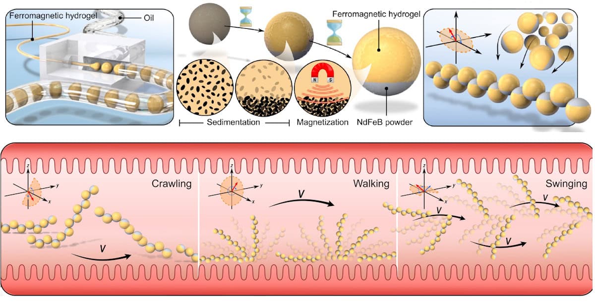
Multimodal locomotion Top panel: fabrication and magnetic assembly of permanent magnetic droplet-derived microrobots (PMDMs). Lower panel: magnetic fields direct PMDM chains through complex biological environments such as the intestine. (Courtesy: CC BY 4.0/Sci. Adv. 10.1126/sciadv.adw3172)
Microrobots provide a promising vehicle for precision delivery of therapeutics into the body. But there’s a fine balance needed between optimizing multifunctional cargo loading and maintaining efficient locomotion. A research collaboration headed up at the University of Oxford and the University of Michigan has now developed permanent magnetic droplet-derived microrobots (PMDMs) that meet both of these requirements.
The PMDMs are made from a biocompatible hydrogel incorporating permanent magnetic microparticles. The hydrogel – which can be tailored to each clinical scenario – can carry drugs or therapeutic cells, while the particles’ magnetic properties enable them to self-assemble into chains and perform a range of locomotion behaviours under external magnetic control.
“Our motivation was to design a microrobot system with adaptable motion capabilities for potential applications in drug delivery,” explains Molly Stevens from the University of Oxford, experimental lead on this study. “By using self-assembled magnetic particles, we were able to create reconfigurable, modular microrobots that could adapt their shape on demand – allowing them to manoeuvre through complex biological terrains to deliver therapeutic payloads.”
Building the microrobots
To create the PMDMs, Stevens and collaborators used cascade tubing microfluidics to rapidly generate ferromagnetic droplets (around 300 per minute) from the hydrogel and microparticles. Gravitational sedimentation of the 5 µm-diameter microparticles led to the formation of Janus droplets with distinct hydrogel and magnetic phases. The droplets were then polymerized and magnetized to form PMDMs of roughly 0.5 mm in diameter.
The next step involved self-assembly of the PMDMs into chains. The researchers demonstrated that exposure to a precessing magnetic field caused the microrobots to rapidly assemble into dimers and trimers before forming a chain of eight, with their dipole moments aligned. Exposure to various dynamic magnetic fields caused the chains to move via different modalities, including walking, crawling, swinging and lateral movement.
The microrobots were able to ascend and descend stairs, and navigate obstacles including a 3-mm high railing, a 3-mm diameter cylinder and a column array. The reconfigurable PMDM chains could also adapt to confined narrow spaces by disassembling into shorter fragments and overcome tall obstacles by merging into longer chains.
Towards biomedical applications
By tailoring the hydrogel composition, the researchers showed that the microrobots could deliver different types of cargo with controlled dosage. PMDMs made from rigid polyethylene glycol diacrylate (PEGDA) could deliver fluorescent microspheres, for example, while soft alginate/gelatin hydrogels can be used for cell delivery.
PMDM chains also successfully transported human mesenchymal stem cell (hMSC)-laden Matrigel without compromising cell viability, highlighting their potential to deliver cells to specific sites for in vivo disease treatment.
To evaluate intestinal targeting, the researchers delivered PMDMs to ex vivo porcine intestine. Once inside, the microrobots assembled into chains and exhibited effective locomotion on the intestine surface. Importantly, the viscous and unstructured tissue surface did not affect chain assembly or motion. After navigation to the target site, exposing the PMDMs to the enzyme collagenase instigated controlled cargo release. Even after full degradation of the hydrogel phase, the chains retained integrity and locomotion capabilities.
The team also demonstrated programmable release of different cargoes, using hybrid chains containing rigid PEGDA segments and degradable alginate/gelatin segments. Upon exposure to collagenase, the cargo from the degradable domains exhibited burst release, while the slower degradation of PEGDA delayed the release of cargo in the PEGDA segments.
Biological environment Delivery of preassembled PMDM chains into a printed human cartilage model. The procedure consists of injections and assembly, locomotion, drug release and retrieval of PMDMs. Scale bars: 5 mm. (Courtesy: CC BY 4.0/Sci. Adv. 10.1126/sciadv.adw3172)
In another potential clinical application, the researchers delivered microrobots to 3D-printed human cartilage with an injury site. This involved catheter-based injection of PMDMs followed by application of an oscillating magnetic field to assemble the PMDMs into a chain. The chains could be navigated by external magnetic fields to the targeted injury site, where the hydrogel degraded and released the drug cargo.
After drug delivery, the team guided the microrobots back to the initial injection site and retrieved them using a magnetic catheter. This feature offers a major advantage over traditional microrobots, which often struggle to retrieve magnetic particles after cargo release, potentially triggering immune responses, tissue damage or other side effects.
“For microrobots to be clinically viable, they must not only perform their intended functions effectively but also do so safely,” explains co-first author Yuanxiong Cao from the University of Oxford. “The ability to retrieve the PMDM chains after they completed the intended therapeutic delivery enhances the biosafety of the system.”
Cao adds that while the focus for the intestine model was to demonstrate navigation and localized delivery, the precise control achieved over the microrobots suggests that “extraction is also feasible in this and other biomedically relevant environments”.
Predicting PMDM performance
Alongside the experiments, the team developed a computational platform, built using molecular dynamics simulations, to provide further insight into the collective behaviour of the PMDMs.
“The computational model was instrumental in predicting how individual microrobot units would self-assemble and respond to dynamic magnetic fields,” says Philipp Schoenhoefer, co-first author from the University of Michigan. “This allowed us to understand and optimize the magnetic interactions between the particles and anticipate how the robots would behave under specific actuation protocols.”
The researchers are now using these simulations to design more advanced microrobot structures with enhanced multifunctionality and mechanical resilience. “The next-generation designs aim to handle the more challenging in vivo conditions, such as high fluid shear and irregular tissue architectures,” Sharon Glotzer from the University of Michigan, simulation lead for the project, tells Physics World.