Work has begun on a major upgrade to CERN’s Large Hadron Collider (LHC) that will see the luminosity increase by a factor of 10. The High Luminosity Large Hadron Collider (HL-LHC), which will be switched on by 2026, will enable the collider’s experiments to boost the amount of data they collect to improve the possibility of detecting new particles. The HL-LHC’s components are expected to be ready in 2023 and installation will take around 30 months. The LHC will be turned off during that process.
It’s a big upgrade and a good investment
Lucio Rossi
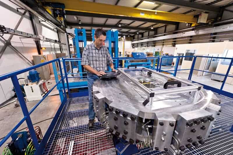
The HL-LHC project began in November 2011 and two years later became a top priority by the European Strategy in Particle Physics. CERN approved the design report in 2015, after which researchers and engineers started building and testing prototypes at the lab. Costing around SwFr1.5bn (£1.1bn), the upgrade will require significant modification to the beam line around the two largest LHC detectors – ATLAS and CMS. This will involve upgrading about 1.2 km of the 27 km ring by including 11-12 T superconducting magnets and superconducting “crab” cavities — that reduce the angle at which the bunches cross — to increase the number of collisions at the two detectors.
Certain parts of the LHC ring will also be upgraded with new dipole magnets so that the LHC can handle the increase of luminosity. “It’s a big upgrade and a good investment,” says Lucio Rossi, the Italian physicist who is HL-LHC project leader. “It will double the lifetime of the LHC up to 2040.”
Rossi adds that as well as testing new collider technologies and improving the precision of existing measurement of known particles, the HL-LHC will also increase the discovery range of new particles by around 20-30% over the current LHC. This will make the LHC more sensitive to heavier supersymmetric particles. “The HL-LHC will be a bridge between the LHC and the next big collider,” says Rossi. “It will tell us where to go next.”
European and Chinese scientists have identified a simple new way to become poor: fossil fuel investment. Not only could it leave you without a penny to your name. It could perhaps precipitate a global financial crash within one generation.
Coal, oil and natural gas are already huge investments. The International Energy Agency foresees price rises until 2040, and investor confidence is high. But researchers from the Netherlands, the UK and Macao don’t see it that way. They warn in the journal Nature Climate Change that, whatever the markets think, and whatever governments do, change is on the way.
Other forces are now driving global power and transportation in directions that suggest a dramatic decline in demand for fossil reserves. These will become what the money markets call “stranded assets”, and their value will slump some time before 2035.
And this bursting of what researchers call “the carbon bubble” – a reference to a three-centuries old financial disaster known to historians as the South Sea Bubble – could wipe between one and four trillion US dollars off the global economy. The financial crash of 2008 was triggered by a loss of a mere $0.25 trillion.
The scientists base their conclusion on a computer simulation known by the migraine-inducing acronym E3ME-FTT-GENIE, which is short for Energy-Environment-Economy Macroeconomic-Future Technology Transformations Grid Enabled Integrated Earth. They say it is the only such model that looks at the big picture: the macroeconomy, energy, the environment and global energy and transport systems according to both sector and geography.
The implication of the latest study is that, unless the world faces this reality, and switches to low-carbon investments, the global economy could suddenly collapse.
“Our analysis suggests that, contrary to investor expectations, the stranding of fossil fuel assets may happen even without new climate policies. This suggests a carbon bubble is forming and is likely to burst,” said Jorge Viñuales, of the University of Cambridge, and one of the authors.
“Individual nations cannot avoid the situation by ignoring the Paris Agreement or burying their heads in coal and tar sands. For too long, global climate policy has been seen as a prisoner’s dilemma game, where some nations can do nothing and get a free ride on the efforts of others. Our results show this is no longer the case.”
There is a catch: suppose nations become aware of the danger. A sudden push to fulfil the 2°C promise, combined with declines in fossil fuel demand but continued high output of fossil fuels, could trigger a collapse that would wipe $4 trillion off the global balance sheets.
Canada, Russia and the US would see their fossil fuel industries collapse. Fuel-importing nations such Japan, China and most EU countries might gain, especially if they had invested in low-carbon technologies to create jobs and boost gross domestic product.
“If we are to defuse this time-bomb in the global economy, we need to move promptly but cautiously. The carbon bubble must be deflated before it becomes too big, but progress must also be carefully managed,” said Hector Pollitt, of the University of Cambridge, and another of the authors.
“If countries keep investing in equipment to search for, extract, process and transport fossil fuels, even though their demand declines, they will end up losing money on these investments on top of their losses due to limited exports,” said Jean-François Mercure of Radboud University at Nijmegen in the Netherlands, and of Cambridge, who led the study. “Divestment from fossil fuels is both a prudent and necessary thing to do.”
Researchers from Belgium have created virtual models of the brain individually tailored to patients’ functional MR images (fMRI), according to an article published online in eNeuro. These models can predict the effects of brain tumours and may help improve surgical planning for their removal (eNeuro 10.1523/ENEURO.0083-18.2018).
When planning for brain tumour resection, clinicians typically examine functional MR images to identify key areas around the tumour as they develop a surgical strategy. But the complex dynamics of the brain make it difficult to predict postsurgical outcome based on the limited information these images provide.
One recent measure to improve the visualization of the brain has been to create more comprehensive brain models that simulate neural activity. These models can integrate fMRI data with the biophysics of the brain to predict brain function and help determine the optimal surgical approach, wrote senior author Daniele Marinazzo and colleagues from Ghent University.
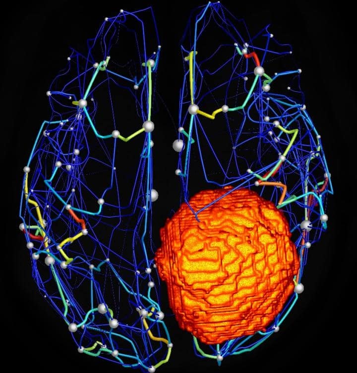
Structural brain network of a patient with a meningioma (orange). (Courtesy: Hannelore Aerts)
To test the viability of this technique, Marinazzo and colleagues used open-source software called the Virtual Brain to construct personalized brain models. They used neuroimaging data from 25 patients who had brain tumours and who underwent an MRI exam at Ghent University Hospital between May 2015 and October 2017. They also made similar brain models of 11 patients without brain tumours.
The investigators found that studying these models allowed the surgical team to assess brain function in all of the patients, as well as accurately predict the effect of tumours on brain function. Specifically, the models revealed that the number of connections in the tumour regions of the brain was much lower (p = 0.0007) and more variable (p = 0.01) than in non-tumour areas.
“Reliable prediction of patient-specific large-scale brain dynamics would open up the possibility … to investigate what types or extent of damage the brain can withstand, and conversely, which kind of distortions can be expected after brain lesions, including those purposively induced by surgery,” the authors wrote.
One of the main limitations of the study was its small sample size, but the authors plan to expand their research in the future and look into whether the virtual brain model can reliably predict postsurgical brain function.
“This [future research] would be a major step toward presurgical virtual exploration of different neurosurgical approaches and to identify an optimal surgical strategy,” they wrote.
“Just like the transistor is the basis of computers, the ion channel is the basic element in many processes in biology,” explains Peter Burke, who heads the nanotechnology group at the University of California at Irvine. He lists some of the aspects of the body they affect, which include neurons (and hence human thought) but also a variety of other processes, such as synthesis of the energy-storage molecule ATP in mitochondria. In fact, ion channels are crucial to so many physiological functions that they are the target of 25% of drugs produced, yet the technology for studying them using external current amplifiers has remained largely unchanged for 30 years. “Since we can manipulate and sense individual electrons in circuits, I thought it would be interesting to apply some of this knowledge in biology,” adds Burke.
Unsurprisingly, Burke and his colleagues are not the first to try to measure the currents of single ion channels. However, these measurements require a wide range of expertise covering both nanowires and physiology. Fortunately, Burke who holds multiple professorships at UCI was able to draw on his expertise in both these fields, as well as the extensive knowledge and experience of his collaborator Mark Reed from Yale University alongside co-authors Weiwei Zhou, LuyeMu, and Jinfeng Li.
“Previously people were looking at the action potential on a membrane, and treating the membrane like a capacitor;” says Burke. “This looks at a single channel so it’s a more difficult measurement.” Their nanowire device is the first to integrate the current measurement into the ion channel, which should ultimately allow much greater measurement sensitivity.
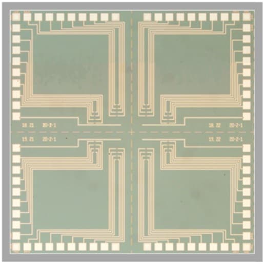
Optical micrograph of a chip containing nanoribbon devices. Courtesy Nano Futures
Sensing success
The researchers first demonstrated the ability to measure single ion channel currents using carbon-nanotube and graphene devices. However, Burke and his colleagues were keen to demonstrate the capability in a top-down fabricated silicon device that could leverage on the decades of development in that industry. This meant finding the right surface chemistry to attach the lipid bilayers, which act as artificial cell walls, to the silicon nanowire. They could then observe the behaviour of ion channel molecules on these bilayers.
They tested the device with two text book ion channels – alamethicin and gramicidin A. Both have antibiotic functions and work by making the bacterial membrane permeable. Ion channels responsible for vision and other neural functions have additional voltage and light dependencies and multiple states making observations of their behaviour more complicated. In their study the researchers observed the opening and closing of the gramicidin A ion channel and three of the four states in alamethicin.
The channels can open and close due to conformational changes in response to thermal fluctuations or interactions between peptides and the cell wall or by admitting some ions and blocking others. “One of the hopes of this work is to be able to apply this technology to answer in more detail: How do ion channels function?” says Burke.
While the behaviour of these ion channels was not affected by being integrated into the sensor, he points out that for larger ion channels that extrude into the space between the nanowire and membrane, this could be an issue. To tackle this the team have been able to demonstrate how to vary the space between nanowire and membrane with a tether.
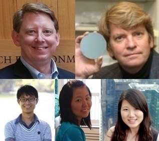
Peter Burke and co-authors Mark Reed, Weiwei Zhou, Luye Mu, and Jinfeng Li (clockwise from top left)
Dynamic modelling
Different ion channels also have different opening times, leading to different “spike widths” in the observed current. “To be able to understand the dynamic behaviour of the ion channel itself we need to understand the behaviour of the ion channel within the circuit it makes with the sensing device,” Burke tells Physics World Materials. “We have a comprehensive circuit model that explains the connected system, which I call the ‘modified Hodgkin-Huxley model’.”
Alan Lloyd Hodgkin and Andrew Fielding Huxley won the Nobel Prize in Physiology and Medicine in 1963 for their mathematical description of how ionic transport gives rise to action potentials and allows them to propagate and carry signals through the neural system, enabling movement and other physical responses to sensory input. In the Hodgkin-Huxley model circuit elements represent different parts of the cell – for example a capacitor represents the lipid bilayer; a voltage- and time-dependent conductance represents the ion channel. Since the capacitance of the nanowire device is comparable to the lipid bilayer, Burke and co-authors could add this as another capacitance in the circuit model of the cell.
Next the team will look into scaling up the system to run millions of devices on a chip in parallel. They are also interested in measuring single ion channels from specific systems such as the heart, brain and other organs. Full details are reported in Nano Futures
The short gamma-ray burst (GRB) that followed gravitational waves from the GW170817 neutron star merger was created in an astrophysical jet pointing 30° away from Earth, according to computer simulations by astrophysicists in the US and Italy.
Carried out by Davide Lazzati of Oregon State University and colleagues in the US and Italy, the simulations of the merger also offer an explanation for why the short GRB was much weaker than expected.
Typically lasting for less than 2 s, short GRBs and their origins have puzzled astronomers for decades. An important clue came in 2005 when X-rays and visible light were detected from the sources of two short bursts. This provided evidence that a short GRB can be created by the merger of two neutron stars to form a black hole.
The idea is that the merger creates two astrophysical jets of fast-moving material that flow out in opposite directions from the poles of the rapidly spinning black hole. Violent interactions that occur within the jet just after the merger create a short GRB. As the jet moves outward and slows down, a radiation afterglow at longer wavelengths is emitted.
However, the GW170817 observations did not fit exactly with what astrophysicists expected from a neutron-star merger. For one thing, the short GRB was much fainter than predicted by theory. Furthermore, the afterglow observed in the days and weeks after the short GRB increased in brightness over time – which was not expected.
Increasing visibility
Now, Lazzati and colleagues have done computer simulations of how radiation is emitted from such a jet and concluded that we are viewing the object 30° away from the direction of the jet. Most of the luminosity of the short GRB is expected to be in a relatively tight beam along the jet, which is why it appeared weak on Earth. Conversely, as the jet blasted out into space it spread out, which means that more of it is visible on Earth. This, say the researchers, is why the afterglow brightened over time.
Writing in Physical Review Letters, the team also estimate that about 5% of short GRBs that are detected in coincidence with gravitational waves will involve jets that point directly at Earth.
Today the FIFA World Cup kicks off in Russia as 32 nations battle it out to be crowned the football champions of the world. In this episode of Physics World Weekly, James Dacey and Matin Durrani consider some of the ways physics interacts with the beautiful game.
Dacey and Durrani discuss how footballs curve in the air, the secret to the perfect throw in, and how physics principles can help in designing safe stadiums. They also take a look at a couple of recent studies that attempt to predict the results of the tournament.
If you enjoy what you hear, then you can subscribe via iTunes or your chosen podcast provider.
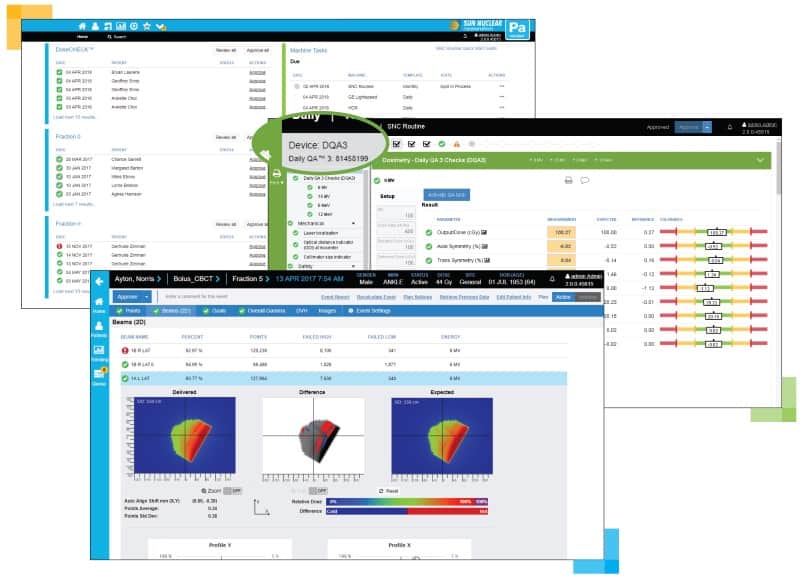
SunCHECK combines routine mechanical and routine patient checks into one user interface.
The delivery of radiation therapy is supported by a wide range of information on the patient, the treatment plan, and the linear accelerator (linac) that manufactures and administers the dose. It’s a branch of medicine that has become highly computerized, involving different combinations of hardware and software, and featuring a wide range of clinical tools and devices.
“One of the biggest challenges facing clinicians is being able to keep up with the rapidly evolving technology and ensure that the treatments are being delivered as intended to the patients,” comments Jeff Simon, CEO of Sun Nuclear – a provider of solutions for radiation oncology quality assurance (QA). “There are a lot of different checks that have to occur in managing, verifying and validating across the whole workflow.”
These steps involve a combination of machine and patient QA to ensure that the whole process runs according to plan.
As part of a multi-year programme, Sun Nuclear has been busy harmonizing each step in the QA process to make radiotherapy much simpler and intuitive for its customers – predominantly medical physicists and radiotherapists working in cancer clinics.
It’s a mission statement that few would argue against, but it’s also a bold undertaking. According to the firm’s figures, more than 4000 cancer treatment facilities worldwide use Sun Nuclear solutions.
“Being able to tap the brakes and create a whole new architecture while keeping current product lines competitive and up-to-date is definitely easier said than done,” admits Simon.
However, the benefits are compelling. “Integration and standardization are really important themes and goals here,” he emphasizes. “With SunCHECK 2.0, we’ve essentially combined the routine mechanical and the routine patient checks into one user interface with a common architecture and a common database.”
Robert Biggar – a principal clinical scientist based at the Clatterbridge Cancer Centre NHS Foundation Trust in the UK – has been involved in beta-testing the software. He sees unified architecture as a major step forward in helping him to do his job more efficiently thanks to the software’s ability to gather and process results in the background.
“It can be difficult to get different pieces of radiotherapy equipment, databases and patient information systems to interact with each other, but Sun Nuclear has managed to get inside the bare bones of how their systems work and integrate all of these information flows into a single platform,” Biggar comments. “One of the biggest benefits is the automation, which helps to focus our resources on the cases that need the most attention.”
Another key requirement was to make the SunCHECK platform agnostic to specific vendors’ technologies. “It doesn’t matter what treatment machine you have or which planning system you have, you’ll get the same results,” Simon explains. “You have standardization across the entire workflow.”
Rather than create a “wrapper” on top of older software, Sun Nuclear decided to re-write and expand its product suite from the ground up, which delivers a number of advantages to users.
“With our software there’s no waiting – the device data show up instantaneously,” Simon highlights. “There’s no need for manual synchronization or mapping of data fields – it’s all automatic.”
The web-based network architecture allows users to connect multiple geographic locations to gain a top-down view in the same dashboard, supported by the same database. What’s more, SunCHECK’s server installation streamlines updates and makes it straightforward to access.
User licenses are based on the number of linacs, rather than having to worry about separate licensing for each workstation.
SunCHECK integrates a number of the company’s QA and dosimetry tools as well as the linacs’ electronic portal imaging devices (EPIDs). “We calibrate the EPID to absolute dose so that you can use it for both pre-treatment QA as well as in vivo monitoring to check each specific treatment delivery,” says Simon. “We can automatically capture the EPID data and bring it into our system for automatic processing to show what dose was delivered and warn the user if there are any deviations from what was expected.”
The EPID can highlight any positioning errors or changes in patient anatomy since it detects the beam after passing through the patient. The EPID also provides a way of verifying the linac’s log files – for example, after routine servicing or the installation of a new component in the setup.
“A lot of the basic routine QA that we used to do with phantoms and ion chambers we are now able to do directly to the imaging panel,” Biggar explains. “Effectively your setup for QA purposes is instant, as you are leveraging equipment that’s already in place on the treatment machine.”
There are other possibilities too, thanks to a combined patient and machine QA environment. “When you have all of those data together, you are now in a position to use machine learning to look for correlations and different predictive analytics that weren’t possible before,” says Simon. “The intention is to take these data and provide decision support to clinicians to help them understand where the issues and opportunities may be.”
Installed across networked cancer centres, the software makes it much easier to spot any deviations between results at one site versus another. “It is a powerful tool for looking across your network,” adds Simon. “We have the broadest range of hardware products, and now with SunCHECK, we have the broadest software platform for comprehensive and independent quality assurance.”
It came out of something called the Biomagnetometer Project, which was set up by Quentin Pankhurst at University College London (UCL). He was looking for applications of magnetic nanoparticles, and a surgeon, Michael Douek, told him that although everybody is terribly interested in new cancer drugs, he and his colleagues would also really like new tools to help them perform operations. One idea they discussed was to use magnetic material to track lymph flow from the area around a tumour through to the lymph nodes, so that surgeons could biopsy the right nodes and thus determine whether a cancer had spread. This information is critical to providing correct treatment for the patient, as over-treatment (unnecessary surgery and chemotherapy) can be almost as damaging as under-treatment. The existing techniques for doing this involved using a blue dye and a radioactive tracer that can be very difficult to obtain, and there’s also a lot of regulatory inconvenience associated with radioactivity. So there was clearly a possibility of using magnetic particles to do the localization instead, and Quentin managed to get a research grant to pursue this. That’s when I got involved.
What had your career been like up to then?
I worked for a very long time at a company called Sira, and I did all sorts of interesting things there: electronics, software, testing instrument systems, developing tools for automatically inspecting things like float glass, and so on. That was very good training for something like the Biomagnetometer Project, and my physics background was incredibly helpful as well because it means you’re up for taking on new challenges and analysing new things. So when Quentin came to Sira in 2004 looking for someone who could replace a researcher he had lost, I jumped at the opportunity.
At what point did the project become a company?
It was quite a long and difficult road. We celebrated a notional 10-year anniversary in May 2017, but the first board meeting didn’t happen until July 2009. The delay was partly because we were spinning out from UCL, and we had considerable issues with doing a deal on the intellectual property. At one point, we thought we had an agreement, but then the proposed deal was withdrawn. That left us in a situation where we really thought the company was going to die, because we couldn’t see how we were going to move forward.
How did you get out of that situation?
Two things happened. The first was that we managed to get a medical device manufacturing company, ITL, to apply to the Technology Strategy Board (now Innovate UK) for a grant to do collaborative development between researchers and industry on a new product. The other was that I worked out a better way of doing the underlying technology, one that was much more practical to commercialize. Our original system for detecting the magnetic nanoparticles was based on a superconducting quantum interference device (SQUID) sensor that operated in liquid nitrogen, and although we managed to cope with supplying liquid nitrogen to the operating theatres during the clinical trials, it was a hell of a nuisance. There were issues even with small things like taking nitrogen up and down in a lift – people get quite exercised about the possibility of spillages in small spaces. Also, SQUIDs are very sensitive to radiofrequency fields, and under certain circumstances they will become completely non-operative if there’s a sufficient level of background interference, however good your screening is – and we spent a long time trying to produce good screening.
On one occasion, the SQUID simply failed to tune in the operating theatre, and it was only after the operation was over (all the trials were done using both the radioactive technique and our magnetic technique) that we worked out that the radiofrequency interference was coming from one of the overhead lights. If I’d known that and I’d been able to turn off that one set of lights, we’d have been able to continue, but that lack of robustness in a system you’re hoping to market everywhere is just not going to be acceptable. Then there’s the fact that not many companies make SQUIDs that work at liquid-nitrogen temperatures – and of course, the low-temperature ones, which require liquid helium, were completely out of the question.
Eventually, I managed to work out a way of doing our measurements with room-temperature electronics by pushing up the frequency to increase the level of sensitivity, using really low-noise amplifiers and developing our correlation techniques a bit more. That realization – together with £400,000 in free money, essentially, from the Technology Strategy Board to do the collaborative venture with ITL – was absolutely key to bringing UCL Business back on board and completing a deal.
Who did you bring in to help get the company started?
In addition to Quentin and myself, we also have a third founder, Audrius Brazdeikis, who is a biomedical physicist at the University of Houston in Texas, US. He got involved as part of a London–Texas initiative for collaborative working in biosciences; he worked at the superconductivity centre in Houston, so the idea was that he would provide advice about the SQUID magnetometry and magnetic sensing. Subsequently, Audrius has produced many beautifully engineered prototype probes during the development process. During the research project, we got considerable support from Michael Douek in getting our equipment into the operating theatre. But when we moved beyond the research project and were trying to set the thing up as a company, we went through about three potential CEOs. The difficulties of completing a deal meant that various people got bored with the process and disappeared, or we decided they were no longer suitable.
It wasn’t until late 2010 that our current CEO, Eric Mayes, joined us. He arrived at quite a critical time, because while our focus had been largely on building a machine that was sensitive enough to detect magnetic nanoparticles, we soon learned there was a real problem with the nanoparticles themselves. We were using a material called Endorem, which is an intravenously-injected contrast agent for magnetic resonance imaging, but then other types of contrast agent, which are gadolinium-based and completely unsuitable for our purposes, essentially took over the market. This was a little bit of a blow, but it was also a very good opportunity, because Eric tracked down an alternative manufacturer of nanoparticles and got an agreement to supply the particles we needed to our specification. He also got a CE mark [European regulatory approval] for nanoparticles that could be injected into tissue. We’d been using the previous stuff “off-label”, so although we could tell surgeons “This is what you have to go out and buy”, we wouldn’t have been able to market it ourselves because it was an unofficial use. And when you do the sums, it becomes quite obvious that it’s the consumable product that makes money for you, not the instrument itself. It’s like printers and ink – you make money out of the ink, not out of selling the printers.
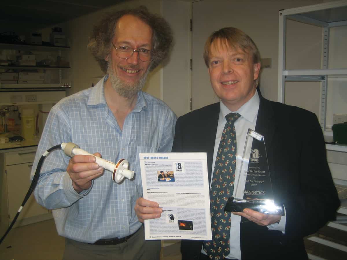
Magnetic tracking: Endomagnetics co-founders Simon Hattersley (left) and Quentin Pankhurst (right) developed a tool to help surgeons determine whether cancer has spread. (Image courtesy: Endomagnetics)
What’s the next step for the company?
Our magnetic nanoparticles have been used to treat more than 25,000 women so far, primarily in Europe, and Endomagnetics are in the process of obtaining approval from the US Food and Drug Administration (FDA). Getting approval for our product in Europe was actually not too bad, but the US process has been more lengthy because we needed to conduct a clinical trial of the products in the US first, and the pre-market approval process is more complex: the result was something like two crates full of paper that needed handling with a forklift. It would be a very big thing for Endomagnetics to get into the US, because it’s such a big share of the world market. Endomagnetics has also launched another product for marking breast tumours, and it is now on the market in Europe and the US. Most breast cancers are identified when they are only a few millimetres in size, which is good for the patient, but means that surgeons require some guidance to find and remove the cancer. The Magseed marker is placed in the tumour under radiological guidance before surgery and the surgeon uses Endomagnetics’ Sentimag probe to locate the marker and remove the tumour. The Magseed marker is in use at some of the top cancer hospitals in the US and featured on the BBC’s “Trust Me, I’m a Doctor” programme in January 2018.
What do you know now that you wish you’d known when you started?
I think I was a bit naïve about how the investment process works, particularly during the technology-transfer stage. The technology-transfer departments of universities are run as investment businesses. They are not the same as venture capitalists, but they are primarily interested in making money, so although they tend to promote themselves as being there to “help researchers commercialize their ideas” (and they do have that role), they can take an extremely hard-nosed attitude. It’s not nearly as straightforward as it is sometimes painted, and you can end up in a situation where you feel you are losing a massive amount of the company to the university. But there are so many things where you just have to live and learn.
Such as?
Well, Endomagnetics has actually gone extremely well in many ways. We’ve never had the dreaded “down round” – we’ve always been able to raise new investment at either the same share price or an increased share price, and that’s really quite important for the position of founders. My other company, Michelson Diagnostics, started off well, with some really cracking technology; the main product is a skin imaging system that can diagnose skin cancers without the need for a biopsy. But it’s never met the level of sales that it needed, and while prospects are now improving, the interim has been absolutely dire. Our larger investors completely lost patience with us: at one funding round they would only put in money on terms that essentially gave them the whole company. This means that the founders have ended up with next to nothing, and the early-stage smaller investors were completely wiped out. It’s a real shame that people who put in so much time, effort and intelligence into the development of something that is fundamentally good can essentially lose the lot. When we set the thing up I was thinking, “Well, it might fly or it might fail, but if it fails, so be it; we’ve given it our best shot.” What I wasn’t psychologically prepared for was that something in-between could happen, where the company keeps going but you’ve got no ownership or control of it anymore.
What led to that situation?
We didn’t realize how difficult it would be to sell our imaging technology to clinicians. It seems that in the field of dermatology, people are very, very conservative, and even with lots of research papers published to show how useful our system is, that doesn’t necessarily make people buy it. Apart from that, there’s the fact that developing medical devices and getting them to market is a long-winded process. Between the really complex technologies and the amount of regulatory stuff that’s involved, you can easily be talking 10 to 15 years from an initial idea through to actually making any money out of it. (Money is not the be-all and end-all, but it is some indication that you are actually getting the product out there.) That is far beyond the time horizons for a lot of investors: even assuming they come in when the idea has got a bit more sorted out, are they really going to want to wait perhaps eight to 10 more years before they’ve got any hope of getting their money back? The answer, probably, is “no”. That’s something particularly difficult with medical devices: finding investors who are prepared to go the distance with you. On a brighter note, Michelson Diagnostics is now selling a device that can image blood-vessel networks within the skin and is developing new applications in the treatment of burns and scars.
Do you have any advice for anyone thinking of starting a new firm in medical devices?
Talk to a lot of good people. It’s difficult to find the good people, but you can sniff them out eventually. There’s so much you need to know, frankly, so it’s very good to find some people who’ve been through it.
A new technique to print soft materials that undergo complex and rapid shape changes when a magnetic field is applied to them can be used to create tiny, untethered, robots capable of useful movement, such as rolling, jumping and grasping objects. Such objects might be used in a host of biomedical applications, like minimally invasive surgery or targeted drug delivery.
“Existing robots are often heavily tethered because they need to be actuated pneumatically, which makes them unsuitable for biomedical applications,” explains study lead author Yoonho Kim of the Massachusetts Institute of Technology. “Soft active materials that change shape in response to external stimuli, such as heat, light, solvents, electric and magnetic fuels, are better alternatives in this context because they can be controlled remotely. Magnetic fields are a particularly good stimulus option because they are a safe, fast and effective way to actuate magnetically responsive soft materials. The problem with most of the materials made so far, however, is that their shape change has been limited to simple bending or elongation.
“Our new way to print ferromagnetic domains in soft materials has allowed us to make far more complex shape-morphing structures that transform between different 3D shapes within fractions of a second.”
Toothpaste-like ink
Current methods to create magnetic materials rely on using “already cured” elastomers containing non-magnetized particles. These elastomers need to be temporarily deformed into the desired shapes and the embedded particles magnetized by applying a strong magnetic field in a certain direction. “One of key differences between these techniques and our approach is that we can directly inscribe magnetic polarity in complex 3D structures from the start to form complicated patterns of magnetic domains,” Kim tells Physics World.
Unlike previous methods, which were limited to simple geometries and simple deformation, the new technique is based on a 3D direct ink writing. “Toothpaste is the best way to describe the inks employed in this technique,” says Kim. “When we squeeze a tube of toothpaste, we in fact apply a shear-yield stress to it, which allows the paste to come out of the nozzle. The paste then maintains its cylindrical shape if we apply no further stress.
“Direct-writable ink materials also possess this rheological property and the ‘paste’ we used in our study is an ‘uncured’ elastomeric composite containing already-magnetized microparticles.”
Specific transformations in a magnetic field
The researchers, led by Xuanhe Zhao, of the Mechanical Engineering Department at MIT, made their ink by mixing microparticles of ferromagnetic neodymium-iron-boron (NdFeB) with silicon resin. To introduce the shear-yielding behaviour, they added fumed silica nanoparticles as a rheological modifier. “These particles form a network based on van der Waals interactions that helps the whole elastomer matrix maintain its shape,” says Kim. “It also helps the embedded magnetic particles to disperse throughout the matrix rather than agglomerating to form large clusters.
“We then apply a magnetic field to magnetize the embedded particles. Each microparticle (which is around 5 microns in size) becomes a strong permanent magnet.”
Before printing (that is, before squeezing the toothpaste-like composite out of a nozzle), the magnetic particles are randomly oriented. Thanks to the applied magnetic field, the particles reorient along the applied field direction during printing. “In this way, we can control the magnetic polarities of the magnetic fibres and thereby programme different regions of the printed material to undergo specific transformations in a magnetic field. For example, they can switch between different static shapes or morph dynamically in response to changing magnetic fields.
“A 3D construct built by arranging and stacking these fibres maintains its shape during the printing process. Once printed, we then cure the structure to make a rubber-like elastic solid, which is encoded with intricate patterns of magnetic domains.”
A hexapedal spider-like grabber
As a proof-of-concept, the researchers printed several structures with programmed magnetic domains capable of performing multiple tasks. One example is a hexapedal spider-like grabber. “By applying magnetic fields in different directions and of different strengths to different parts of its structure, this robot can be made to crawl, roll over, carry a drug cargo and even catch and release a fast-moving ball,” says Zhao.
“These demonstrations prove that our shape-morphing structures are strong and agile enough to interact with fast-moving objects. We hope our technology will allow us to develop untethered magnetically-remote-controlled magnetic soft robots that can operate in confined and enclosed spaces, like the human body.”
Indeed, the team, reporting its work in Nature 10.1038/s41586-018-0185-0, says that it is now focusing on developing specific biomedical applications for its technology. “This line of work will also require us to improve our materials and fabrication platform and advance magnetic field control for actuating such soft robots,” adds Zhao.
A bill aimed at restructuring the US’s approach to quantum computing research has been introduced by Kamala Harris, who is a US senator representing California. If passed by Congress, the Quantum Computing Research Act of 2018 would form a centrally-coordinated Defense Quantum Information Consortium that would include researchers from the academia, government and the private sector.
Elsewhere in Washington DC, Lamar Smith of the US House Science, Space and Technology Committee says he will introduce a bill to create the National Quantum Initiative. The member of the US House of Representatives from Texas says the initiative “will promote greater quantum research, standards, federal coordination, and collaboration among the key quantum players – laboratories, industry and universities”.
Harris says that the Defense Quantum Information Consortium will provide grants and assistance to scientists, with the goal of establishing the US as a global leader in quantum computing research. This, she believes, would give the nation a competitive edge in areas ranging from healthcare to national security.
Early stages
Developments in quantum computing may still be in their early stages, but experiments involving small numbers of qubits have already promised significant future advances in the technology. Much of this research has been done in California; carried out by institutions such as Stanford University and the University of Southern California, as well as companies including Google and Rigetti Computing.
Quantum computing is the next technological frontier that will change the world and we cannot afford to fall behind
Kamala Harris
Harris argues that coordinated support for these entities will accelerate the development of advanced quantum technologies. “Quantum computing is the next technological frontier that will change the world and we cannot afford to fall behind,” she says. “It could create jobs for the next generation, cure diseases, and above all else – make our nation stronger and safer.”
Smith adds, “Quantum computing could work up to millions of times faster than our conventional computing systems and solve problems we thought were unsolvable. The United States must get there first.”
The Defense Quantum Information Consortium would include officials from the Office of Naval Research, the Army Research Lab, the Office of Science and Technology Policy as well as the proposed National Quantum Initiative. The consortium would award competitive grants to researchers and administer research collaborations. It would also coordinate research in the US to ensure that different institutions do not compete with each other but work on separate tasks to accomplish larger-scale goals. The bill also seeks to ensure that research results are published at the lowest possible level of secrecy classification.
Harris says that the US faces a need for increased collaboration within quantum computing research. “Without adequate research and coordination in quantum computing, we risk falling behind our global competition in the cyberspace race which leaves us vulnerable to attacks from our adversaries,” she says. “We must act now to address the challenges we face in the development of this technology – our future depends on it.”