Skyrmions have attractive and repulsive tendencies

Interactions between individual 3D skyrmions have been measured by physicists in China, Sweden, Russia and Germany. Their study shows that the magnetic quasiparticles feel both attractive and repulsive forces, depending on the strength of an applied magnetic field. As well as providing insights into the fundamental physics of magnetic materials, the research could lead to the development of devices that store data using skyrmions.

Skyrmions were first proposed as a new type of fundamental particle in the 1950s by British physicist Tony Skyrme. While these hypothetical particles have never been seen, certain collective particle-like excitations (quasiparticles) in magnetic solids have been shown to behave much like skyrmions. These solid-state skyrmions resemble vortices and have topological stability, which means that they persist for very long times and are resilient to external perturbations such as noise. Skyrmions can be extremely small and be manipulated using relatively small amounts of energy. Together, these properties suggest that skyrmions could be used to make dense and energy efficient computer memories.

In this latest work, Mingliang Tian at the University of Science and Technology of China and colleagues studied a type of skyrmion that is created when a magnetic field is applied to a “nanostripe” of iron germanide (FeGe). These 3D skyrmions are tubular magnetic vortices with diameters of about 40 nm. They extend below the surface of the nanostripe and can move around in directions perpendicular to the applied magnetic field.

Edge effects

Using Lorentz transmission electron microscopy, the team observed the motions of individual skyrmions and then worked-out how the skyrmions interact with each other. They also studied how the skyrmions interact with the edges of the nanostripe, which was about 430 nm wide, 120 nm thick and 1600 nm long.

The team first looked at a nanostripe that contained tens of skyrmions. At relatively low magnetic fields (260 mT), the skyrmions formed chains or clusters at or near the edges of the nanostripe. As the field strength was increased to 390 mT, the clusters and chains moved away from the edges to the centre of the nanostripe – where the cluster and chain configurations were maintained. When the field was turned up to 480 mT the clusters and chains broke apart and the skyrmions were distributed across the centre of nanostripe.

Writing in Physical Review Letters, Tian and colleages surmise that the chain and cluster formation at low magnetic fields is the result of an attractive interaction between skyrmions. The migration of the skyrmions away from the edges and the subsequent break-up of the chains and clusters suggests that both the skyrmion-skyrmion and skyrmion-edge interactions become repulsive at higher magnetic fields.

Pair potential

However, the team points out that clustering can also occur in systems of particles with repulsive interactions and so to get a better understanding of the interaction they looked at the behaviour of individual pairs of skyrmions.

Their second experiment began at low magnetic field and with two pairs of skyrmions – one pair at each end of the FeGe nanostripe (see figure). As the magnetic field was increased from 200 mT to 500 mT, the team measured the distance between the two skyrmions in a pair. They also measured the distances between individual skyrmions and the edge of the nanostripe.

The initial separation between skyrmions in a pair was about 75 nm and this increased very slowly until the magnetic field reached about 450 mT. Then, the separation jumped to about 200 nm where it saturated by the time the magnetic field reached 470 mT. The distance between a skyrmion and the edge of the nanostripe was about 50 nm at low fields and increased to 200 nm and saturated there at about 420 mT.

Similar behaviour was seen in reverse as the magnetic field was reduced back down to 200 mT. This, the team says, shows that the observed interactions are real – rather than the result of skyrmions being pinned by defects in the FeGe nanostripe.

Theoretical agreement

The experiments reveal that the skyrmion-edge interaction switches from attractive to repulsive at a significantly lower field than the switch that occurs in skyrmion-skyrmion interaction. The team also did theoretical calculations, which suggest that the observed behaviour can be explained using our current understanding of 3D skyrmions.

The research could lead to a better understanding of the possible density at which skyrmions could be packed together in a memory device, and how data could be stored and retrieved from such devices.

In the audio interview below, Mohit Randeria of the Ohio State University tells Hamish Johnston why physicists are interested in skyrmions.

Interview with Mohit Randeria


Insects face calamitous habitat loss

Habitat loss may soon mean half the world’s insects, and many plants and animals as well, could find themselves without their familiar home ranges.

Right now, climate scientists warn, global planetary temperatures are on course to rise 3.2 °C above the average for most of human history. They have already risen by about 1 °C in the last 100 years.

And if they do, then 49% of insects, 44% of plants and 26% of vertebrates could lose more than half of their ranges.

If the 195 nations that agreed in Paris in 2015 to take steps to restrict global warming to a target of 1.5 °C keep their pledges, only 6% of insects, 8% of plants and 4% of vertebrates will experience severe reductions in their ranges. Even half a degree makes a huge difference.

“Insects are particularly sensitive to climate change. At 2 °C warming, 18% of the 31,000 insects we studied are projected to lose more than half their range. This is reduced to 6% at 1.5 °C. But even at 1.5 °C, some species lose larger proportions of their range,” said Rachel Warren of the University of East Anglia, who led the study.

“The current global warming trajectory, if countries meet their international pledges to reduce CO2, is around 3 °C. In this case, almost 50% of insects would lose half their range.”

These figures are projections based on a sample of animal and plant studies: the sample is however one of the largest undertaken.

Professor Warren and colleagues from Australia report in the journal Science that they studied data involving 34,000 insects and other invertebrates, 8000 birds, 1800 reptiles, 1000 amphibians and 71,000 plants, and took into account the capacity of each species to move to new habitat as the world warms.

Pattern of alarm

Such studies build on evidence assembled piecemeal, sometimes over many decades, about the impact of humanity on its fellow citizens of the planet. This evidence confirms a consistent pattern of alarm.

Researchers have established repeatedly that ecosystems already under pressure from human invasion are made more vulnerable by global warmingand climate change. More precisely, German scientists have established that the sheer numbers of insects that used to make a living around European farmlands have fallen dramatically, and even those insects that seem to survive almost everywhere could be under threat.

The new study found that a small number of species will extend their range in a warming world. Most will not. Many will have fewer places to go.

“This is really important because insects are vital to ecosystems and for humans,” said Professor Warren. “They pollinate crops and flowers, they provide food for higher-level organisms, they break down detritus, they maintain a balance in ecosystems by eating the leaves of plants, and they help recycle nutrients in the soil.”

Options narrowing

And, writing separately in the same journalGuy Midgley of Stellenbosch University in South Africa warned that the options for humanity are becoming severely restricted.

Humans depend on plants, insects and other animals to deliver water quality, soil conservation, flood prevention, crop pollination and natural pest control. All this is now threatened, not just by the clearing of forests and the growth of the cities, but by the profligate use of fossil fuels which release greenhouse gases such as carbon dioxide into the atmosphere, to drive global warming.

Researchers know, through a detailed study of the geological past, what higher concentrations of carbon dioxide can do to global climate. “There is way too much debate about the issue of climate change and whether or not it is real. What we really need to be doing is debating how we solve this problem,” said Professor Midgley.

“Those very high CO2 concentrations could well change the ecosystems of the world irrevocably. If we increase CO2 to over a thousand parts per million, over the next 50 to 60 years, which we are quite capable of doing if we fail to reduce our dependence on fossil fuels, we could literally move the world back 20 to 30 million years in the space of a century. It is like moving ecosystems backwards in time at the speed of light.”

Bioengineered gel helps repair brain tissue

Stroke is one of the leading causes of death and disability worldwide and an estimated six million people in the US alone are living with its debilitating after-effects. The brain only has a limited capacity to regenerate following a cerebrovascular attack, as it is also known. This is because stroke damages brain tissue and produces a “dead” cavity devoid of blood vessels, neurons or axons (the thin nerve fibres that protrude from neurons). Researchers at the University of California, Los Angeles, have now developed a bioengineered angiogenic gel that promotes new brain tissue growth as well as the formation of axonal networks along regenerated blood vessels when it is injected directly into the stroke cavity of mice. The gel contains molecules that stimulate the growth of these blood vessels, explain team leaders Tatiana Segura and S. Thomas Carmichael.

The UCLA researchers made their biomaterial from an amorphous non-fibrous hydrogel composed of hyaluronic acid, the vascular endothelial growth factor (VEGF) and heparin nanoparticles (to bind the growth factor molecules). Once injected, the gel thickens to mimic the properties of brain tissue so creating a “scaffold” for tissue growth.

Promoting tissue repair following stroke

“This Clustered VEGF Nanoparticle (CLUVENA) Hydrogel, as it is called, promotes tissue repair following stroke through at least two mechanisms,” says Carmichael. “It induces angiogenesis and subsequent in-growth of neuronal connections in the stroke cavity. The heparin nanoparticles also reduce inflammation and scarring around the damaged stroke site and enhance the vascular and neuronal growth processes.”

“Both features of the material are required and necessary for brain repair to occur,” adds Segura, who is now at Duke University.

In a normally developing body, blood vessels and nerves grow together into tissues as they mature, grow larger and begin their adult function, continues Carmichael. There is a complex signalling system between nerves and blood vessels in this process. By stimulating angiogenesis and then maturation of new blood vessels, local brain cells are “attracted” to these vessels and grow connections along them. This is known as a neurovascular interaction.

Potential new therapy for stroke

“Our work importantly shows that the nanoparticle clusters containing VEGF induce a specific and very different pattern of VEGF signalling than that seen when VEGF is simply injected by itself into a stroke cavity,” he tells Physics World. “This clustered VGEF promotes organized and mature vessel networks that are likely key in secondary processes of nerve in-growth,” adds Segura.

The study, which is detailed in Nature Materials, also shows that post-stroke tissue is not irreversibly degenerated, she says. “A biomaterial like the one we have developed could thus be injected into the stroke cavity following an attack to regenerate brain tissue and promote recovery. This could make for a potential new post-stroke therapy.”

In the experiments on mice, the researchers found that stroke cavities in the animals contained regenerated brain tissue after 16 weeks, including new neuronal networks. This has never been seen before, they say. “The mice with new neurons also showed improved motor behaviour, but we are unsure of the mechanism behind this,” say Carmichael and Segura.

“The new axons could actually be working, or the new tissue could be improving the performance of surrounding, unharmed brain tissue.”

This study focused on the period immediately following chronic stroke (five days in mice, which translates to two months in humans). The UCLA team will now be looking into whether the biomaterial works as well after longer delays.

Immuno-PET tracer offers hope for early cancer treatment

© AuntMinnie.com

Researchers are developing a PET radiotracer designed to adhere to and illuminate immune cells in cancer patients. They believe it could offer an earlier look at treatment response compared with tracers such as FDG that focus on cancer cells, according to a study published in the Journal of Clinical Investigation.

The immuno-PET tracer is designed to attach to T cells - a type of white blood cell - when they become active and attack cancerous tumours. By viewing their progress on PET images, the approach opens a molecular window to what is going on inside cancer patients, and it could potentially help them avoid toxic radiation and chemotherapy (J. Clin. Invest. doi: 10.1172/JCI98509).

"With a lot of therapies and diseases, we cannot visualize what is going on deep within the human body. It is not the same thing as taking a CT or MRI scan and utilizing anatomy," said senior author Sanjiv "Sam" Gambhir, radiology department chair at Stanford University. "We need to visualize molecules, and that is what a PET scan does best. By visualizing molecules, we can elucidate the underlying mechanisms of different diseases and different treatments. Otherwise, we are shooting blindly."

Immuno-PET tracers

Gambhir has been actively involved in the development and testing of novel PET radiopharmaceuticals and immuno-PET tracers to determine how well they can identify molecules in cancer cells that might prevent a person's natural immune system from attacking the disease.

In an April 2017 paper, Gambhir and colleagues discussed how immune checkpoint inhibitors have emerged as a promising tool for monitoring cancer treatment. However, the lack of imaging methods to noninvasively assess immune checkpoint expression has been a major stumbling block to predicting and monitoring response to therapy.

Gambhir eventually came to collaborate with Ronald Levy, a professor of oncology at Stanford: By coincidence, both men were investigating uses for a protein known as OX40, which can be activated on T cells.

Levy was working with OX40 to create a tracer that would activate T cells to attack tumours. Gambhir, meanwhile, was looking at OX40 and its role as a checkpoint inhibitor and potential biomarker to assess the efficacy of immunotherapy.

"He wanted to use that target to rev up the immune system," Gambhir told AuntMinnie.com. "We wanted to use OX40 as a marker for highly activated T cells, so we could predict whether a given therapy would work or monitor whether it is working."

OX40 factor

Levy's PET tracer features two stimulating agents: One part coaxes T cells into producing OX40 on the cell surface, while the other binds to OX40 and enables the T cells to interact with tumour cells. Overall, the tracer energizes immune cells to destroy tumours.

PET scan

When the PET tracer is injected, it seeks the cancer-killing T cells. As it binds to the OX40, the accumulation glows on PET images, showing the T-cell activation. Little or no such activity on a PET scan would indicate that immunotherapy is not working and the treatment should be adjusted or changed.

"It is one way to grade the immunotherapy and look to see if the tumour changes," Gambhir said. "We can see what the immune cells are doing and whether they are even going to the tumour site so they can destroy the tumour. If not, where are the immune cells going?"

This use of PET differs from conventional scans with FDG, which is taken up by tumours.

"Because tumour cells love to eat sugar, they also love to eat FDG," Gambhir said. "That is what produces a hot signal on a PET scan. On a PET scan with FDG, we are looking at where tumours are, not where the immune cells are. So this new scan is not measuring tumour metabolism with FDG, but is measuring activated T cells in the body. By doing that, we can know much earlier whether someone is responding to immunotherapy."

Naturally, if patients are not responding to immunotherapy, it is advantageous for them to find out sooner rather than later.

"If they are not responding, you don't want to wait months to find out because then we are losing time," Gambhir said. "A tumour might grow and become more heterogeneous and that much more difficult to treat.

Follow the T

The primary objective of immunotherapy is to activate T cells and prompt them to kill tumours. From a patient's perspective, immunotherapy also can be advantageous because it relies on natural processing within the body. When successful, a patient might avoid having to undergo radiation or chemotherapy.

"[Immunotherapy] potentially would be less toxic in that we don't have to give all kinds of things that not only hurt tumour cells but also hurt regular cells," Gambhir said. "The whole idea of immunotherapy is giving something that is less toxic and is not dependent upon some very toxic approach that, unfortunately, hurts the tumour but also the patient."

In about four months, Gambhir and colleagues plan to begin a phase I trial to see how well the immuno-PET approach works in humans. Thus far, they have experimented solely on mice.

"Just because things work in mice, of course, does not mean they will work in humans. So there's a big jump now to go to humans," he said. "This approach of using OX40, which sits on T cells, is a good way of therapeutically approaching cancer and, from an imaging perspective, letting us track these immune cells in a new way."

If the combination of PET, OX40 and tracking activated T cells is not successful for cancer, Gambhir speculated that the approach might be helpful for other disorders such as rheumatoid arthritis and multiple sclerosis.

  • This article was originally published on AuntMinnie.com. © 2018 by AuntMinnie.com. Any copying, republication or redistribution of AuntMinnie.com content is expressly prohibited without the prior written consent of AuntMinnie.com.

Unenlightened thinking

Are you incensed when Deepak Chopra, the US alternative-medicine advocate, promotes “quantum healing”? Are you infuriated by people throwing around abstract concepts like relativity, energy and evolution without understanding what they mean to scientists? Then you may understand how I feel when scientists make ignorant assertions about philosophy and the humanities.

My current source of annoyance is Enlightenment Now: the Case for Reason, Science, Humanism, and Progress – a new book by the Harvard University cognitive psychologist Steven Pinker. Life is wonderful, Pinker says, and claims he has data to show it. The book has more than six dozen graphs demonstrating that good things such as life expectancy, literacy, income, education, human rights, leisure time and tourism are on the way up, and that bad things such as wars, violence, poverty, crime, disease, plane crashes and death by lightning are on the way down.

A “war on science”?

The chief menace Pinker sees in the modern world is an “intellectual war on science” that is “wreaking havoc in universities and jeopardizing the progress of research”. The rightful rule of “Enlightenment optimists” like himself, Pinker says, is threatened by anti-science “Romantic declinists”, whose leaders are philosophers such as Nietzsche, Heidegger, Foucault and Derrida.

Imagine that! Four dead white males, one of them long gone from this planet, are leading an army of humanists that threaten to bring down science! Pinker’s on shaky ground, however, given that scientists like him get by far the lion’s share of grant money and public adulation. His ground is even shakier given that he reveals he knows as much about what these four said as Chopra does about quantum mechanics.

Let me give just one example. Pinker claims Nietzsche recommended that people become – he’s quoting Nietzsche now – “hard, cold, terrible, without feelings and without conscience, crushing everything, and bespattering everything with blood”.

You can hardly get Nietzsche more wrong. Pinker plucks these words from Nietzsche’s book On the Genealogy of Morals. Published in 1887, it is, as its subtitle suggests, a polemic (ein Streitschrift). Polemics, especially by authors who are well known for their use of metaphor, irony and hyperbole, cannot be read as if they were conventional journal articles. As the philosopher Robert Scharff likes to say, one might just as well cite the famous beginning of The Social Contract (“Man is born free…”) to mock Jean-Jacques Rousseau for believing that everyone is walking around wearing chains.

Humanities scholars like to read carefully and know context is important. Pinker’s lifted phrase comes from the 11th section of the first essay of The Genealogy. Nietzsche, a philologist by training, is scrutinizing the origins of the concepts of “good” and “bad”. He notes that the masters and cultural victors – the top 1%, we would say – are likely to have one understanding of these concepts; the oppressed and cultural losers another.

As an example, he cites the Greek epic poet Hesiod’s classification of history chronologically into five Ages of Man. Two of these ages are actually the same age, Nietzsche says, but one represents the perspective of the winners, the other of the losers. Hesiod’s “Heroic” age is the world seen from the perspective of the likes of the heroes of Thebes and Troy, while the “Iron” (Erz) age is that world as seen from the perspective of “those who have been crushed, despoiled, brutalized, sold into slavery.” That latter age, Nietzsche writes, is the one with leaders whose actions are “hard, cold, terrible”, and so forth.

Nietzsche is not recommending we behave the way the wretched see their oppressors acting. Nor is he saying the two perspectives are equal. He is holding up a mirror to the conventional Christian morality of his time, trying to jolt readers into reflecting on its impact on their lives. Nietzsche is also showing that, if you simply banter about abstractions without connecting them to the life source from which they arose, you can say anything you damn well please, because you have lost track of life itself. You can say the world is terrific or terrible, depending on your perspective.

It’s ironic that Pinker has misunderstood a passage in which Nietzsche was illustrating the misleading use of abstractions

Robert P Crease

It’s ironic that Pinker has misunderstood a passage in which Nietzsche was illustrating the misleading use of abstractions. For one can imagine an anti-Pinker writing a book packed with graphs illustrating the rise of inequality between rich and poor, numbers of refugees, data breaches, industrial-scale political lying, mass shootings, genocide and so on. Such graphs would not capture the world as seen from the privileged perch of a tenured position at Harvard. Yet the fact that abstractions can channel privilege or oppression is not the worst of it. Nietzsche’s point is that when we live by abstractions, we forget how we live. Using only psychological theories to, say, guide our behaviour towards others, we forget what human relationships are.

In Pinker’s case, the forgetting is coupled with a false confidence that expertise in one thing makes you an expert in everything, such as how to read Nietzsche. Later in his book, Pinker quotes more passages from Nietzsche and labels them “genocidal ravings”. He’s culled these passages from secondary sources. To understand them in context, you’d have to learn how to read a brilliant writer who uses metaphor and irony to connect and transform people in ways that literal language does not. That would require taking a humanities course. And if you can read better, you can appreciate many things better, including the value of science.

The critical point

Thank you, Steven Pinker, for showing the harm when scientists don’t take enough humanities courses. A humanities education helps provide a deeper grip on one’s experiences and on the world in ways other than through graphs and abstractions. Isn’t it more likely that the very lack of humanities education is what fosters thoughtless about the world and the dangerous sway of science denial today?

China launches ‘Queqiao’ lunar satellite

China has successfully launched a satellite to the Moon that will perform radio astronomy as well as communicate between Earth and a separate lunar lander, which is set for launch later this this year. Dubbed Queqiao, or Magpie Bridge from an ancient Chinese folklore tale, it took-off from the Xichang Satellite Launch Center on 21 May. It will now be put at Langrange Point 2 “L2” - a gravitational-balance point about 65000 km behind the Moon - where it will stay visible both to ground stations on Earth and the future lander.

Due to tidal locking between the Earth and the Moon, only one side of the Moon is visible to Earth. This “far side” remains of enormous interest to scientists as studies have indicated that there is a very different world on the far side, being geologically more ancient and dominated by highlands, unlike the planar landscape that prevails on the near side.

A radio antenna behind the moon will open up a new window on the universe

Marc Klein Wolt

The far side is also of interest for the radio-astronomy community. While almost all celestial radio wave frequencies can be received on Earth, those that are below 30 MHz are blocked by the atmosphere. Yet such frequencies contain important information about the early universe and can only be measured from a special vantage point like the back of the moon, which is free from atmospheric and man-made interference. This means the far side is one of the best places to measure the 21 cm hydrogen emission line that can be used to study the mass and dynamics of galaxies and will allow scientists to peer into the “cosmological dark ages” – a period between the Big Bang and the birth of the first stars.

Queqiao will include a Dutch-built antenna - the Netherlands-China Low-Frequency Explorer (NCLE) - that is designed to measure radio waves between 1-80 MHz. "A radio antenna behind the moon will open up a new window on the universe,” says NCLE project leader Marc Klein Wolt, who is managing director of the Radboud Radio Lab at Radboud University. According to Albert-Jan Boonstra from the Netherlands Institute for Radio Astronomy in Dwingeloo, the Dutch antenna is especially designed to receive low-frequency radio waves over a larger range. “We have found ways to avoid the electromagnetic interference of the satellite itself and successfully developed a broadband receiver,” he says.

Limited observations 

Also riding on Queqiao is a pair of microsatellites that will be released into an elliptical lunar orbit for similar radio astronomy experiments as the Dutch antenna. The twin microsatellites, developed by Chinese scientists, will carry out interferometry tests to demonstrate the feasibility of a future microsatellite array, which would be more sensitive than a single probe in detecting faint-radio signals from afar. However, due to the size of the microsatellites, their observation times will be limited to 10 minutes from the far side and 20 minutes of data transmission from the near side every orbit.

As well as performing radio astronomy, another key aim of the Queqiao mission is to enable the transmission of commands and data from Earth to the Chang’e-4 lander that will launch later this year. Chang’e-4, which will land in the South Pole-Aitken Basin area, will be the first mission to land on the far side of the Moon and it will also have a low-frequency radio-spectrum analyzer, which has been developed by scientists from the Institute of Electronics, Chinese Academy of Science in Beijing.

Rising temperatures could boost antibiotic resistance

Current forecasts of the burden of antibiotic resistance could be significant underestimates in the face of a growing population and climate change, according to a team from the US and Canada.

The researchers found that the common bacterial strains Escherichia coli, Klebsiella pneumoniae and Staphylococcus aureus showed a higher degree of antibiotic resistance where local temperature and population density were higher.

"The effects of climate are increasingly being recognized in a variety of infectious diseases, but so far as we know this is the first time it has been implicated in the distribution of antibiotic resistance over geographies," said Derek MacFadden of Boston Children's Hospital, US. "We also found a signal that the associations between antibiotic resistance and temperature could be increasing over time."

MacFadden and colleagues looked at antibiotic resistance data from 2013–2015 for a total of 1.6 million bacterial pathogens. Areas with higher antibiotic prescription rates tended to exhibit increased antibiotic resistance.

"Estimates outside of our study have already told us that there will already be a drastic and deadly rise in antibiotic resistance in coming years," said John Brownstein of Boston Children's Hospital and Harvard Medical School. "But with our findings that climate change could be compounding and accelerating an increase in antibiotic resistance, the future prospects could be significantly worse than previously thought."

A local average minimum temperature increase of 10°C was associated with a 4.2% increase in antibiotic resistant strains of E. coli, a 2.2% rise in antibiotic resistant K. pneumoniae and a 3.6% rise in S. aureus, the team found.

What’s more, the study linked an increase of 10,000 people per square mile to a 3% increase in antibiotic resistance in E. coli and a 6% increase in K. pneumoniae. The antibiotic resistance of S. aureus did not appear to change with population.

"As transmission of antibiotic resistant organisms increases from one host to another, so does the opportunity for ongoing evolutionary selection of resistance due to antibiotic use," said MacFadden. "We hypothesize that temperature and population density could act to facilitate transmission and thus increases in antibiotic resistance."

The team published the study in Nature Climate Change.

One step closer for kidney tissue engineering

Dutch researchers have engineered 3D cellular constructs called organoids for kidney research using human induced pluripotent stem cells (iPSCs) and embryonic stem cells (ESCs). The organoids were successfully vascularized once implanted in vivo (Stem Cell Reports 10 751).

The kidney has multiple functions: homeostasis (balance of many body functions), control of blood pressure, production of red blood cells, and elimination of waste (from food, medication or toxic substances), while keeping a balance of essential substances, fluids and minerals in the body.

One kidney is composed of more than 1 million nephrons, the functional unit of the kidney. The nephron filters the blood, processes nutrients and passes out waste from the blood. The blood is first filtered by the capillary network (glomerulus). The filtrate is collected in the Bowman’s capsule and passes through a series of renal tubules (proximal tube, loop of Henle and distal tube). These absorb water, minerals and glucose. Filtered fluids leave the nephron by the collecting duct and enter the renal pelvis.

The cells composing the kidney can be developed in the lab. However, the classic way to cultivate cells in vitro is using a 2D structure, which does not represent the complexity of the organ. Instead, researchers are investigating ways to cultivate cells in a 3D system, as organoids. Organoids are cellular 3D constructs that replicate fully or partially the structure, cellular organization and composition of the in vivo organ. One limitation of organoids is their incapacity to undergo morphogenesis, meaning that they can’t be physically modified to achieve a complete maturation. For example, functional vascularization is not achievable yet.

Now, Ton Rabelink and his team at the Leiden University Medical Center in the Netherlands have developed a kidney organoid which, once implanted in vivo, becomes vascularized and starts to become more mature.

The researchers were able to generate kidney organoids from ESCs and iPSCs obtained by reprogramming of somatic cells to the pluripotent state. Both cell types were able to develop into kidney organoids.

Observed structures

They identified many structures in these kidney organoids, including the glomerulus surrounded by the Bowman’s capsule, the renal tubules and the collecting duct. They also observed more specific features, including the presence of podocytes in the glomerulus (cells involved in the retention of plasma proteins from going into the urine), interstitial cells, which act as the kidney scaffold, endothelial cells (that form blood vessels) and pericytes, which are cells localized around the vessels.

Upon implantation of the organoid under the renal capsule of mice, the researchers observed glomerular vascularization by the mouse endothelial cells, without needing further stimulation. Also, organoid maturation and organization was more advanced upon transplantation in vivo, compared with prolonged culture in vitro.

Vascularized glomerular structures

This study presents the development of a more mature kidney organoid that is more comparable to adult kidneys. This research will be useful in many applications, such as drug screening, disease modelling and studying kidney regeneration.

Butterfly wings inspire nanostructured medical implants

Glaucoma is the second leading cause of blindness worldwide, with numbers expected to increase every year, according to the World Health Organization. Though the cause of glaucoma remains unclear, research suggests that the disease damages eyesight via random increases in pressure inside of the eye. Medication can alleviate the pressure to prevent long-term damage, but currently, no cure for the disease exists. Developments from the Choo lab at Caltech and Sretavan lab at the University of California, San Francisco, however, could ease the lives of glaucoma patients by providing them with real-time readings of their intraocular pressure.

The team recently published the successful fabrication and testing of a new microscale implantable intraocular pressure (IOP) sensor in Nature Nanotechnology (doi:10.1038/s41565-018-0111-5). Five years in the making, the device relies on nanostructures with optical properties the team first discovered in the wings of the longtail glass butterfly species Chorinea faunus.

A flutter of inspiration

Certain sections of the wings of the butterfly are coated in nanostructures about 100 nm or 150 nm apart depending on the location on the wing. This distribution gives the wings of the species an optical property called angle-independent anti-reflection, in which their wings scatter light uniformly in all directions during transmission. The wings therefore appear transparent.

“Nature often evolves with multifunctional nanostructures for their diverse biological functions,” said co-author Radwanul Hasan Saddique, a post-doctoral fellow in the Choo lab. “The longtail glasswing was not an exception.”

The angle independency of the wings’ transparency inspired the Choo group because existing IOP sensors prior to the work reported here heavily depended on the readout angle for signal, limiting the range of detection to 10 degrees. The group therefore sought to fabricate nanostructures with the same transparency as the wings to see if they could eliminate angle dependency in IOP sensors. They found success using a silicon nitride (Si3N4) membrane, increasing the readout range to 30 degrees.

Beyond its optical properties, the membrane also prevented bio-fouling, or buildup of biological material, on the device through strong structurally mediated hydrophilicity. According to Vinayak Narasimham, a PhD student in the Choo lab, prevention of biofouling can improve the lifetime of the in vivo sensor.

Now that the team has performed initial animal studies, they are pursuing long term in vivo studies in animals. They hope to see the technology available to glaucoma patients within five years.

Deep neural networks synthesize full-dose PET images

For individuals receiving multiple PET scans, especially children, there is a particular incentive to reduce the doses they receive to minimize the long-term risk of radiation-induced cancers. However, low-dose scans lack diagnostic power due to higher levels of noise. An international collaboration is using deep neural networks as a potential solution to the problem.

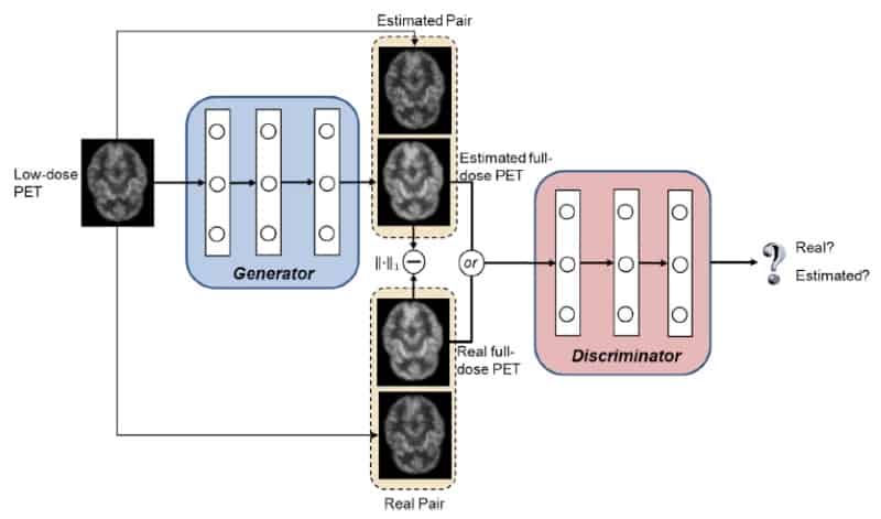
“Our technique uses unique machine learning algorithms – known as 3D conditional generative adversarial networks (or 3D c-GANs) – to estimate the high-quality full-dose PET images from low-dose ones,” said co-author Luping Zhou from the University of Sydney. Developed by Zhou, first author Yan Wang from Sichuan University, and co-authors in China, the US, South Korea and Australia, the new technique performed well against other methods used to synthesize full-dose PET images (NeuroImage 174 550).

GAN models use two deep neural networks, a generator and a discriminator, which achieve the best possible result by competing against one another. In the new application, the generator’s goal is to synthesize a full-dose PET image of sufficiently high quality to convince the discriminator that the image is genuine. The discriminator’s goal is to spot that the output of the generator is not a true full-dose image.

Each network is trained using a database of pairs of low- and full-dose PET images from the same individuals. Once trained, a new individual’s low-dose PET image is then fed into the generator for it to synthesize the corresponding full-dose, higher quality PET image.

Image synthesis framework

In a key feature, the technique handles 3D image data sets. Many other, previously reported techniques handle 2D axial slices independently which, though less data intensive, leads to the loss of information in the coronal and sagittal planes.

To train and validate the 3D c-GANS technique, the researchers acquired 18F-FDG brain scans of eight individuals with normal uptake and eight with mild cognitive impairment (MCI). Conventional full-dose clinical scans, delivering an effective dose of 3.86 mSv, acquired counts for 12 minutes. They were immediately followed by three-minute acquisitions that were used as low-dose scans in the 3D c-GANS model.

Following the common "leave-one-out" cross-validation approach, the researchers used data from 15 individuals to train the model and data from the remaining individual to test the model’s capabilities. The process was repeated such that the model was tested using data from all 16 individuals.

To maximize the training data set, 125 individual sub-volumes were extracted from each scan, making 1875 training samples and 125 test samples for each leave-one-out case. The final entire synthesized full-dose PET volumes were then constructed by merging the individually synthesized sub-volumes generated by the model. By maximizing the number of training samples, the likelihood of overfitting by the model was minimized, improving its potential performance in a wider clinical population.

Quantitative technique comparison

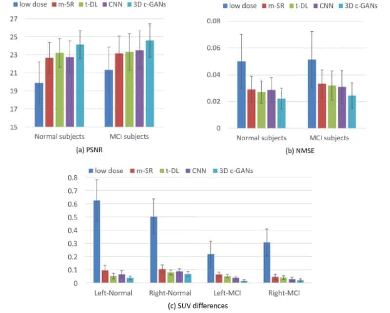
The researchers found the image quality achieved with 3D c-GANS compared favourably against three existing methods used to synthesize full-dose PET images. In a quantitative comparison, for example, the normalized mean square error (NMSE) - a measure of the difference in voxel intensities between synthesized and true full-dose PET images - was lowest using the 3D c-GANS technique. This was seen both in individuals with normal scans and those with MCI. Standard deviations in the parameter did, however, overlap between the different methods. The new technique performed similarly well when standardized uptake values (SUVs) and peak signal-to-noise ratios were examined and in a further, qualitative comparison.

Amongst several lines of further research, the authors plan to investigate a multi-modality approach to full-dose PET synthesis, incorporating clinical CT or MRI scans that are acquired alongside the PET scans. They also plan to increase their training database size to improve 3D c-GANS generalizability - its ability to work effectively in the clinical patient population at large.

Copyright © 2026 by IOP Publishing Ltd and individual contributors