Nanosilicates stimulate growth of bone and cartilage tissue

2D nanoparticles known as nanosilicates can be used to grow bone and cartilage tissue from human mesenchymal stem cells, according to new gene sequencing experiments by researchers at Texas A&M University in the US. The finding could help in the development of next-generation bioactive materials for regenerative medicine.

Human stem cells show much promise for regenerative medicine because they can transform into various specialized cell types, including bone and cartilage cells. At the moment, such specialized cells are obtained by subjecting stem cells to specific instructive protein molecules, known as growth factors. However, these factors can produce harmful effects in the body, such as unwanted tissue growth and tumours.

A team led by Akhilesh Gaharwar has now found that 2D nanosilicates (or nanoclays) can be used to grow bone and cartilage tissue in the absence of growth factors. The researchers obtained their result by looking at how the nanoparticles interact with human mesenchymal stem cells (hMSCs) at the whole transcriptome level using a technique called high-throughput sequencing (RNA-seq).

Snapshot of cell gene activity

2D nanomaterials are emerging as ideal structures for making electronics devices for a host of applications, including in energy and optics. However, until now, we did not know how they interact with human stem cells and if indeed, they could be used in biomedical applications.

RNA-seq is a next-generation, highly sensitive sequencing technique that reveals how much RNA there is in a biological sample at a given moment and under a certain condition – in the presence of nanoparticles, for example. “Cell-nanoparticle interactions can produce significant change in cellular behaviour that we can observe using this technique,” explains Irtisha Singh, who is one of the senior authors of this study. “Put simply, RNA-seq takes a snapshot of the gene activity of the cell. It’s rather like taking a high-resolution photo during the Super Bowl and identifying the reaction of every fan during the touchdown.”

Transcriptome dynamics

The researchers employed so-called transcriptome dynamics to discover the biophysical and biochemical pathways that are triggered when 2D nanosilicates interact with hMSCs. “We observed widespread changes in gene expression profiles in more than 4000 genes following nanoparticle exposure, something that had not been reported on previously,” say first authors Jake Carrow and Lauren Cross. “What is more, the technique identifies key genes and enriched gene ontology pathways, as well as categories related to stem cell differentiation – and specifically toward osteochondral lineages (that is, related to bone and cartilage tissue growth).”

The team says that it validated its RNA-seq findings with in vitro studies that back up this result – that nanosilicates can indeed direct the differentiation of hMSCs towards bone and cartilage tissue.

Bone regeneration therapy applications

“Our study shows that hMSCs present signalling typically observed during regeneration of bone and cartilage in the presence of nanosilicates,” Gaharwar tells nanotechweb.org. “This means that these nanoparticles might be used as a possible bone regeneration therapy for osteoarthritis and osteoporosis, among other diseases. We believe that the cellular response to nanosilicates comes from these particles’ unique physical and chemical composition, and their highly bioactive nature, opening the way to a new field of ‘mineralomics’ as an alternative to current stem cell treatments that rely on growth factors.

“Osteoporosis, for example, causes bones to become weak and brittle – so brittle that even mild stresses (such as bending over or coughing) can cause a fracture,” he adds. “2D nanosilicates could come into their own here and help stimulate bone growth. If we can deliver these particles to bone cells, they could encourage bone cells to deposit bone matrix that would strengthen the surrounding weak bone.”

Towards in vivo trials

The nanosilicates could also be used to treat arthritis/osteoarthritis, he says. “Osteoarthritis progressively degrades cartilage and often specifically attacks the spaces between joints,” explains Gaharwar. “Because these intra-articular spaces lack blood vessels, systemic delivery of morphogens and growth factors supporting cartilage matrix synthesis usually results in limited improvement. These nanoparticles could be delivered to this space to stimulate chondrocytes to synthesize new cartilage matrix.”

The present study, which is detailed in PNAS https://doi.org/10.1073/pnas.1716164115, was performed on human cells in vitro. The researchers say that they would now like to show that bone and cartilage tissue grows in the presence of nanosilicates in vivo (in animals such as mice and rats to begin with). “In particular, we are looking to develop implantable scaffolds containing these nanomaterials to improve local tissue growth,” says Gaharwar. “We will also be using computational biology and techniques like genomics to design a next-generation of bioactive materials for regenerative medicine.”

Falcon’s high-speed dive generates forces needed to catch agile prey

Peregrine falcons dive from great heights and at extreme speeds when hunting to generate high aerodynamic forces that enable them to execute precise manoeuvres and catch agile prey. Using a physics-based computer simulation, researchers in the Netherlands and the UK have explained why the raptors have evolved an attack strategy that puts extreme physical and cognitive demands on them. The research could also help with the development of autonomous flapping-wing drones.

The peregrine falcon is the fastest diving bird in the world and the fastest animal on the planet. According to Guinness World Records, in 2005 one was recorded travelling at speeds of more than 380 km/h while stooping – diving after prey.

Dropping out of the sky

“Witnessing the high-speed stoop of a peregrine falcon is impressive: a falcon may first soar at altitudes of several hundred metres, up to several kilometres above their prey,” says Robin Mills, a biologist at the University of Groningen, in the Netherlands, and academic visitor at the University of Oxford. “Then, within the blink of an eye, it dives down – first flapping forcefully and then folding its wings to decrease drag. It basically appears to drop out of the sky. When the falcon nears its prey, it may knock it with a massive blow, or catch it with its talons.”

Mills’ colleagues at the University of Oxford previously demonstrated – using onboard GPS loggers and cameras – that stooping peregrine falcons use the same steering laws as many man-made, guided missiles. Using these “proportional navigation” rules, the falcons remain on a collision course with their prey by simply tracking changes in their line-of-sight on the target. If the angle of the line-of-sight changes the falcon turns at a rate proportional to the speed of that change. With a constant of proportionality, known as the navigation constant, determining the falcon’s turn rate and how quickly it hits its target.

Simulated attacks

In the latest research, described in PLOS Computational Biology, the team built a physics-based simulation of falcon attacks on aerial prey to investigate why they stoop from great heights and at extreme speed.

The simulation modelled the aerodynamic forces on falcons during stoops, and how they target and react to prey using proportional navigation. It was run millions of times, with the researchers varying the falcon’s starting position, its navigation constant, and assumptions about errors and delays in its vision and control. They also modelled different patterns of prey flight.

Previously it had been suggested that falcons’ high-speed dives surprise prey, which then struggle to respond in time. But it had also been presumed that the speed of the attack would reduce precision, particularly if the prey turned sharply.

The researchers discovered, however, that when prey moves erratically the extreme speed of a falcon’s stoop maximizes aerodynamic forces that enable precise manoeuvring and increase catch success compared to slower, low-altitude attacks.

Intercepting prey

“By folding the wings appropriately, the stooping falcon is able to reach the lateral acceleration (of over 15 g), and roll acceleration – agility – required to meet its steering demands and by using the same mathematical steering rules as man-made missiles, the falcon is able to intercept sharply turning prey without turning sharply itself,” explains Mills.

According to the simulation, this only works if the falcon’s guidance law is precisely tuned, and if the birds have a high degree of steering control and visual precision. The researchers say that this shows that the stoop is a highly specialized attack strategy, adding that the optimal navigation constant for the simulation was close to that observed in peregrine falcons.

The team also found that high-speed stoops do not require faster reaction times than slower attacks. “By increasing the falcon’s agility, the stoop compensates for the decrease in catch success otherwise resulting from a slow response,” explains Mills. “If the falcon’s decision making was essentially instantaneous, it wouldn’t require a high-speed stoop to catch most prey.”

Replacing falcons

Ultimately, Mills hopes to use the data to develop fully autonomous flapping-wing drones. One practical application of such technology would be to replace the costly falconers and falcons currently used at airports to chase away birds, to prevent them flying into plane engines.

“What is great about our simulations is that it bridges the gap between artificial flying machines and biology: by uniquely combining theoretically derived optimal control laws with empirically verified aerodynamic modelling, attack behaviour and physiological constraints of real birds, we learn precisely learn what birds are doing and why – but also how to better control man-made flapping-wing drones,” Mills says.

Re-gating mitigates motion in scanned proton therapy

The use of motion mitigation strategies is of critical importance when delivering proton therapy to a tumour affected by organ movement. If motion is not adequately addressed, radiation delivered by pencil-beam scanning (PBS) could miss the precise tumour target, thus not delivering the planned dose and potentially hitting adjacent healthy tissue or organs-at-risk.

A study conducted in Switzerland comparing motion mitigation performance among different PBS delivery systems has shown that, for treatment of liver tumours, re-gating is an effective motion mitigation approach for all the PBS systems studied. Re-gating, the combination of gating (a system that tracks a patient’s normal respiratory cycle and monitors the movement of a tumour) and rescanning, was more effective and robust than either single technique (Radiother. Oncol. doi: 10.1016/j.radonc.2018.01.019).

For any PBS system, the target volume of a tumour is scanned both laterally and longitudinally by a sequence of narrow proton pencil beams. The maximum applicable beam current used to deposit a planned dose at a pre-defined location differs between cyclotron- and synchrotron-based systems. Modulation of beam intensity during radiation delivery may also vary among PBS systems. Because of these factors, the exact timing of the delivery of each pencil beam from the same treatment plan can be very different depending upon the PBS system used.

Ye Zhang

Lead author Ye Zhang, a research scientist of the Center for Proton Therapy at the Paul Scherrer Institut (PSI), and co-authors evaluated potential differences in the effectiveness of re-gating as a function of different PBS systems. They also evaluated variations in delivery time-lines using varying clinical assumptions.

The research team used four-dimensional CT (4DCT) and 4D MRI datasets from six patients with liver tumours with irregular motion greater than 10 mm. They performed 4D dose calculations for seven different field doses, simulating four different starting phases of the delivery of each field. For each case, they created 16 two-field plan combinations considering all possible starting phases.

In total, the researchers simulated 54 motion mitigation approaches for each of eight PBS scenarios, patient, prescribed dose and field for single field-plans. These were combined with 1-9x layered and volumetric rescanning and three gating window sizes. The PBS scenarios included spot or raster scanning, layered or volumetric rescanning, gating, constant or varying beam current, and cyclotron or synchrotron beam sources.

For both layered-and volumetric-rescanning, an adapted re-scanning approach was used to assure that all beam weights were deliverable. Gating signals were determined by tracking the external surface motion from the 4D-MRI data sets used for modelling the motion and by applying a linear, internal-external motion correction.

The researchers compared the qualities of all the 4D plans to determine dosimetric impacts and treatment efficiency. Differences in the magnitude of interplay effects were analysed. They also selected 13 typical motion mitigation approaches from the 54 options to perform a more comprehensive analysis.

The authors reported notable differences in dose distribution between plans delivered among the PBS models. However, combining gating with either rescanning mode achieved considerable improvements in mean homogeneity index and its uncertainty band. Robust results were attained across all PBS delivery scenarios, and a large range of fraction prescription doses and motion parameters.

The choice of raster or spot lateral scanning had insignificant dosimetric impact. Raster scanning reduced delivery time by 25-53% compared with spot scanning for all accelerator types and beam current models for the re-gating scenarios. Variable beam current models reduced treatment time for both cyclotron (by between 2-10%) and synchrotron (by 3-28%) PBS systems. The major advantage of cyclotron-based systems was a reduction of up to 70% in treatment time for all investigated field doses, especially when a cyclotron was combined with variable layer-by-layer beam current. However, treatment time reduction occurred with all motion mitigation techniques.

“For many years at PSI, we have been developing a comprehensive 4D dose calculation and optimization platform, taking into account many dedicated models of PBS scanning dynamics and deformable motion variability,” said Zhang. “With the help of this tool, we are able to explore many dimensions for the unrelated uncertainties in 4D PBS treatments, to investigate their dosimetric impacts, to evaluate the plan robustness, and eventually to suggest the optimum motion mitigation solution for individual patient treatments.”

The past, present and future of 3D bioprinting

Bioprinting has emerged as one of the most promising techonologies for fabricating artificial tissues and organs that could revolutionize the diagnosis and treatment of many different medical conditions. In a recent review article for the journal Biofabrication, researchers at Stanford University in the US discuss the current status of bioprinting research, and assess its future potential for drug screening and toxicology studies, as well as tissue and organ transplantation.

“Just as the printing press allowed massive amounts of information to be accessed at low cost for the first time in mankind’s history, so bioprinting could potentially provide a high-throughput and affordable way to assemble cells to make complex tissue constructs that are widely available to a very large number of researchers and scientists,” says team leader Utkan Demirci. “We aim to provide real solutions to many clinical problems that exist today.”

Demirci defines bioprinting as the process of using advanced additive manufacturing technologies to pattern biological materials, such as cells, biomaterials and biomolecules, for the fabrication of tissue-mimicking constructs. This novel approach requires biocompatible materials called bioinks to act as the matrices for printed cells, which can then be grown in bioreactors to further develop and become functionally mature.

Control at the cellular level

In their own research, Demirci and his colleagues exploit various micro- and nanoscale technologies, including 3D bioprinting and assembly, to create artificial tissues for biomedical applications. A key focus for their research is to control the cellular micro-environment and push the limits of cell manipulation through dedicated nano- and microscale technology platforms. Creating architectures that mimic the complexity of native tissues, as well as the functions and structures of specific cells, are expected to have important applications in precision medicine.

“Our research group aims to build complex cellular systems that mimic nature, but we also want to create systems from scratch that direct self-assembly,” explains Demirci. “Externally applied forces can actually trigger the act of complex self-assembly; these forces can be magnetic, electrical or even acoustic.”

Demirci says that a number of significant new approaches have been developed by research groups around the world since the team’s review was published in March 2016. “One example that comes to mind is an innovative label-free magnetic levitation platform that has been developed for 3D assembly of cells in complex living architectures,” he says.

Out of this world

Demirci and his team have focused their efforts on biofabrication through the self-assembly of cells, rather than growing constructs on tissue scaffolds. More recently, however, they have become interested in biomanufacturing cellular constructs in the absence of a gravitational force field – with the aim of exploring whether tissue could be engineered in space.

“All existing bioprinting methods rely on the presence of droplets (for example, in the drop-by-drop or drop-on-demand bioprinter) or on extruding materials landing on a surface, which is the one of the most affordable ways to bioprint,” comments Demirci. “We are trying out alternative approaches, such as assembling cells in 3D using a controlled magnetic field, to direct cells to form organoids.”

Demirci explains that cells in nature self-assemble at the microscale into complex functional configurations and microarchitectures, and researchers are increasingly exploiting this mechanism to assemble biomimetic systems in vitro. “However, we would ultimately like to precisely code 3D multicellular complex living materials, which is an exciting challenge given their architectural complexity and spatiotemporal heterogeneity.”

“Such techniques, which would build living materials with 3D control over geometry and organization, might be able to create model systems that mimic the physiological as well as pathological behaviour of native tissues,” he continues. “Such models might be extremely valuable in cancer for precision medicine to better care for patients.”

Methane from permafrost melt more than thought

A key source of a powerful greenhouse gas, methane emissions from permafrost, is producing much more than realised, scientists report.

Methane emissions are the source of the greenhouse gas which, after carbon dioxide, probably causes climatologists more sleepless nights than any of the other gases. And now it appears they have quite a lot more to bother them than they had realised.

Methane is reckoned to be at least 30 times more powerful than CO2 at warming the Earth, with some estimates putting its potency much higher still. The good news, research has suggested, is that there is far less methane than CO2 in the atmosphere to worry about.

The bad news, announced by an international research team, is that previous calculations may have been seriously wrong, and that thawing permafrost is likely to be producing appreciably more methane than anyone had thought.

The researchers were headed by Christian Knoblauch of the Centre for Earth System Research and Sustainability (CEN) at the University of Hamburg, Germany. Their findings, published in the journal Nature Climate Change, make it possible to predict better how much of this significant gas may be released by the thawing of the Arctic permafrost.

Methane and carbon dioxide are both produced in thawing permafrost as dead animal and plant remains decompose. But methane is formed only in the absence of oxygen. Until now, scientists had also thought that more greenhouse gases were formed when the ground was dry and well-aerated – in other words, when oxygen was available.

So they did not expect much methane to be produced by the thawing permafrost. What Dr Knoblauch and his colleagues have now shown is that water-saturated permafrost soils without oxygen can be twice as harmful to the climate as dry soils – which means the role of methane has been greatly underestimated.

They have, for what they say is the first time, measured in the laboratory the long-term production of methane in thawing permafrost. The team had to wait for three years before their roughly 40,000-year-old samples from the Siberian Arctic finally produced methane.

They observed the permafrost for a total of seven years and found that, without oxygen, equal amounts of methane and CO2 were produced.

Gigaton estimate

A co-author of their report, Susanne Liebner, from the Helmholtz Center Potsdam – GFZ German Research Center for Geosciences, said: “By combining process-based and molecular-microbiological methods, our study shows for the first time that the methane-forming micro-organisms in the thawing permafrost have significant influence on the greenhouse gas budget.”

The team used the new data to improve a computer model that estimates how much greenhouse gas is produced in permafrost in the long term – and they compiled a first forecast: the permafrost soils of northern Europe, northern Asia and North America, they say, could produce up to one gigaton (one billion tons) of methane, and 37 gigatons of carbon dioxide, by 2100.

Earlier studies have expressed concern about the interplay between permafrost and global warming, and this latest research will be exhaustively scrutinised as other teams try to corroborate, modify or contradict it. That is how science works. And there are certainly uncertainties that need resolving.

For example, how deep will the soil actually have thawed by the end of the century? Will it be wet or dry? What is certain, the team concludes, is that the new data will enable more accurate predictions to be made about the impacts of thawing permafrost on the climate. – Climate News Network

• This report was first published in Climate News Network

EGU 2018: robot boats assess the Danube

Every morning this week, thousands of geoscientists have taken the U1 train from central Vienna across the Danube to the European Geosciences Union 2018 General Assembly. On Wednesday morning, in what may have been a meeting first, some of them ventured just a few minutes away from the conference centre to the banks of the Danube to test out a couple of robot boats.

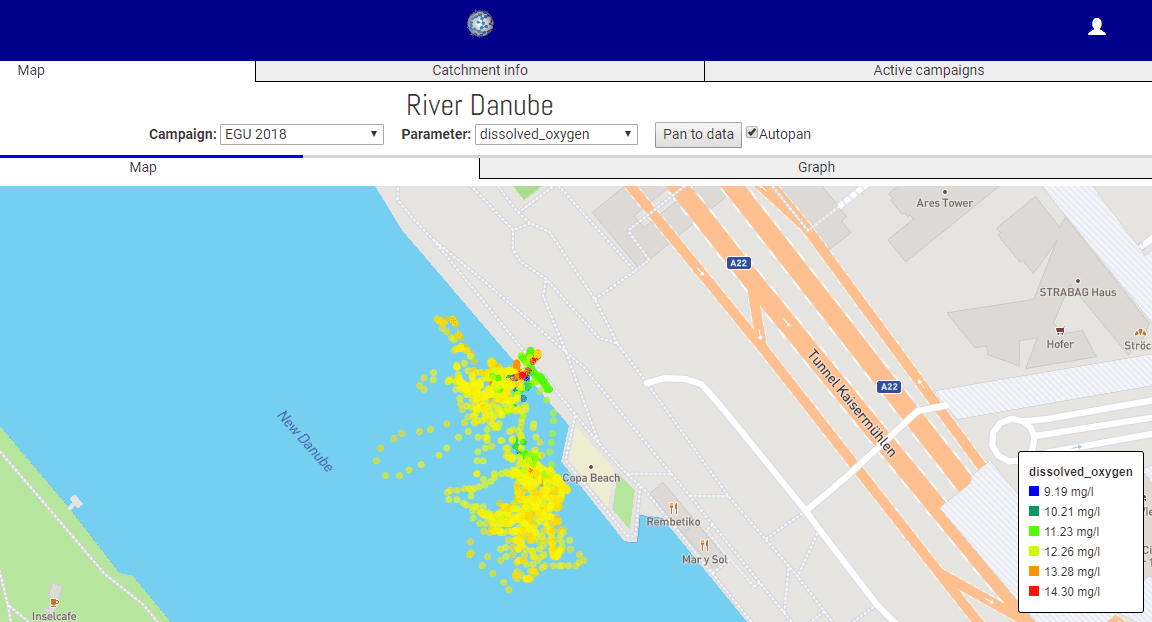
The boats – one large, one small – offered complementary measurements. The smaller is under development by IntCatch 2020 for citizen-science projects and can be run autonomously, automatically spiralling or motoring in a grid to take data on water quality. EGU delegates were also able to drive the boat by remote control and see the data it was collecting via a weblink on their smartphones.

Demonstration data collected on the Danube

The boat, which is fitted with GPS, could help citizen-scientist data make more impact with policy makers – it’s currently underused. Since the boat can take measurements and collect water samples autonomously it doesn’t need an expert to be present and its georeferencing means citizen scientists can prove, for example, that they didn’t fix their measurements by taking them directly from a sewage outlet. What’s more, as Nathalie Gilbert of Thames21, UK, put it, the boat can “get to places you can’t go with a bucket”. The IntCatch 2020 project is halfway through and looking for collaborators. So far, test sites have included Lake Garda in Italy and the river Ter in Spain.

The larger boat, some 2 m long, held an acoustic Doppler current profiler with up to 9 beams to measure river flow and sediment load. Using this ARC-Boat is much faster than taking measurements using traditional flow measurement techniques, as Nick Everard of the UK’s Environment Agency explained. Better still, since it’s remote controlled, nobody puts their life at risk in a manned boat.

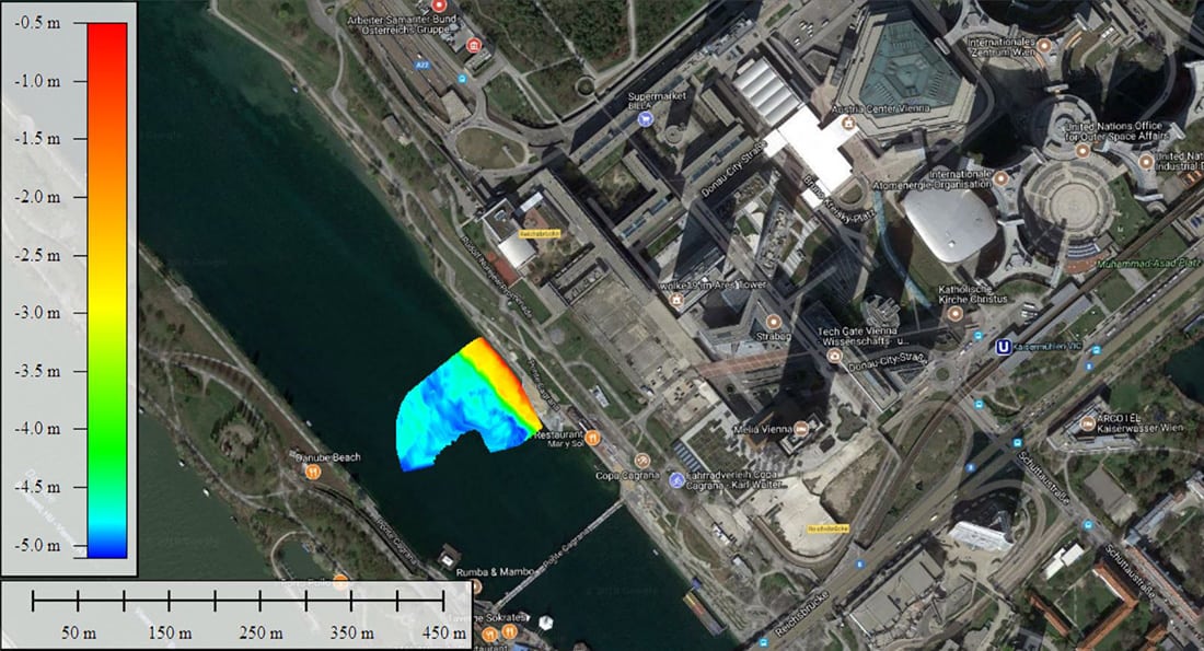
Bathymetry measurements taken on the Danube

Older techniques such as rotating element current meters take data only at specific points in the river and can require up to five people (if using a boat rather than suspending the kit off a bridge) to measure a site in one day. The acoustic Doppler current profiler on the ARC-Boat, on the other hand, profiles a river in just a few minutes. Two people can assess up to six sites in one day, making this method some 15 times more efficient. The boat is big enough to deal with floods but splits into two parts for transport in cars. And if you include GPS on board, it can also map a river’s bathymetry as well as velocity. This “lifts the lid on the river”, Everard said. Next the team wants to add water quality measurements and autonomous control. “Don’t just model, measure,” said Everard. “Measuring is more fun”.

EGU 2018: could eagle ‘eyes’ measure urban boundary layer?

Rick Thomas is an experimentalist, he likes to test the models. Normally, he does that using sensors attached to drones and planes. But you’re not allowed to fly drones over cities and to measure the urban boundary layer you must stay lower than an aircraft can go. The answer? A tame white-tailed eagle. Complete with sensor pack and GPS tag, the eagle can fly above the city and record the temperature of the atmosphere at the heights Thomas needs. Just one snag: first the Birmingham University, UK, researcher had to check if the eagle’s body would heat up the sensor and give false air temperature measurements.

Fortunately, tests above the Scottish countryside for the Cityflocks project revealed that if the eagle flies above a certain speed, the sensor will provide an accurate temperature reading. It helps that when the eagle is flying with its head down, the sensor faces directly into the airflow without obstructions. When the eagle’s at rest, however, that’s another matter – Thomas’s readings show the measured temperature rising when the bird takes a break.

Trained eagles are hard to come by so Thomas has also tested a smaller, lighter version of the sensor pack on homing pigeons, as yet only in rural areas. He’s teamed up with the Royal Pigeon Racing Association, who are interested in finding out more about the routes their pigeons take. There are also plans afoot to put the sensors on gulls in Birmingham, which could have the side benefit of revealing more about their behaviour. Thomas is keen to stress that all the necessary precautions were taken to ensure the welfare of Victor and the other birds.

As well as presenting his findings in a couple of posters at the EGU 2018 General Assembly in Vienna, Thomas has also published some of these results in an early online version.

Traumatic brain injuries could be caused by low-frequency vibrations

Numerical simulations have revealed how motions of the brain during head impacts can be characterized by just a few low-frequency modes of vibration in brain tissue. The study, led by Kaveh Laksari at the University of Arizona and Mehmet Kurt of the Stevens Institute of Technology in New Jersey, used sensors in the helmets of American football players to record head impacts. The motion of the brain was then recreated in computer simulations. Their research gives new insights into the mechanics responsible for traumatic brain injury (TBI).

The simple dynamics of head impacts have been well understood for decades. As the skull accelerates rapidly during an impact, the brain lags behind because its motion is damped by the various membranes and fluids connecting it to the skull. However, the brain is a remarkably complex and heterogeneous material and its many different parts have varying mechanical properties that respond differently to forces. As a result, established models of brain motion during a head impact are too simple to be able to predict whether a TBI has occurred. This is an important shortcoming because TBI is currently the single largest cause of injury-related deaths in the US.

Experiments on volunteers

Previous attempts to improve our understanding brain motion have involved experiments on volunteers subjected to mild head accelerations while inside a magnetic resonance imaging (MRI) machine. Meanwhile, studies of more violent injury-inducing impacts are done on cadavers. Computer simulations have also provided important insights into brain motion, allowing for predictions of the motions of the brain as it responds to different accelerations.

To improve these simulations, the team used a relatively new technique called “dynamic mode decomposition” (DMD) to analyse data from impacts. DMD bears some similarity to Fourier analysis, but calculates a new set of vibrational modes from every set of data gathered. This allows the motion of the brain to be represented using fewer modes than Fourier analysis, which is particularly useful for understanding the damped movements of a bound, heterogeneous material like the brain.

Football helmets

The team gathered their data by attaching motion sensors to the helmets of American football players to measure 187 head impacts that occurred during games and training. They then used the measurements as input to a head impact simulation, using DMD to break down the resulting strain and displacement fields in the simulated brain into a few damped, oscillating modes of vibration. To their surprise, they found that the impact fields could be closely approximated using just a few modes, with frequencies around 40 Hz.

The researchers also found that milder impacts were characterized by a single mode across the entire brain, while stronger impacts required larger numbers of modes. They team believe they have shown for the first time that brain injuries are caused by vibrational modes induced by impacts, rather than the peak acceleration experienced by the head. This could help explain why their is a low correlation between peak acceleration and brain injury in patients. However, the team does emphasize that more clinical studies are required to back up the connection between vibration and injury.

The research is described in Physical Review Letters.

Inspiring through games

Ask any scientist why they do science and they often say it’s because of a deep passion to learn and understand nature. Ask a scientist why they do outreach and usually the response is to inspire more young people into science. After all, what better way to inspire someone than by sharing your ­passion and knowledge?

But sometimes there can be disadvantages to scientist-led outreach. While scientists are very knowledgeable, they may not have the necessary skills to engage with some audiences. It can then be hard for them to simplify their work to a level that is inspiring, rather than perplexing. Those drawbacks can sadly be amplified for audiences that are most under-represented in science – such as those from low socio-economic backgrounds or those that live in geographically remote areas. These audiences may switch off when meeting a scientist, whom they assume cannot be related to as “someone like me” – instead assigning a stereotypical caricature.

Informal learning

While some scientists are indeed excellent communications, what if we could also use groups that have the same interest of raising career aspirations in science but already provide informal learning opportunities? I am thinking of staff at organisations supporting young homeless people and children from disadvantaged backgrounds, say, as well as librarians, activity organisers for semi-retired and retired people, and employment consultants from local chambers of commerce. How could we go about working with these people to engage their communities with aspirational activities and opportunities?

Sam Illingworth and Paul Wake, the co-directors of the Games Research Network at Manchester Metropolitan University, UK, suggest that one avenue is through table­top games – anything played on a flat surface, such as a board game or card game. Such activities not only provide a highly effective way to engage different audiences with complex topics, but also offer the opportunity for further discussion and debate.

Such games, in addition, allow for shared social experiences and can be easily modified and adapted depending on the targeted audience. Given they are so flexible as a communication tool, we decided that they would provide the perfect platform to introduce community leaders to physics-based activities and opportunities that could be used with their audiences.

During a training session for community leaders that we ran on 1 December 2017 at Manchester Metropolitan University, four commercially available tabletop games were played: Math Fluxx, Women in Science, Lab Wars and Terraforming Mars. After playing each of these games, the organizers led a discussion about the pros and cons of each, exploring the ways in which they might be used to address key issues in physics and, more widely, within science.

For example, while Women in Science provided a wonderful opportunity to discuss the way that women have been perceived and continue to be perceived in science, Terraforming Mars prompted a more extended discussion of physics, climate change, wildlife conservation, and energy production. Playing as teams of two, participants enacted the role of powerful corporations vying for control of the red planet, experiencing some of the ethical and economic dilemmas associated with humankind’s plans to colonize other planets.

Following the success of the session, plans are now under way to implement tabletop games as a science communication tool by all of the community leaders. For example, Women in Science will be used at the Dukes Theatre in Lancaster, running a community project with an audience of young girls from the travelling community. Most girls from this community leave school at age seven or eight, so by also involving their parents in the activities we hope it will boost their children’s education and career aspirations. Terraforming Mars will feature in school-based projects across Cheshire in northern England led by the local chamber of commerce.

Of course, some people might be asking why we should bother doing this for audiences that might not traditionally see themselves as future scientists? For example, if young women in the travelling community have already left school, the chances of them returning and studying to be scientists might be slim. Would our efforts not be better spent with school students? We would argue not.

Supporting aspirations

The Institute of Physics, which publishes Physics World, also wants to work towards a society in which parents, grandparents and community leaders support a child’s aspirations in science. So raising the value of science in those audiences is important too. We are working with the University of the Third Age – an international organization to educate older members of the community – to support members to encourage their grandchildren’s science aspirations and in a similar session for parents with social-services involvement.

Overall, this work has led to non-scientists going away with the confidence and skills to inspire new audiences in science in a way that best works for them. We should do more of it.

Non-invasive patch measures glucose through the skin

Diabetics are required to regularly monitor their blood glucose levels, conventionally via an invasive finger-prick blood test. Now, scientists at the University of Bath have created a non-invasive adhesive patch designed to measure glucose levels through the skin, potentially removing the need for this painful and unpopular procedure (Nature Nanotechnology doi: 10.1038/s41565-018-0112-4).

The patch does not pierce the skin, instead it samples glucose from interstitial fluid via extraction through hair follicles. The follicles are individually accessed via an array of miniature sensors using a small electric current. The glucose collects in tiny reservoirs and is measured every 10 to 15 minutes over several hours.

An important advantage of this device is that each sensor in the array can operate on a small area over an individual hair follicle, which significantly reduces inter- and intra-skin variability in glucose extraction and increases the measurement accuracy. As a result – and in contrast to other proposed non-invasive approaches, such as detection of glucose in sweat, tears or saliva – the patch does not require calibration with a blood sample.

“A needle-less method to monitor blood sugar has proven a difficult goal to attain,” explained Richard Guy. “The closest that has been achieved has required either at least a single-point calibration with a classic ‘finger-stick’, or the implantation of a pre-calibrated sensor via a single needle insertion. The monitor developed at Bath promises a truly calibration-free approach, an essential contribution in the fight to combat the ever-increasing global incidence of diabetes.”

In this study, Guy and colleagues tested the patch on pig skin, where they showed it could accurately track glucose levels across the range seen in diabetic human patients. They also tested it on healthy human volunteers, where again the patch was able to track blood sugar variations throughout the day.

They hope that the patch could eventually become a low-cost, wearable sensor that sends regular, clinically relevant glucose measurements to the wearer’s phone or smartwatch wirelessly, alerting them when they may need to take action.

“The specific architecture of our array permits calibration-free operation, and it has the further benefit of allowing realization with a variety of materials in combination,” explained Adelina Ilie. “We utilized graphene as one of the components as it brings important advantages: specifically, it is strong, conductive, flexible, and potentially low-cost and environmentally friendly. In addition, our design can be implemented using high-throughput fabrication techniques like screen printing, which we hope will ultimately support a disposable, widely affordable device.”

The next steps in this project – a collaboration between the University of Bath’s departments of physics, pharmacy and pharmacology, and chemistry – include refining the patch design to optimize the number of sensors in the array, demonstrating full functionality over a 24-hour wear period, and undertaking a number of key clinical trials.

Copyright © 2026 by IOP Publishing Ltd and individual contributors