Quasiparticles called rotons have been seen for the first time in a Bose–Einstein condensate (BEC) of ultracold atoms. The research was done by physicists in Austria, Germany and Italy and could lead to new insights into superfluids and supersolids.
Almost 80 years ago, the Soviet physicist and Nobel laureate Lev Landau developed the mathematical underpinnings of superfluid helium, which flows without loss of kinetic energy and exhibits other bizarre behaviour due to quantum mechanical effects that dominate at extremely cold temperatures. His ground-breaking work revealed that some of this odd behaviour is the result of phonons and rotons. These are two types of particle-like collective excitations (quasiparticles) in the superfluid.
These quasiparticles are distinguished by their differing dispersion relations – the relationship between the momentum and energy of a quasiparticle. Phonon energy typically increases with momentum, but rotons combine large momentum with low energy.
Long-awaited quasiparticle
BECs are ultracold atomic gases in which the majority of atoms are in the same low-energy quantum state. BECs share some properties with superfluids, but before 2003 it was believed that the relatively low density of a BEC would preclude the existence of rotons. However, subsequent calculations suggested that roton excitations might occur in BECs with special types of interactions – specifically, magnetic atoms with long-range anisotropic dipole–dipole interactions.
Now, Francesca Ferlaino at the University of Innsbruck and colleagues have created a BEC with these properties by cooling approximately 100,000 erbium atoms in a cigar-shaped trap made from laser light. In this geometry, the atomic dipoles attract each other when they sit along the short axis of the trap and repel when they sit along the long axis. The team was then able to detect roton peaks in the momentum spectrum of the BEC.
Supersolids
The team now plans to investigate the interplay between phononic and rotonic modes in BECs as well as the role of roton excitations in the formation of quantum droplets. Ferlaino and colleagues also plan to put their experimental system to work on supersolids. These are rigid crystalline structures that have superfluid properties and have been made using BECs.
A group of astronomers in the US has made a new and more precise measurement of the universe’s rate of expansion by using NASA’s Hubble Space Telescope (HST) to observe miniscule shifts in the apparent position of stars known as Cepheid variables. The group’s results reinforce a disagreement over the value of the Hubble constant as measured directly and as calculated via observations of primordial radiation – a disparity, say the researchers, which likely points to new physics.
In his pioneering work of the 1920s Edwin Hubble observed that galaxies further away from Earth recede more quickly, as measured by their red-shifted radiation. This implied that the universe was expanding, and that expansion has since been described by the Hubble constant, which states how many kilometres per second faster galaxies move apart from one another for every megaparsec, or 3.25 million light-years, of distance between them.
Measurements of the famous constant were imprecise until the launch of the HST, which allowed scientists to pin down a value of 72±8 in 2001. That result has since been improved upon by Adam Riess at the Space Telescope Science Institute in Baltimore, US, and colleagues, who from 2009 have reported a series of improved values thanks to data from the HST’s Wide Field Camera 3 – arriving at 73.2±1.8 in 2016.
Shortly after the Big Bang
The Hubble constant can also be deduced by calculating the universe’s rate of expansion shortly after the Big Bang using data from the cosmic microwave background (CMB) and then extrapolating to the present assuming certain properties of dark matter and dark energy. This CMB-derived value is in clear disagreement with the HST value. In 2016, the European Space Agency’s CMB-measuring Planck satellite reported a value of 66.9±0.6, implying that the cosmos ought to be expanding more slowly today than is observed.
The mismatch has now been reinforced by new results from Riess and colleagues, who have looked at Cepheid variables. These stars pulsate at a rate fixed by their intrinsic brightness, which means their apparent brightness can be used to work out how far away they are. They can also be used to calibrate the (known) brightness of type 1a supernovae, given that both are visible in some nearby galaxies, with such supernovae in turn being used to establish the distance to further-flung galaxies. This process creates a billion-parsec long “distance ladder” used to calculate the Hubble constant.
Since astronomers must initially calibrate the Cepheids themselves, the first (and hardest) rung on the ladder involves independently measuring the distance to these objects. This is done using parallax, the apparent change in position of an object compared to the background stars as seen by a moving observer. The distance between object and observer is obtained via triangulation – combining the (apparent) change in the object’s position with that of the observer.
More distant objects
Previously, Riess and colleagues had measured the parallax of Cepheids lying just a few hundred light-years from Earth. They have now turned their attention to more distant objects – eight Cepheids situated between 6000-12,000 light-years away (although still within the Milky Way). These are particularly well suited to the distance ladder since they pulsate at the lower rates characteristic of Cepheids found together with type 1a supernovae in other galaxies.
Riess’s group measures parallax by observing each Cepheid twice a year, with the Earth (and with it the HST) on opposite sides of its orbit around the Sun. But because this change in position is tiny compared to the distance separating the stars and Earth, the parallax is correspondingly minute – amounting to just one hundredth the size of a single pixel on Wide Field Camera 3.
To get around this problem, rather than taking a snapshot of each Cepheid the researchers instead scanned the camera across it as the HST moved in its orbit, so spreading the light over 4000 pixels. As Riess explains, doing so overcomes the fact that each pixel is like a well and fills up after receiving a certain number of photons. “You get more photons altogether by scanning,” he says.
“Conspiracy of errors”
Using this approach, the group calculate a Hubble constant of 73.5 ±1.7, which is a 3.7σ disagreement with the Planck results. This means that there is a 1 in 5000 chance that the disparity is a statistical fluke. What is more, Riess points out, over the last couple of years independent probes have confirmed both the distance-ladder and CMB results – gravitational lensing and baryon acoustic oscillations, respectively. “There would have to be a series of systematic errors in techniques that have nothing to do with each other,” he says. “And once you start to think about a conspiracy of errors that doesn’t look very likely.”
As to what new physics might be responsible for the disparity, Riess says that it could be caused by hypothetical “sterile neutrinos”, interactions with dark matter, or a strengthening over time of dark energy (which accelerates the universe’s expansion). He adds that the team will use the HST to measure more Cepheids and that data from ESA’s Gaia satellite, due to be released in April, should contain parallax information from around 200 such stars – thus further reducing the Hubble constant’s uncertainty and potentially narrowing down the source of the disparity, he says.
Chuck Bennett of Johns Hopkins University in the US, who led the team on Planck’s predecessor WMAP, is cautious. He says that the new result “places even further stress on some potential cracks in the standard model of cosmology” but argues that more work needs to be done. “Unfortunately, none of the commonly discussed potential modifications to the standard model seem to solve the tensions while also being compelling.”
Full-thickness skin wounds, especially those caused by burns, require skin grafts to fully heal and prevent infections. An ideal skin substitute for such grafts should have similar mechanical properties to human skin, support cell attachment and proliferation, degrade at a comparable rate to the formation of new skin, and prevent infections. To date, no such product exists.
To address this shortfall, a research collaboration from Iran, UK, USA and Portugal is developing a novel skin substitute based on decellularized human amniotic membrane (AM). AM is a natural bio-scaffold that offers high elasticity and structural integrity, antibacterial activity and support for cell growth. While AM is widely used to manage burn wounds, its weak mechanical properties and rapid degradation make it less than optimal. By coating AM with silk protein, the researchers hope to overcome these disadvantages (Biomed. Mater.13 035003).
“Burn injury – from fire, battlefield or acid attack – has been reported as an important cause of morbidity and mortality and is still considered an unmet clinical need,” explained Alexander Seifalian, from the Nanotechnology and Regenerative Medicine Commercialisation Centre, The London BioScience Innovation Centre.
Stable structure
Seifalian and co-workers – including first author Mazaher Gholipourmalekabadi – fabricated the artificial skin by electrospinning nanofibrous silk fibroin solution onto decellularized AM. After electrospinning for 20 minutes, scanning electron microscopy (SEM) revealed that the electrospun silk fibroin (ESF) nanofibers had successfully collected on the AM. For further experiments, the team created AM/ESF bilayer membrane samples by electrospinning for 3 hr, followed by ethanol treatment for 1 hour to induce transition into an insoluble β-sheet conformation.
The researchers first evaluated the biomechanical behaviour of AM and AM/ESF samples. They found that the AM/ESF bilayer showed significantly improved mechanical and viscoelastic properties – including maximum load value, suture retention strength, strain deflection at break and thickness – compared with AM samples.
The degradation rate of tissue scaffolds can profoundly affect healing effectiveness. If a biomaterial degrades too quickly the scaffold may disintegrate before the damaged tissue is healed. The team tested the in vitro degradation rates of AM and AM/ESF, and observed that coating the AM with ESF slowed the degradation rate of the resulting bilayer membrane. “ESF keeps the biological structure in place for regeneration of skin while the AM gradually bioabsorbs,” Seifalian explained.
Stem cell seeding
One of the most important characteristics of scaffolds used in tissue engineering is their ability to support cell attachment and growth. As such, the researchers examined the growth of adipose tissue-derived mesenchymal stem cells (AT-MSCs) on AM samples and on AM/ESF before and after ethanol treatment.
The researchers first used SEM to analyse the morphology of AT-MSCs cultured on the various substrates. Three days post-seeding, they clearly observed the spindle morphology of the AT-MSCs on all samples, demonstrating effective cell-substrate attachment. All samples exhibited similar cell density, implying that both AM and AM/ESF offer desirable cell adhesion properties for tissue engineering applications.
They also examined the long-term cell viability and cytotoxicity, using MTT and LDH assays, respectively. They found that neither AM nor AM/ESF affected the viability of the AT-MSCs after specific incubation intervals, and that neither sample conferred any cytotoxic effects on the cells.
Finally, the team investigated whether AM/ESF could accelerate blood vessel growth after injury, following previous reports that AM possesses anti-angiogenic properties. They seeded AT-MSCs on AM and AM/ESF and incubated the samples in 5% CO2 and 95% air for seven days. They observed that AM/ESF significantly increased expression of the pro-angiogenic VEGFa and bFGF from the AT-MSCs, compared with the AM, indicating that the ESF coating enhanced angiogenesis in vitro.
In vitro angiogenesis
“We have shown that the AM/ESF bilayer membrane provides a good microenvironment for growth and attachment of AT-MSCs,” said Seifalian. “We suggest this membrane as a promising cell delivery system for scaffold/cell-based therapy.”
Next, the team plan to evaluate the AM/ESF artificial skin in vivo in a rodent model, then if the outcome is satisfactory, they will start GLP (good laboratory practice) preclinical evaluation. “At this stage, we will also talk to the regulatory body, MHRA in the UK, with regard to their requirements for a clinical feasibility study,” Seifalian told medicalphysicsweb.
Europe’s vehicles could be an “urban mine” for scarce and critical metals, with around 20 tonnes of gold discarded in scrapped vehicles each year. That’s according to the Prosum project, which created the Urban Mine Platform database of metals in vehicles and electronic and electrical equipment.
Europe is highly dependent on imports of metals such as gold, cobalt and lithium for batteries, mobile phones, electronic gadgets and vehicles. In 2015 Europe’s vehicle fleet contained around 400 tonnes of gold.
“These metals are required for the ongoing transition to greener technologies, such as electric cars, solar cells, LED lighting and wind power, so any supply risks are a strategic and economic problem for the EU,” said Maria Ljunggren Söderman of Chalmers University of Technology, Sweden. “What’s more, these are finite resources that must be used in a sustainable way.”
Ljunggren Söderman assessed the 260 million light-duty vehicles in Europe’s vehicle fleet. The quantities of critical and scarce metals have increased substantially, she found, and vehicles also now include many new metals.
“This is mainly because we are constructing increasingly advanced vehicles, with a great deal of electronics, lightweight materials and catalytic converters,” she said. “The increase in the numbers of electric vehicles adds to this development, even though they so far represent a small proportion of the vehicle fleet.”
By 2020, for example, there could be nearly 18,000 tonnes of the rare earth metal neodymium in vehicles, nine times the amount in the year 2000.
“Our calculation shows that the quantity of gold in end-of-life vehicles is now in the same order of magnitude as the quantity in electrical and electronic scrap,” said Ljunggren Söderman. “This is an increase that cannot be ignored.”
In the EU, Norway and Switzerland about 10 million tonnes of electrical and electronic equipment and 2 million tonnes of batteries are disposed of as waste each year, while 14 million tonnes of vehicles leave the fleet.
The EU has regulations for recycling precious metals from electrical and electronic equipment but not from vehicles. It’s difficult to recycle scarce metals from cars as they tend to be spread out in small quantities – a new vehicle may contain a couple of grams of gold in total, in several tens of components.
“There are clear economic values here that I don’t think people have realised the extent of,” said Ljunggren Söderman. “Automotive manufacturers and the recycling and material industries need to work together to ensure that something happens. It must be possible to do more than at present – after all, this has been achieved with electrical and electronic equipment.”
The researcher added, however, that gold is a comparatively low-hanging fruit and the prospects for recycling other critical and scarce metals are significantly less favourable – from both electrical and electronic equipment and vehicles. “If we want to alter this, policy changes may be necessary,” she said.
Researchers from Imperial College London have developed a new technology to grade tumour biopsies. The method, which uses mid-infrared imaging to map out chemical changes that signal the onset of cancer, could significantly reduce the subjectivity and variability in grading the severity of cancers (Converg. Sci. Phys. Oncol.4 025001).
The majority of cancers are still diagnosed by doctors taking a biopsy of the tumour, then slicing it thinly and staining it with haematoxylin and eosin, vegetable dyes that have been used for over 100 years. They then examine this H+E stained sample under a microscope and judge the severity of the disease by eye alone.
Life-changing treatment decisions are based on this grading process, yet if the same biopsy is graded by different practitioners, they typically only agree about 70% of the time. This results in an overtreatment problem that constitutes a massive unmet need worldwide.
The team’s new “Digistain” technology addresses this problem by using mid-infrared imaging to map the nuclear-to-cytoplasmic ratio (NCR) – a recognized biological marker for a wide range of cancers – across an unstained biopsy section.
“Our machine gives a quantitative Digistain index (DI) score, corresponding to the NCR, and this study shows that it is an extremely reliable indicator of the degree of progression of the disease,” explained team leader Chris Phillips “Because it is based on a physical measurement, rather than a human judgement, it promises to remove the element of chance in cancer diagnosis.”
Phillips and colleagues performed a double-blind clinical pilot trial using two adjacent slices taken from 75 breast cancer biopsies. The first slice was graded by clinicians using the standard H+E protocol. It was also used to identify the region-of-interest (RoI) containing the tumour. The team then used Digistain to acquire a DI value averaged over the corresponding RoI on the other, unstained, slice and ran a statistical analysis on the results.
“Even with this modest number of samples, the correlation we saw between the DI score and the H+E grade would only happen by chance one time in 1400 trials,” said Phillips. “The strength of this correlation makes us extremely optimistic that Digistain will be able to eliminate subjectivity and variability in biopsy grading.”
The NCR factor that Digistain measures is common to a wide range of cancers. It is elevated by increased mitotic activity, because cells divide when they are younger and, on average, become smaller as the disease progresses. Also, extra DNA and RNA are generated as the nuclear transcription machinery goes awry. As such, it is likely that in the long run, Digistain will help diagnose a wide range of cancers.
The authors note that the Digistain imaging technology can easily and cheaply be incorporated into existing hospital labs.
3D printing opens the door to the engineering of complex tissues, but its success depends on making the right materials available to developers. When printing any material, one of the challenges is to match the material properties to the printing process.
“For extrusion-based printing, the ink must flow during printing and then be rapidly stabilized after extrusion to maintain the desired print structure,” explains Jason Burdick of the University of Pennsylvania’s Department of Bioengineering.
Burdick‘s team – which includes researchers Yi-Cheun Yeh, Christopher Highley and Liliang Ouyang – is looking to expand the number of materials that can be 3D printed, particularly elastomers. These flexible polymers are needed by developers to better match the mechanical properties of tissue-repair scaffolds to the elasticity of target locations in the body. In addition, the deformable designs must be strong enough to withstand cyclic loading, which is essential for scaffolds targeting the repair of certain parts of the body, such cardiac tissue or cartilage.
In a recent study, published in the journal Biofabrication, Burdick’s group carefully altered the viscosity of a biocompatible elastomer so that it could be extruded during printing. At the same time, the scientists formulated their ink to ensure that the material could still be cured effectively with light. If the viscosity was too low, the ink would run too rapidly – which would compromise the fixing stage of the process.
Tests showed that the printed filaments supported cell growth and proliferation.
“Until this study, there were few examples of 3D printed elastomers, so it was encouraging to show that photocurable acrylated polyglycerol sebacate is a promising material for the fabrication of elastomeric scaffolds for biomedical applications,” said Burdick. “We hope that other groups will be inspired by the work to print new materials and to find new applications for 3D printed structures.”
Back in the lab, the team has followed up on its study by printing elastomers using additional cross-linking processes to give further control over the material properties.
This article is one of a series of reports reviewing progress on high-impact research originally published in the IOP Publishing journal Biofabrication.
Known as the Cybathlon, it took place in October 2016 at the SWISS Arena in Zurich. Organized by ETH Zurich – one of Switzerland’s top universities – the event was designed to showcase the latest technologies available for disabled people. But rather than concentrating on conventional sporting success, the Cybathlon instead centred on everyday activities that are not always easy for those with disabilities, such as climbing stairs, hanging up washing or setting the breakfast table.
Using equipment designed and built by 56 teams of scientists and engineers from 25 different nations, these state-of-the-art devices were piloted by 66 drivers with disabilities, who were pitted against each other in six different disciplines. Think Formula One racing for assistive devices – from prosthetics to brain-computer-interface technologies – with the drivers as the Lewis Hamiltons of the Cybathlon. They might not be living the glamorous lifestyle of a professional racer, but each driver’s expertise was vital to their team’s success: winning meant exploiting the technological innovations to the full.
The idea for the Cybathlon was the brainchild of Robert Riener, who heads the motor-systems lab in the Department of Health Sciences and Technology at ETH Zurich. He had been wondering how to bridge the gap between the capabilities of devices designed in labs such as his and what people with physical disabilities really need. “He wanted to shift new developments out of the lab and into the lives of people with disabilities,” says Roland Sigrist, who co-directed the event with Dario Wyss (both having done PhDs with Riener).
But Riener hadn’t envisaged something on the scale of the 2016 competition. “We didn’t know if it would be interesting for people and how they would react,” admits Sigrist. “Would they cheer or be quiet? Would they leave the stadium because it was so boring?” What Sigrist and Wyss eventually organized turned into an exciting and sometimes tense public sporting event. “For me it was overwhelming,” says Sigrist. “To see the whole audience and the pilots and the teams very enthusiastic [made] it a unique experience.”
It was exciting, it was stressful, it was amazing
Ana Matran-Fernandez, University of Essex, UK
With 4600 spectators watching on, the Cybathlon showed the public how assistive technology can improve the quality of life. “It was exciting, it was stressful, it was amazing,” says Ana Matran-Fernandez, an engineer from the University of Essex, who led one of four UK teams to take part in the Cybathlon. Dubbed the “BrainStormers”, her team won a bronze medal in the brain–computer interface event, in which an avatar is manoeuvred around a computer game through thought only.
The devices on display at the Cybathlon covered a wealth of cutting-edge science and engineering – and developing many of them needed a careful understanding of the mechanics of human motion. Prosthetic legs, for example, have to support movement on a range of surfaces with varying forces. Assistive devices also increasingly deploy electronic signals to control and instruct movement, which involves finding methods to interface them efficiently with the user. As a result, the teams are becoming ever more interdisciplinary, with physicists, engineers and computer scientists all involved in the design.
Faisal’s Team Imperial entered several events, including the powered-arm prosthesis race. The races in this discipline required pilots who do not have arms to manipulate a series of objects, such as opening and closing clothes pegs to simulate someone hanging out their washing. As the latest bionic designs aim to transfer an intended motion from the user to the prosthesis automatically, the most natural solution would be to detect electronic activity in the remaining arm muscle of a user’s damaged limb.
Skilled operator: Kevin Evison, who was the pilot representing Team Imperial in the powered-arm prosthesis race, is a below-elbow amputee as well as a keen cyclist and motorbike enthusiast. (Courtesy: ETH Zürich/Cybathlon/Alessandro Della Bella and ETH Zürich/Cybathlon/Nicola Pitaro)
Based on the work of many different student projects in physics, maths and engineering, Team Imperial’s arm prosthesis took a new approach. It avoided one of the biggest problems in powered-arm prosthetics, which is that the electrical contact between the user and the prosthetic can break down if, for example, the user starts to sweat. If that happens, the signal from the user does not then get received by the bionic arm. The team instead used a novel way of controlling a prosthetic arm designed at Imperial, which exploits the fact that when muscles tense up, they vibrate.
Instead of detecting electrical signals, their system detects mechanical signals from the user’s bicep muscles with an acoustic microphone, known as mechanomyography. These tiny vibrations are then converted back into electric responses at the microvoltage level and the signal is sent to the robotic hand, which moves in response. “Not only can we tune a better data-to-noise ratio, but it’s also about a hundred times cheaper than expensive high-gain amplifiers for each muscle,” Faisal claims.
Exploiting exoskeletons
The Cybathlon also included a race involving powered exoskeletons, which is a less established type of technology. These rigid, powered “gait-restoration” frames, which are worn or strapped on to the body, allow people with little or no mobility to walk upright, often using crutches for balance. While they may sound like science fiction, exoskeletons are already available in some spinal-chord-injury rehabilitation units. But current devices are slow – users typically go at barely a third of normal walking pace – and, like the Daleks in Doctor Who, they find stairs difficult.
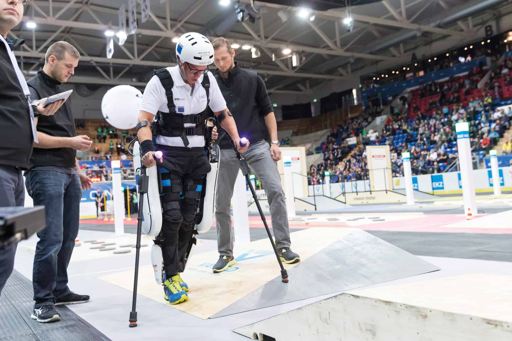
In the exoskeleton race, pilots with complete paraplegia, who cannot control their legs, were required to climb stairs, sit down on a chair and manoeuvre across ramps. Researchers from ETH Zurich’s Rehabilitation Engineering Lab, who competed in the VariLeg team, approached the challenge by starting their exoskeleton design from scratch, seeking to replicate human movement as closely as possible. That’s harder than it sounds. If you’re an able-bodied person and, say, collide with an object, it doesn’t affect your gait: step on a stone, and your leg will become “compliant” and stiffen or bend as required. “We humans do that automatically and our research is now trying to imitate this natural human behaviour,” explains team member Volker Bartenbach.
Replicating human movement: The VariLeg team from ETH Zurich took part in the powered exoskeleton race at the Cybathlon for pilots with complete paraplegia. (Courtesy: ETH Zürich/Cybathlon/Nicola Pitaro)
In robotics, the term “compliance” means flexibility or suppleness. It’s the property that allows able-bodied humans to grasp an egg without crushing it. A non-compliant robot, in contrast, has predetermined positioning and will follow the same path despite any impediment. Compliance allows a variation of positioning and is what the VariLeg team tried to design into their exoskeleton – essentially allowing a user to deal with the ups and downs of uneven ground.
“Human muscle is very complex, but [we] have attempted to imitate it,” says Bartenbach, whose team has essentially integrated a spring into the leg joint so that it is compliant. The knee joint is driven by a variable stiffness actuator (VSA) that can change the stiffness of the knee so that it can adapt to different surfaces. This stiffness is controlled by modulating the tension of the spring, which is currently preset, though staff in Bartenbach’s lab are working on how an exoskeleton user could change the setting at will or even automatically.
VariLeg came fifth out of seven teams in the exoskeleton race, which was eventually won by ReWalk – a German firm that makes commercial exoskeleton systems for people to use at home and in rehabilitation clinics. Mirroring the Formula One analogy, the event not only required technical innovation, but also a skilful driver. “This is not easy to do, especially with the harder obstacles,” says Bartenbach. One of the obstacles in the race was a series of uneven stepping stones, which required a really high mobility, “We do not have [that] at the moment with our system, but maybe will in the future,” adds Bartenbach.
Brain waves
Another assistive technology that featured at the Cybathlon involved brain–computer interfaces (BCI). It’s a new area of research, which involves placing electrodes over the scalp to measure electrical activity in the brain. Known as electroencephalography (EEG), the technique can turn characteristic signals associated with particular thoughts into commands that let people with paralysis control a computer or device. In the Cybathlon race, the pilots controlling the BCI were required to move an avatar in a computer game through a series of obstacles using only their thoughts.
According to Matran-Fernandez from Essex’s BrainStormers team, a key part of any BCI design is to identify the distinct brain patterns that could constitute commands. It’s a bespoke effort, which in her case required asking the pilot to think different things many times over and then using a computer algorithm to find any signals that are different enough to be used in the game. “You are trying to find different thinking activities that produce a different pattern on the brain of the person who is going to wear the BCI,” Matran-Fernandez explains.
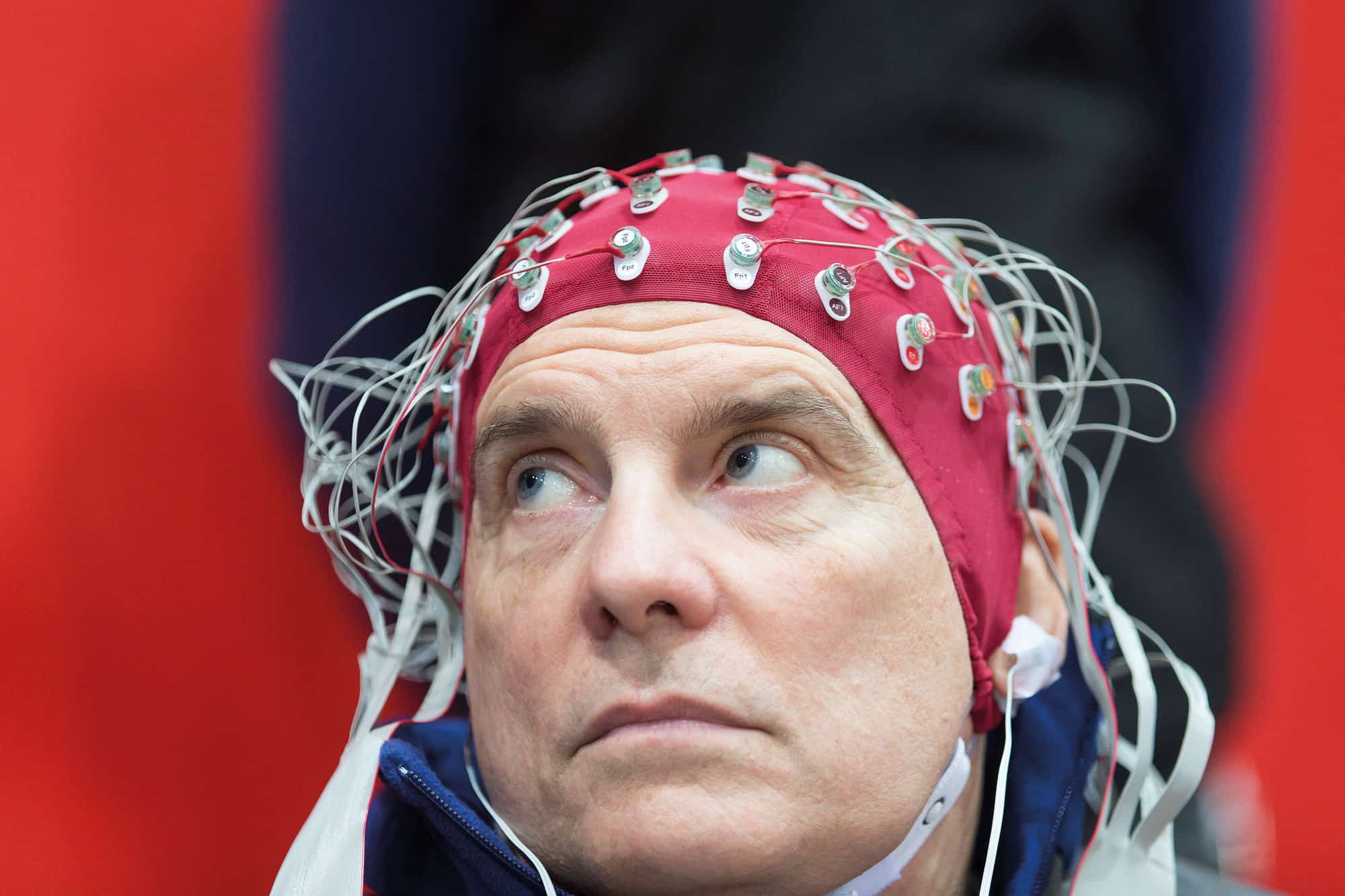
Deep thinker: Former rugby player David Rose representing the University of Essex’s BrainStormers team, in which the electrical activity accompanying certain thoughts in his brain was used to move an avatar in a computer game. (Courtesy: ETH Zürich/Cybathlon/Nicola Pitaro)
The pilot in question was David Rose – a former rugby player who Matran-Fernandez found in an online forum and was keen to be part of the BrainStormers team. Once Rose had been tested, Matran-Fernandez and her colleagues came up with four usefully distinct thoughts from him: three to move the avatar in different ways and one for it to stay put. The thoughts they ended up using were: moving your right hand; moving your feet; thinking of a telephone ringing; and playing a word game in your mind. These all produced identifiable signals that could be differentiated by the BCI as they came from different parts of the brain.
The BrainStormers’ system was set up specifically to work with Rose’s brain and, unfortunately, would need to be reprogrammed if it were used by a different competitor as there are slight variations in everyone’s brain pattern. As Matran-Fernandez explains, when people with long-term paralysis think about moving their limbs, they are no longer used to using them. The brain area in which the signal is observed may therefore have shifted from the specific part of the motor cortex used by able-bodied people to somewhere new.
As with the exoskeleton race, the pilot has a huge role to play in the BCI events too. “If you start tensing the muscles in your neck the signal [this creates] is much bigger than what you get from the BCI and that will mess up the whole system,” says Matran-Fernandez. Luckily Rose was an experienced competitor and the BrainStormers bagged a bronze medal.
The future starts here
Key to the success of the Cybathlon was that it allowed different technologies to be directly compared and observed in a race. As Faisal points out, researchers working on assistive technology usually do things in isolation and rarely compare themselves against each other. “Everyone does their own thing and never benchmarks themselves against the competition,” he says. “Here we had a level playing field and could objectively see how good the different approaches are.”
Everyone does their own thing and never benchmarks themselves against the competition. Here we had a level playing field and could objectively see how good the different approaches are
Aldo Faisal, Imperial College London
The Cybathlon also allowed people to assess the maturity of each discipline – to see what is and is not possible – and revealed where people have misconceptions. “For example, BCIs just don’t work particularly well and give the impression you can read people’s minds,” Faisal says, “[but] we are very far away from that.”
Even for arm prostheses, which have been commercially available for more than 50 years, there are still challenges for what is a mature technology. The team that won the powered-arm prostheses race in Zurich, for example, used relatively old technology based on the movement of shoulder muscles to open and close a claw hand. “In real life, we know that 30% of bionic prosthetics are abandoned within six months of first use,” Faisal points out, with users finding that the devices either don’t work well, are uncomfortable or aren’t easy to use, meaning that the challenge is to find something that feels more intuitive. “It needs to be seen as part of your body and that requires thinking about the cognitive and psychological aspects of the design,” Faisal says.
As for exoskeleton technology, it is accelerating rapidly in terms of the number of scientific publications as well as companies, more than 50 of which were listed on the exoskeleton.com website in 2015. Researchers at ETH Zurich, for example, have set up a firm called MyoSwiss to develop wearable muscle supports to improve the mobility of people with weak muscles. Bartenbach, who is not part of the firm, says that while it’ll be at least a decade before paraplegics can throw away their wheelchairs, it’s likely they’ll find themselves using an exoskeleton in certain situations, such as getting out of a chair. “An exoskeleton won’t heal your injury, but it can help your body remain active. The task now is to make current devices faster, lighter and cheaper.”
An exoskeleton won’t heal your injury, but it can help your body remain active. The task now is to make current devices faster, lighter and cheaper
Volker Bartenbach, ETH Zurich
Matran-Fernandez, however, is not as optimistic about the future for BCI. Current communications technology allows the user to spell two or three words a minute, “which is really slow for a person who can type or speak” she says, and it is impossible to combine the technology with movement. “No system is going to be able to filter out the [electrical signal] noise that you create by moving,” she adds.
One area where BCIs are actively being pursued is for people suffering from “locked-in syndrome” – when they have awareness but are completely paralysed and sometimes cannot even move their eyes. In 2017 researchers from the University of Utrecht in the Netherlands implanted a BCI in a locked-in patient who was suffering from motor-neurone disease. The electrode was positioned over their brain’s motor cortex, connected to a transmitter implanted in the chest. When thinking about moving their hand, the patient was able to control a typing programme, at a speed of two letters a minute.
With so many promising research avenues, another Cybathlon in Zurich is already being planned for May 2020, with organizers envisioning a longer, two-day competition next time around. “We have space for 96 teams, which will be around 30 teams more than in 2016,” says Sigrist. And while the disciplines will stay the same, co-director Wyss promises extra challenges for teams in some disciplines. Smaller, satellite events in separate disciplines are also planned before the main event, hosted by universities across Europe, including one in Graz, Austria, next year.
Veterans of the 2016 Cybathlon are now planning their strategies for the 2020 rematch and eyeing up yet further innovations. Matran-Fernandez, for example, is trying to persuade her colleagues to create a new team to take part in Zurich and also to develop a practical BCI system. And that’s the key. If the Cybathlon can speed up the development of assistive technologies, it will have shown the value and rewards of research – and benefit people in everyday life.
A new way of creating femtosecond, quasi-monoenergetic gamma-ray pulses has been proposed by Serge Kalmykov at the University of Nebraska-Lincoln and colleagues. Their scheme involves using a stack of lasers to create pulses of electrons, which then produce gamma rays through Thomson scattering. Laser-driven gamma-ray sources could have a wide range of applications including nuclear forensics and radiation physics.
When a relativistic electron collides with a near-infrared photon, some of the electron’s kinetic energy can be imparted to the photon via Thomson scattering. For a 900 MeV electron colliding head-on with a 1.5 eV near-infrared photon, for example, the photon is converted to a 19 MeV gamma ray.
Compact, low-cost solution
As a result, pulses of gamma rays can be created by firing electron pulses at pulses of near-infrared laser light. Gamma-ray pulses lasting just a few picoseconds have already been made using electrons from conventional accelerators. However, these accelerators are large and expensive and, because of their design, cannot create pulses of sub-picosecond duration
Writing in the New Journal of PhysicsKalmykov and colleagues point out that laser-plasma accelerators (LPAs) can create sub-picosecond (femtosecond) pulses of electrons, and therefore offer a way of making ultrashort gamma-ray pulses. LPAs involve firing ultrashort, intense laser pulses at a plasma cavity, accelerating electrons to energies normally associated with conventional particle accelerators.
While LPAs offer a way forward, the physicists point out that there are challenges that must first be overcome. In the Thomson scattering process, the electrons are much more energetic than the gamma-ray photons they produce. Therefore, the energy spectrum of the gamma rays is highly sensitive to the energy profile of the electron pulses. This is a problem because the electron pulses produced by LPAs tend to develop a broad range of energies as they travel through the plasma cavity. This results in gamma-ray pulses with broad energy distributions – something that is incompatible for practical applications. Another important challenge is that the lasers of today are simply not capable of delivering the power required to create a practical gamma-ray source.
Stack of lasers
Using simulations, Kalmykov and his colleagues have shown that these problems can be solved by using a stack of several different pulse lasers – each generating light at different wavelengths with modest energy outputs of about a joule per pulse. The research suggests that having tight control over the time delay, frequency difference, and energy ratio of each individual laser as it fires into the cavity, the system can produce focused gamma-ray pulses with a narrow spectrum of photon energies.
If such a system could be built, it would allow for applications including nuclear forensics, which require monoenergetic gamma radiation to investigate the atomic constituents of samples. It could also be useful for use in radiation physics, which is concerned with the interactions between radiation and matter.
BEAMSCAN™ is the world’s first all-in-one water phantom featuring wireless auto set-up and operation.
BEAMSCAN™ reduces manual interaction to the touch or click of a single button. Its innovative “Wireless Auto Setup”, which includes a unique patented three-point auto levelling function, uses advanced mathematical algorithms and computations rather than semi-automatic tools which require no screws, no tank shifts and no manual adjustments. An intuitive “Auto Setup Wizard”, which is easily accessed from a smart device or PC guides users step by step through the entire set-up process.
Use of a gamma camera to image sentinel lymph nodes during cancer surgery can help the surgeon assess the extent of cancer spread, and could help reduce mortality rates. Portable gamma camera systems for this application are already commercially available, but one key challenge of this approach is determining the exact spatial location of the recorded signal.
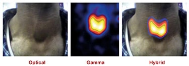
Speaking at the recent MediSens conference in London, John Lees from the University of Leicester described how combining gamma and optical imaging could solve this localization problem. “We can use hybrid gamma and optical imaging to improve diagnosis,” he explained. “The idea is to take a small camera into the operating theatre and help improve treatment outcomes for patients.”
Lees and colleagues have created a handheld hybrid gamma camera (HGC) based upon a high-resolution CCD chip coated with a columnar caesium iodide scintillator, located behind a pinhole collimator. The columns in the scintillator act as light pipes, channelling the light onto the CCD and maintaining a high spatial resolution. The device integrates an optical camera aligned to provide the same field-of-view.
The team tested the HGC using a hot-spot phantom filled with radioactive solution, and observed that the camera could resolve features as small as 1 mm. They also determined that the sensitivity was good enough: “We could detect down to 25 kBq in about one minute,” said Lees.
Lees described a contrast-to-noise ratio (CNR) analysis performed on a head-and-neck phantom, using cameras with 0.5 and 1 mm pinhole collimators. After imaging for just 10 s, the HGC could identify inserts with 0.1 and 0.2 MBq activity, at the parotic gland level, and 0.5 and 1.0 MBq signals at the submandibular level. In another example, imaging a thyroid phantom filled with 99mTc demonstrated that the HCG could visualize the thyroid after just 200 s, with greater detail appearing as scan time increased.
Clinical transition
The next step involves testing the HGC in the clinic. “Last year, we got ethical approval to undertake a clinical evaluation of the camera with patient volunteers,” Lees explained.
Clinical thyroid gland imaging
The team has already demonstrated that the HCG can perform combined optical/gamma imaging of the thyroid gland in patients. “We’ve got sensitivity, portability and spatial resolution,” said Lees.
He also presented a recent lymphoscintigraphy image in which the HGC clearly visualized the lymph nodes.
Lees also shared an image from a lacrimal drainage study. A couple of hours after administering 1 MBq of the radiopharmaceutical 99mTc DTPA, the hybrid image (using a 5 min acquisition at 7 cm from the patient) clearly showed the path of lacrimal drainage.
“We are also looking at preclinical imaging and pushing our camera to higher resolution,” said Lees. With this aim, the researchers have demonstrated that the HGC can image radiopharmaceutical in a mouse, with a 5-minute acquisition at a distance of 12.5 cm. He noted that the image quality was comparable to that recorded by the U-SPECT preclinical imaging system.
The next steps
Looking further ahead, one potential evolution of the hybrid camera could be to combine radionuclide imaging with near-infrared (NIR) fluorescence imaging, using an integrated gamma/NIR camera. “There is a lot of interest in taking a radiolabelled tracer and combining it with a fluorescent tracer,” said Lees. Such an approach could be employed in both preclinical or surgical applications.
He presented images of a four-hole phantom recorded using optical, gamma, hybrid optical/gamma and NIR fluorescence. Combining all three modalities should provide exceptionally good spatial resolution. He also showed the first examples of in vivo radio-NIR fluorescent imaging in a mouse.
Finally, Lees described the use of two cameras to perform depth estimation. He presented an image of a breast phantom containing two radioactive 99mTc sources. The positions of the sources were apparent, but a single image could not provide information as to their depths. By using two cameras, and taking a sequence of optical and gamma images, it was possible to estimate the depth of the two sources. “We believe that we can also do this inside the body,” he noted.
Lees was asked by an audience member as to the possibility of adding another modality, such as ultrasound, to the camera. “Ideally, I would like to combine optical, gamma, NIR and ultrasound,” he responded. “But the technology needed to combine them is not straightforward.”