What if gadolinium-based contrast agents (GBCAs) could be used at just 10% of current dose levels without significantly degrading image quality or contrast enhancement? Artificial intelligence (AI) could make that possible, according to research published in the Journal of Magnetic Resonance Imaging (J. Magn. Reson. Imaging 10.1002/jmri.25970).
Researchers from Stanford University have trained a deep-learning algorithm that can create synthesized images that are approximations of full-dose, contrast-enhanced brain MRI studies; however, they are based only on pre-contrast and post-contrast acquisitions at just 10% of the standard GBCA dose. In testing, the algorithm’s synthesized full-dose images were found to offer a significant quantitative improvement in image quality over the low-dose images, and they were deemed to be qualitatively non-inferior to the actual full-dose images.
“This [algorithm] will benefit clinical practice, especially for the populations that are more vulnerable to contrast risks, such as paediatric patients and [multiple sclerosis] patients who have routine contrast-enhanced MRI more frequently,” lead author Enhao Gong told AuntMinnie.com.
Gadolinium deposition
Gadolinium deposition within the brain and body from GBCA use has become a major issue in medical imaging and has led to concern about – and greater regulatory scrutiny of – these agents worldwide. Higher doses of GBCAs have also been linked to the development of nephrogenic systemic fibrosis (NSF). However, these agents have also been shown to be useful and are widely applied in contrast-enhanced MRI to assist diagnosis, Gong said.
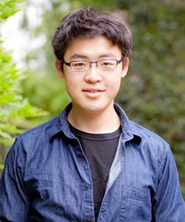
Enhao Gong
“For a lot of clinical applications such as for cancers and [multiple sclerosis], contrast-enhanced images are key for diagnosis,” he said. “Therefore, a solution is needed to reduce the risk of the potential side effects while still ensuring the benefits of having contrast information for diagnosis.”
Gong noted that radiologists read contrast-enhanced images by visually and subjectively comparing the pre-contrast/non-contrast T1-weighted images and the contrast-enhanced T1-weighted images, and then identifying pathology from the differences.
“Gadolinium or other type of contrast dyes are used since they significantly boost the differences for subjective evaluation,” he said. “However, with the help of an AI/deep-learning algorithm, it is possible to boost the differences without a large-dosage injection because the algorithm is actually much better at extracting subtle differences and constructing visualizations in quantitative approaches.”
The researchers, which included senior author Greg Zaharchuk, sought to determine if it was possible to reduce the gadolinium dosage and risk to patients while still maintaining the image quality and contrast information of full-dose contrast images. Previously, Stanford researchers had trained a deep-learning algorithm that could enable PET imaging to be performed using only 1% of current radiotracer dose levels.
In this AI project, the researchers retrospectively gathered MRI data from 60 patients who had received perfusion imaging with bolus contrast-enhanced dynamic susceptibility contrast (DSC) for evaluating suspected or known enhancing brain abnormalities. The patients, which included 36 men and 24 women with an average age of 49.7, had received 3D non-contrast axial T1-weighted imaging using an inversion-recovery prepped fast spoiled gradient echo (IR-FSPGR) sequence on 1.5- or 3-tesla scanners (MR750, MR750w, and Signa HDx; GE Healthcare). Prior to perfusion imaging, a 10% dose (0.01 mmol/kg) of MultiHance (Bracco Diagnostics) was given to the patients to reduce the effects of leakage on cerebral blood volume measurements.
As part of a quality control study, the same IR-FSPGR sequence was also performed immediately following the 10% dose to confirm compliance with the protocol. The patients then received a standard DSC study with the remaining 90% (0.09 mmol/kg) of the contrast agent dose, followed by a post-contrast 3D T1 IR-FSPGR sequence with the same parameters, according to the researchers.
Training the algorithm
As the three scans at different dose levels were collected separately, the researchers then co-registered and normalized the images to enable quantitative extraction of contrast information from the subtle differences in signal. With the full-dose, post-contrast MRI scan used as the ground truth, a 2D convolutional neural network was trained to approximate the full-dose study using the post-processed, pre-contrast MRI and the 10% dose postcontrast MRI as inputs.
Greg Zaharchuk
Standard deep-learning data augmentation techniques such as 90° rotations and flips were applied on a training set of 10 patients with mixed clinical indications. After this training was completed, the model’s performance was tested on an additional 20 patients with mixed indications. Finally, the researchers performed quantitative evaluation of the algorithm on a separate dataset with 30 patients with known gliomas to determine if its performance varied for a specific clinical indication.
The algorithm can produce a synthetic full-dose, 512 x 512-image in 0.1 seconds, according to the researchers. After repeating the process for each slice, the algorithm generates an entire 3D volume.
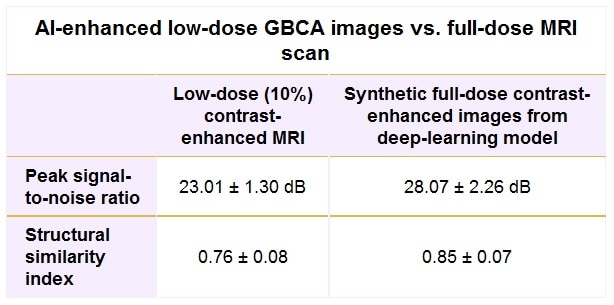
The group conducted several performance evaluations, including using quantitative metrics – peak signal-to-noise ratio (PSNR) and structural similarity index (SSIM) – to compare the low-dose contrast images and the synthetic full-dose images with the truth full-dose images for all 50 patients. Two experienced neuroradiologists also blindly performed qualitative assessment of the test set of 20 patients with mixed indications.
The deep-learning algorithm’s synthetic full-dose images were found to be quantitatively and qualitatively superior to the low-dose images, as determined by comparison with the actual full-dose contrast-enhanced images and measured by PSNR and SSIM.
The more than 5 dB gain in PSNR and more than 11% improvement in SSIM for the synthetic images were statistically significant (p < 0.001), as were the qualitative assessments for image quality (p = 0.003) and contrast enhancement (p < 0.001).
When qualitatively compared with the true full-dose images, the algorithm’s synthetic full-dose images were deemed to have slightly – but not statistically significant – lower image quality (p = 0.083) and contrast enhancement (p = 0.068). However, the researchers did find that motion artefact suppression was slightly better in the synthesized images, an improvement that reached statistical significance (p = 0.039).
In addition, a non-inferiority test rejected the inferiority of the synthesized images to true full-dose images for image quality, artefact suppression and contrast enhancement, according to the researchers.
“While larger datasets, better experimental design, and more advanced models may lead to further improvements, the current study suggests that [deep learning] is a promising solution to reduce gadolinium dose used for brain MRI exams while preserving image quality and avoiding significant degradation in contrast enhancement, which could be of great benefit to patients,” the authors wrote.
The algorithm has now been trained and evaluated on approximately 80 cases, and the researchers are working on a large, in-depth evaluation of the model using several hundreds of cases, Gong said. The researchers also believe the algorithm will be applicable to body MRI studies.
How much does energy cost? LCOEs – Levelised Costs of Energy – are widely used as a comparative measure. They give an estimate for the cost of energy generation from specific plants, but do this by averaging out the investment and running costs over the plant’s lifetime and comparing that with the value of the electricity generated. However, the costs and earnings can and do vary over time and are hard to predict. LCOEs also omit any associated grid balancing/backup costs. So they have big shortcomings. Can we do better?
Certainly there are many weaknesses in the LCOE approach. Finance costs will depend on interest rates and inflation both of which can change over time, sometimes dramatically. So may fuel and labour costs. Energy output may also vary for many reasons and in the case of renewables will vary with the weather. In that case, use is usually made of ‘capacity factors’ to reflect average likely delivered outputs, but in reality these variations are dealt with by balancing capacity and services, the cost of which, arguably, should be added to the cost of generation.
However, doing this is not simple. Studies by Imperial College London have suggested that in order to capture the full costs and benefits of variable renewables, we have to look at total system costs, including the cost of full grid balancing/backup, not just the individual component LCOE of generation and of individual backup plants. Robert Gross from Imperial has pointed out that “Demand response, flexible generation, storage and interconnection offer benefits to the system as a whole and building them as if they need to be dedicated to each specific variable renewable installation will result in over-investment. System costs should be charged to generators as cost-effectively as possible, but on the proviso that they are assessed at a system wide level rather than on an assumption that variable renewable installations need to self-balance.”
Similar views have emerged from other studies. A World Bank study of variable renewable energy (VRE) system optimisation said that it was not helpful just to look at individual balancing or supply options in isolation, focusing just on the lowest cost ones: “Policy, planning and regulatory interventions should be designed to minimize overall system costs subject to meeting performance targets, rather than minimizing the costs of VRE generation alone.”
A study of optimal approaches to managing high renewable energy mixes in the US similarly stressed the need to select generation options appropriately, looking at total system costs, not individual component costs, and where possible, choosing supply options with output profiles that complemented each other.
Rough estimates of the extra balancing costs imposed by renewables on the energy system have been made and range from 10% to as much as 50%, depending on the assumptions and data used. For example, the 50% estimate is from the Potsdam Institute. They include what they call “profile costs”, the extra costs faced by conventional generation companies given that renewables, with their low marginal costs, can at times push conventional generation out of the market, undermining the company’s profits. Are their losses strictly a cost or just part of commercial reality?
Even if agreement can be reached on what to include in the costs, it is hard to identify future optimal mixes and the exact extra costs, given that the whole system may change – it’s a moving target, with many unknowns as to the future mix. The UK Energy Research Partnership (ERP) notes that “The value to the system is highly dependent on the technology mix on the system, and the effect of diminishing returns reduces the value of all technologies as they are added, but especially so of variable renewables which generate an increasing proportion at times of surplus energy.” Certainly, the ERP says, “Using a fixed number (like LCOE) to characterize a technology’s economic value is quite unhelpful in these circumstances.”
So the ERP, along with Imperial College and others, all agree, although sometimes for different reasons: LCOE is not a good enough measure. Some analysis has consequently tried to come up with new measures to include the extra costs. However, there is no consensus on how this should be done. For example, the Potsdam Institute makes use of ‘overall system costs’, including integration and, as noted above, more contentiously, profile costs.
Clearly, even leaving aside issues like that, it is not easy to assess the full likely costs: all generators (including nuclear) can need backup at times, not just renewables, and it is hard to assign fair shares of the costs of this provision to each generator in the overall supply mix. In the UK, a Capacity Market has been set up with auctions for balancing capacity. That provides a way to quantify costs, across the system, but doesn’t allocate them to specific generation projects. And the costs, as with generation costs, are just passed on to consumers.
The last point, of course, suggests another approach – start from the other end, the costs to energy users. In the UK, new renewable and nuclear projects are being supported by a Contract for Difference (CfD) subsidy system, backed up by the Capacity Market, with the extra cost being passed on by the power companies to energy consumers. In a review of Tidal Lagoons by the Tidal Lagoon Power Ltd, use was made of measures of costs to consumers, calculated not on the basis of LCOE, but on “equivalent CfD contract” costs, with the net present value of actually delivered electricity calculated over the lifetime of the project, taking capacity factors into account. However, grid costs were not included: while it was noted that some may increase the total cost, any flexibility provided (e.g. via storage) may offset that. The end result was some dramatically different cost figures. Whereas on an LCOE basis, lagoons look to be the most expensive option, on this consumer cost basis some lagoons came out cheaper long-term than all other energy options.
It is not immediately obvious that this is a fair rendition of reality, but then neither is an approach based on LCOEs, especially if it is just used for individual projects. For example, in the case of extra gas plants used for back-up power, their capacity factors may be very low (they only run occasionally), so their LCOE will be very high, but they can make a valuable contribution to balancing the overall system. A wider cost valuation framework is needed. I will be looking at some new attempts to take variability into account, including one from Imperial College, in my next post.
Researchers from France have combined traditional PET-CT and ultrafast ultrasound imaging (UUI) to create a new hybrid imaging modality that can identify metabolic activities while capturing rapid phenomena with high resolution. This approach should yield simultaneous anatomical, metabolic and functional information while being relatively low in cost. In a proof-of-concept study, the team details how the technology could benefit the fields of oncology and cardiology (Nature Biomed. Eng.2 85).
Strength in numbers
The current trend in medical imaging consists of coupling imaging modalities to increase the number of parameters obtained during scans. This has led notably to the emergence of PET-CT and SPECT-CT imaging, or more recently to the development of PET-MRI scanners. The advantages of such hybrid modalities, however, are counteracted by their additional cost and added complexities in image processing.
PET imaging allows observation of the metabolisms of radio-tagged molecules such as fludeoxyglucose (FDG), and its combination with CT has long been a reference for cancer imaging as the combination provides both metabolic and anatomical information. However, PET-CT cannot detect dynamic phenomena and is hence only used as a static imaging modality. Conversely, UUI has the unique ability to capture thousands of images per second of 3D volumes, and the development of super-resolution transducers enables the capture of microscopic details such as tissue microvasculature. Combining the three modalities would hence offer unique anatomical, molecular and functional insights.
Merging PET-CT and UUI
A team from Institut Langevin, INSERM and Université Paris Descartes, led by Bertrand Tavitian and Mickael Tanter, tackled the challenge of designing the first prototype. Their initial work consisted in coupling the UUI transducer probe with the PET-CT scanner. The transducer is positioned over the organ to be imaged and the ensemble is inserted inside the scanner gentry.
Preliminary results showed that the presence of the transducer had negligible impact on the quality of the PET-CT scan, while its location within the gantry-volume yielded a markerless co-registration of the body-of-interest. The next step was to resample the CT, PET and UUI volumes to the same voxel size and fuse them together to obtain the desired representation of the organ.
Providing new insights
The researchers then tested the functionalities of this new modality. For example, they implanted tumour tissues in mice and focussed on the animals’ in vivo metabolism and vascularization, two major hallmarks of cancer used for tumour stratification. The PET-CT scan captured the uptake of the contrast agent FDG by the tumour, and the ultrasound imaging tracked the vessel perfusion. While the analysis revealed an increase in both tumour volume and perfused vessel region over time, their pace of growth differed: the ratio of perfused vessel volume over tumour volume decreased continuously, from 51% on day 14 to 28% on day 32 after implantation.
The hybrid imaging modality also facilitated the identification of tumour status based on metabolic and vascular profiles. Indeed, tumours can sometimes affect cells by deregulating their energy metabolism – a feature that can be detected by tracking FDG uptake – while in earlier stages, the metabolism is not altered yet. This difference can guide the choice of treatment given to patients.
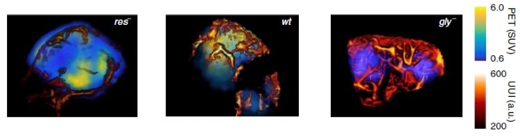
The experiment showed that the proposed modality could unambiguously differentiate three different metabolic statuses often found in tumours: an exclusively anaerobic metabolism (res–), and exclusively aerobic metabolism (gly–), and a mixed anaerobic and aerobic metabolism (wt).
Different metabolisms observed by the PET-CT-UUI setup
Finally, UUI can observe phenomena, such as beating hearts, more easily, and the addition of PET maps of FDG uptake makes delineation of the cardiac anatomy even more precise.
Weighting on the benefits
Combining imaging modalities usually comes with extra development complexity and cost. In this respect, the integration of a relatively inexpensive and portable ultrasound transducer probe in a PET-CT scanner is appealing. The fact that PET-UUI is simultaneous allows co-registration of the volume imaged and the establishment of a topographical relationship between multiple imaging parameters.
It is too early to know whether such an imaging combination will power new breakthroughs, but its potential for linking physiology and pathophysiology is promising. It could become a new standard for cancer imaging, where its ability to characterize both metabolic and vasculature can be particularly helpful.
Fermilab’s Don Lincoln is back with yet another entertaining video, this time about what people get wrong about time dilation. In particular, he asks whether the famous “twin paradox” is actually paradoxical. You can watch the video above, enjoy.
Have you taken a stunning photograph of the sky lately? If so, you still have a week to enter the Insight Astronomy Photographer of the Year, which is run by the Royal Observatory at Greenwich and sponsored by Insight Investment. This is the tenth year of the competition and to celebrate, the 2018 winners will have their work displayed at a new gallery at the National Maritime Museum in Greenwich – which will also show some of the best images from past contests.
Triumph of alliteration
Finally, in a triumph of alliteration there is a paper on the science of science in Science. According to the authors, he science of science (or SciSci) is “a transdisciplinary approach that uses large data sets to study the mechanisms underlying the doing of science – from the choice of a research problem to career trajectories and progress within a field”.
Among other things, the paper analyses the publication record of three physics Nobel laureates. This shows that these leading physicists have published high-quality work throughout their careers – and not just at moments of prize-winning brilliance.
Another interesting plot in the paper shows that the average number of authors on science and engineering papers has increased from just a shade more than one in 1900 to nearly five today. What’s more, today the average number of authors on papers judged to be of high quality is six – suggesting that bigger research collaborations may be better.
A dense, ultracold atomic gas has been created using blue-detuned laser light with a frequency that is higher than the atomic transition used to cool the atoms. This feat was achieved by Kyle Jarvis and colleagues at Imperial College London and their technique could offer a new way of trapping ultracold molecules – something that has proven very difficult to achieve.
Magneto-optical traps (MOTs) are particularly useful pieces of equipment for physicists wishing to trap samples of atoms at ultracold temperatures. MOTs use lasers with frequencies lower than certain atomic transitions (red-detuned frequencies) to cool the atoms. This “Doppler cooling” process involves atoms absorbing and emitting light in such way that their motion is reduced. It can be used to cool atomic gases to microkelvin temperatures – allowing the atoms to be used in a wide range of applications such as simulating quantum solids, measuring tiny changes in gravity and creating highly accurate clocks.
Avoiding rotation
Most MOTs achieve Doppler cooling through “type-I” atomic transitions, whereby the angular momentum of the excited state atomic state is higher than in the ground state. However, type-I transitions are of little use when trapping molecules because the trapping process must avoid exciting a molecule’s rotational and vibrational states. Instead, molecules need to undergo type-II transitions in which the excited-state angular momentum is lower than the ground state.
Attempts at using red-detuned lasers in MOTs to cool atoms and molecules using type-II transitions have resulted in gases that are too warm and too diffuse to be of any practical use. In their experiment, Jarvis and colleagues used blue-detuned lasers – with a frequency higher than that of the transition frequency. They describe this as a “counterintuitive” change in the cooling process because the absorption of red-detuned light by an atom is normally what causes the atom to slow down.
Despite this unconventional approach, the team could trap and cool rubidium-87 atoms using type-II transitions and achieved a density of 10¹¹ atoms per cubic centimetre – which is about one million times higher than achieved by red-detuned lasers. They also managed to cool the gas to below 30 μK.
When an ecosystem changes, those creatures most precisely adapted become the most vulnerable – and that is bad news for king penguins.
King penguins – one of the most charismatic species of the Southern Ocean – are under threat from climate change.
More than a million breeding pairs will either shift to new colonies or perish before the century’s end, as conditions in the Antarctic begin to change. The species Aptenodytes patagonicus could lose 70% of its population, according to a new study in Nature Climate Change.
The problem – increasingly familiar to conservationists and biologists – is one of mismatch engineered by climate change driven by human-induced global warming, itself a consequence of profligate combustion of fossil fuels that spill ever greater ratios of greenhouse gases into the atmosphere.
King penguins have adapted to a precise habitat range: they favour a scatter of islands in the Southern Ocean, away from the sea ice but close to an oceanic upwelling called the Antarctic Polar Front, which concentrates colossal quantities of fish into a small area.
And because of climate change, this front is in retreat. So penguin parents must swim ever further to find food for their chicks.
The new research predicts that – for most colonies – the distance to find fresh food will mean longer absences. At some point, the chick’s capacity to withstand starvation will be exceeded. The population will crash.
This is a reprise of an increasingly familiar story: human-induced climate change, along with other human bequests such as pollution and habitat destruction, have begun to threaten the wild things everywhere.
And even though the southern hemisphere is relatively less disturbed, and with a smaller human population, there have been signs of change. There have been separate cases of concern for Adélie and other penguins, for crested and rockhopper penguins, and even for the great survivor of the frozen south, the emperor penguin.
The entire region faces problems, in many cases driven by overfishing, and there has even been alarm about the condition of the albatross as it circles the southern ocean.
Repeated recoveries
The conclusion is based at least in part on evidence from the king penguin’s genome. The enormous string of DNA that encodes a creature’s machinery for survival is also a record of inheritance: it tells the story not just of the individual’s parentage but also of the entire species over many millennia.
And, so far at least, the king penguin DNA tells a tale of species survival: king penguin populations have crashed, and then recovered, more than once in the last 50,000 years.
But the bird faces new challenges. Humans have begun to alter the global climate, as well as to bring fishing on an industrial scale to a once isolated ocean. So hungry birds face high-technology competitors.
And faced with the choice of starve or migrate, the birds have only a limited range of potential breeding colonies. Around half of the population, on the Crozet and Prince Edward Islands, could lose their breeding colonies completely.
Poor prospects
Around a fifth of the population, based on the Kerguelen, Falkland and Tierra del Fuego Islands, will face ever greater foraging distances.
Climate change may create new opportunities for some, but overall, the outlook is bleak. “The main issue is that there is only a handful of islands in the Southern Ocean and not all of them are suitable to sustain large breeding colonies,” says Robin Cristofari, of the University of Strasbourg in France.
“There are still some islands further south where king penguins may retreat but the competition for breeding sites and for food will be harsh, especially with the other penguin species like the chinstrap, gentoo or Adélie penguins, even without the fisheries,” said his co-author Céline Le Bohec, also of the University of Strasbourg.
“It is difficult to predict the outcome, but there will surely be losses on the way. If we want to save anything, proactive and efficient conservation efforts but, above all, coordinated global action against global warming should start now.” – Climate News Network
Hadron therapy shows potential to improve the effectiveness of cancer radiotherapy. By exploiting the characteristic depth-dose distributions of hadrons in tissues (the Bragg peak), the technique offers precise delivery of radiation dose. The large gradients of the highly conformal dose distribution, however, make it critical to precisely and accurately plan the radiation field, align it with the target, and determine the stopping power of the ions. Current methods to do this have an uncertainty on the predicted range of 3–4%.
Advanced methods for absolute range verification are in development throughout the world. Researchers at Heidelberg University Hospital and the German Cancer Research Center (DKFZ) have developed a prototype detection system for helium-beam radiography (αRad) using silicon pixel detectors. The long-term goal of this research project is to develop a range verification method based on αRad.
The team has successfully demonstrated a proof-of-principle of αRAD, based on energy deposition measurements of single ions in thin layers, ion tracking and identification. In one key result, they showed that the rejection of secondary hydrogen ions originating from nuclear interactions distinctly improved the contrast-to-noise ratio (CNR) of the radiographs (Med Phys.45 817).
Lead author Tim Gehrke and colleagues created a prototype detection system consisting of three parallel silicon pixel detectors. The team selected pixel detectors (the CERN-developed Timepix chip) because they can measure the position of an ion in a single thin layer, and also provide unambiguous position determination in cases where multiple ions hit the detector in the same time window. These capabilities facilitated the construction of the ion track, and result in small multiple Coulomb scattering (MCS) in the tracking system.
The team selected helium ions instead of protons because they reasoned that helium ions would generate a better spatial resolution and undergo less pronounced MCS in the object being imaged. Moreover, they expected to achieve this resolution gain without any disadvantage in terms of CNR or applied radiation dose compared with proton radiography.
Detector verification
The authors used a phantom comprising a 160 mm thick block of acrylic glass, approximately the size of a human head. The phantom had a mean water-equivalent thickness (WET) of 192 mm and contained maximal WET-variations of ±6 mm. They imaged the phantom with a 173 MeV/u helium ion beam at the Heidelberg Ion-Beam Therapy Center.
The detection system was placed behind the phantom to register the ions leaving it. The system enabled detection of single particles, so that any with energy deposition higher than a user-defined threshold (in this case 5 keV) could be identified. Three detector layers were deployed: two to measure the arrival time of the ions for tracking and one to measure the energy deposition of the single ions.
When a heavy charged particle impacts the detector, the signal spreads over several adjacent pixels, forming a cluster. The so-called cluster volume, which is the sum of the pixel values in a cluster, was converted into the energy deposition of the incident particle by pixel-wise calibration. The authors note that desirable signals for image formation are clusters caused by single incident ions traversing all three detector layers, creating a matched event. They selected clusters corresponding to helium ions that did not have artefacts or degraded information.
The authors performed data processing, tracking and backprojection of the ions, calculated CNR, estimated the delivered dose and evaluated spatial resolution. They reported that this particular set-up achieved a spatial resolution of at least 1.15 lp/mm for, as well as a resolution of relative thickness differences of 1.2%, at a dose level typical for diagnostic radiography.
Range verification
Helium-beam radiography can be applied as an image-guidance tool for proton, helium and carbon-ion radiotherapy. Gehrke explained that a goal of αRad is to measure WET maps that might be used for in vivo range verification.
“Since the WET measured by αRad retains its validity for proton and carbon-ion beams, αRad is in this respect equally useful for proton, helium and carbon-ion therapy,” he said. “From a practical standpoint, αRad is especially interesting for helium-beam therapy, because this combination of imaging and treatment modality would not require any ion-type switching during the whole process. The applicability of αRad for intra-fractional imaging in proton or carbon-ion treatments will depend on the speed of the ion-type switching of a particular facility.”
The researchers have also recently performed an in-depth comparison of proton and helium-beam radiography, using the silicon pixel detectors and the data processing procedures that they developed. The detection system could be successfully applied to perform αRad, as well as proton radiography (pRad). This research showed theoretically and experimentally that helium ions provide a higher spatial resolution compared with to protons, retaining the same CNR at a comparable dose (Phys. Med. Biol.63 035037).
Gehrke says that the team is currently focusing on new ideas and concepts to overcome two major limitations of the method. They are working on the extension of the sensitive WET range (currently about 12 mm) in which a high CNR can be achieved. They are also developing methods to decrease the acquisition time of radiographs measured by their prototype detection system, therefore requiring silicon pixel detectors with a higher readout speed.
The history of physics is littered with people affected by mental-health conditions, and too often their stories have sad endings. Isaac Newton, Wolfgang Pauli and David Bohm, to name a few, are all thought to have suffered to some degree, and Ludwig Boltzmann took his own life after struggling with bipolar disorder for years. But mental illness does not just strike the elite. It is something that can affect anyone and you will undoubtedly have friends, family or colleagues facing such problems. Unfortunately, there is very little awareness of mental health in academia and few practical resources or material to draw on.
I suffer from bipolar disorder, which has affected me throughout the various stages of my academic career. Nowadays, I am working within my university to try and establish an effective support network and to create awareness around mental health. As part of this effort I have decided to share the story of how my own mental health has affected me as an academic in physics.
I hope that by sharing my experiences it might help start a dialogue in our community. People should not feel embarrassed to talk about these issues, and those suffering from mental illness need to know they are not alone and there is always help available – although more options on that front would be beneficial. We also need to improve the stigma around mental health. In the past, people have said some awful things in front of me, directly to me and behind my back, whether intentional or not. Others, meanwhile, tend to laugh things off, especially when they’re uncomfortable. But mental illness isn’t funny or a joke. By generating awareness of the issues, I hope people will think twice before making light of matters.
Before I start my story, one of the first things you will have noticed is that I have chosen to remain anonymous. While this decision runs counter to the ideal culture of openness, it is a necessity for me. Many of my friends, family and colleagues are incredibly supportive of me, but unfortunately, some people still have a bizarre and outdated view of mental illness. It’s sad that I must think this way – and it makes me angry sometimes too – but it’s hard to express the discomfort it could cause me if my mental-health problems were publicly known.
The beginning
Bipolar disorder, formerly known as manic depression, is mainly characterized by experiencing periods of depression, where you feel low and lethargic, and periods of mania – feeling very high and overactive, sometimes in an unpleasant and disorientating way (such as having thoughts that don’t stop racing). Bipolar disorder is a wide-ranging term, covering varying degrees of severity and symptoms, and I fall on the “schizoaffective” side. This means that on top of the typical bipolar symptoms, I have also suffered from those associated with schizophrenia, including audible and visual hallucinations and delusions.
I was not diagnosed with bipolar disorder until my early 20s, but my therapists and psychiatric team have looked back at my past, and we think the symptoms started when I was around 15 or 16. During sixth form (age 16–18), I began to experience severe mood swings that could last weeks or months, disrupting my attendance, focus and levels of motivation.
These symptoms worsened during my undergraduate degree in theoretical physics. Depression made it hard for me to do basic things – even getting out of the house was a challenge, let alone attending lectures. Yet, if I was heading towards a manic episode, I felt like the most confident person in the world. The mania meant I needed little sleep. I could plough easily through the learning material, and would do well in exams and assessments. Although experiencing such mania seemed “useful” at times as an undergraduate, it ultimately caused chaos – the feeling never lasted and was often followed by a crash into depression.
Depending on how intense the mania was, I could lose touch with reality, suffer from delusions and become uncharacteristically arrogant
As well as impacting my studies, such extreme highs and lows tested and strained my personal relationships, as friends and family would become concerned with my eccentric and erratic behaviour. Depending on how intense the mania was, I could lose touch with reality, suffer from delusions and become uncharacteristically arrogant. Sometimes I even experienced psychotic episodes, which were upsetting for all involved.
The mania could sometimes wreak havoc on my finances. I planned many unrealistic scientific projects, often involving expensive equipment and exotic locations, all of which never got done due to a crash in mood, because I was off to plan “more important” things, or because they were just plain nonsense. There were some seriously chaotic times.
This was all about 15–20 years ago, at a time when people were even less aware of mental-health issues than now. As far as I knew, there were very few support services available at my university – although, given they were so hard to discover, there may have been more. Meanwhile, my undergraduate peers and many of the academic staff appeared to be uncomfortable around me. I often felt ignored and saw a lack of empathy in the community towards people who were clearly suffering. I can understand this a bit though, as my behaviour, depending on the mood swings, could be difficult to deal with.
My undergraduate tutor did take notice, however, and was particularly concerned about me. He tried to get me to go to some of the limited available services but I did not listen. The problem with being manic is that you feel great at first and why would you want to stop that? However, when I felt depressed I couldn’t even think of talking to someone new, and unfortunately, I didn’t have much down time in-between. In hindsight, I wish I had listened to my tutor more.
A downward spiral
Although I faced problems during my undergraduate degree, my ambition to study physics remained strong, and I got a PhD studentship in theoretical physics at a different university. Unfortunately, despite all my enthusiasm for physics, my illness continued to worsen.
It’s hard to understand why. I’ve wondered if getting older meant my symptoms got worse, or if the open nature of research work lends itself to mood swings. There are big pressures during a PhD, such as managing your time, meeting deadlines and planning your future career, and I also had the legacies of my illness to carry, including friendship issues and financial debts. It did not help either that I had less direct supervision, leaving less chance for someone to intervene.
My illness continued to get worse and I had some severe bouts of mania during these times (I felt I was really going mad) and some serious depressive crashes. Being manic felt like it helped me do amazing research, but once things calmed down, I would re-read my notebook from the previous few days or weeks and often find utter nonsense in there despite me feeling I had done something amazing at the time.
Courtesy: iStock/agsandrew
In stark contrast, the depressive episodes were harder and darker than ever, and led to time off, disruption and, most distressingly, my first suicide attempt. Thankfully, the city I had travelled to to do this had some wonderful members of the public who prevented me from going through with it. Meanwhile, back at home the police had been called and had I think (I still don’t know fully what happened) been to my place of work. It felt humiliating at the time, but this forced me into my first proper treatment and I was put on some serious medication to try and lift my mood.
I hated taking medication – it made it hard to concentrate and I had difficulty performing the problem solving and logical thinking I needed to carry out research. I felt it was impacting my ability to work and ruining any chance I had of a career in research and academia. So, I would come off the medication – a disastrous decision. I usually ended up in a period of mania or hypomania – a milder version of the condition – that then resulted in a bigger crash, and ultimately, I would be forced to go back on the medication by friends and my medical team.
I felt at this point that the people around me, at work and at home, did not understand the illness or the effects of managing it. I was feeling very low and had a year remaining of my PhD. I needed to finish my doctorate and look for a job, but I also needed some time off to get my health under control. When I informed my department, I was told, however, that I would have to have my stipend suspended if I wanted to take time away from my PhD – essentially, I would be forced to take unpaid leave.
I still cannot believe this was how they handled someone who was suffering from severe depression. I was a good student – I had been awarded prizes in my first and second year and was otherwise on track to successfully get a PhD. Yet the university did not try and find a solution that would help get me through this horrific time. They had no protocols or systems in place to help students in bad situations, and they did not look for alternative funding. There was a total lack of effort from my department and supervisor, and I wasn’t even given details of support programmes that could help while I continued to work – I had to find them myself. It was worse than during my undergraduate degree – there was no sympathy and no empathy towards my case. I was shocked and hurt, but had no other choice than to plough on with the work.
New place, old problems
Fortunately, the National Health Service (NHS) was fantastic, and with its support I got through the final year of my PhD. I even managed to submit my thesis on time, which was pretty good considering everything that was going on. In spite of my troubles, my mental-health problems had once again not dampened my love of studying physics and doing research. Not wanting to leave academia, I amazingly found my dream job – a two-year postdoc in the US. Working abroad felt like it would be a fresh start and the institute I was going to be at was one of the best in the world for my chosen field.
I needed to have a good crack at this new job but, once again, I felt my medication was limiting my ability to work properly. So, I decided to stop taking it, which led me to some very scary and dark times in my life.
It’s often hard for people to understand why I would go on and off my tablets like this: surely controlling the bipolar disorder was the priority? Had I not learnt from past experiences? But all I wanted was a fair chance to do something I love, and I’ve always felt the medication was stopping my academic brain functioning to its full capacity, holding me back from succeeding and developing in the field.
This is when the schizoaffective element to my illness started to become very apparent. I was becoming delusional and having psychotic episodes. I thought I was being followed or attacked by creatures. I became scared to go out of my apartment, too frightened to even leave the bedroom. My colleagues started to worry as I became erratic around them.
The university offered no sympathy and no empathy towards my case. I was shocked and hurt
The medications I was then prescribed in the US were much stronger than my previous tablets and the impact was horrendous. I was sleepy all the time and it became impossible to function normally, but they kept me out of hospital. A real battle began and this period of work was a disaster for me as I couldn’t find a balance – I seemed to be either manic or too depressed to get to work or knocked out by medication.
Difficult decisions
At the end of the placement I returned to the UK and took up a two-year postdoc with my previous university. It was a very generous offer and this was due to my record of good performance – at times – as a PhD student. At this point my UK medical team changed my prescription again, as they were shocked by how many tablets the US doctors had put me on. Unfortunately, going off one type of medication and onto another was as bad as when I had stopped taking them completely. I started to become manic but was keen to hide it from people as it felt good – I felt like my old self again. But I began to hear voices, my behaviour became erratic, and it ended how it usually ends – in stronger medication and a lot of time off work.
I felt like I was failing. I really wanted to work in physics research but it was looking increasingly impossible – the bipolar disorder was ruining my career in academia. At this point, I started to become suicidal again. Thankfully, though, things calmed down, and towards the end of the postdoc I made the difficult decision to find a different career.
I felt like I was failing. I really wanted to work in physics research but it was looking increasingly impossible
I chose teaching – something I had always been interested in – but my heart was still in academia. I became very depressed again and found managing my illness through the teacher training and first year to be very tough. Thankfully, I had outstanding support from my mentors and lecturers. It was very different from my experience in academia – the education staff showed empathy and it was an unusual experience to feel supported. During this time, I was also incredibly lucky to meet the amazing person I would later marry. I suddenly had the support I never really had before, and I learnt to understand and manage the disorder much better.
I worked as a teacher for three years until a perfect opportunity came up to re-join my old university physics department on a permanent basis in a position involving both research and teaching. And this is where I am still – doing the research I love and enjoying teaching at a university level.
I am in a much better position with my mental health than I ever was, but the bipolar disorder will never truly go away. I have been on medication for about 13 years and still occasionally suffer from mania and depression, and the desire to reduce my medication. Fortunately, I have the support and love of my partner to keep me on the right track and, with the help of friends and learning from my experiences, I have managed to get through relapses without too much disruption.
Combating the stigma
My story highlights the struggles of dealing with mental illness while pursuing a career in academia – but whether this is a problem specifically within physics, or whether it is a wide-spread problem across the academic community, I cannot say. Without seeing reliable statistics, all I can offer is my own personal evidence, and what concerns me from that is the lack of awareness and understanding of such issues within the academic workplace.
I hasten to say that I don’t think this is a problem solely limited to academia – the stigma that surrounds mental health is rooted in our society’s attitude and culture. But this has been improving a lot in recent years, and there are many initiatives striving to improve awareness and provide support (see box opposite). Indeed, I have friends working for companies that have a supportive and open culture when it comes to mental health. I wish this was the case everywhere. Even my own time as a teacher highlighted the comparative lack of support in academia.
Again, I can only give my own anecdotal evidence, but there seems to be more stigma towards mental health in academia and improvements don’t seem to be keeping pace with the wider world. I know colleagues who have mental-health issues but refuse to come forward about them because they are worried about the stigma. Throughout my own career I have experienced some terrible attitudes from people, which I have found hurtful and sometimes cruel. It worries me greatly that our community lacks empathy towards individuals who are unwell.
I often sensed that people thought less of me and would not take a chance on me, or even did not trust my work
So why is academia worse? In my opinion, it partly comes down to the nature of academic work. The career paths are target-based, stressful and very competitive. There are limited pots of money and contracts are often short. It’s not surprising that working in such an environment can trigger mental illness. This backdrop means people who are unwell for a long period of time can be viewed as a liability, and working with them could be seen as detrimental to a project’s progress. I worried about this constantly when I was unwell – I felt I was wasting money and stopping a project from moving forward. I often sensed that people thought less of me and would not take a chance on me – at stages I even got the impression that my colleagues did not trust my work. I have seen this in how others suffering from mental illness have been treated too, and these attitudes mean you also begin to doubt your work and capability.
The reasoning behind these attitudes could be numerous – perhaps some people think that having a mental illness means you are not as intelligent, or maybe they think avoiding people with mental illness is simply logical and practical for the benefit of their project. Whatever the reason, I believe it comes down to ignorance and a lack of awareness, and this is something I would like to address. I think it is vital that we generate awareness of mental-health conditions and highlight the problems that some of us have to go through, ultimately improving the support offered in the academic workplace.
By making sure the community is informed and aware, you can also reach and educate those suffering from mental-health problems, ensuring they know they are not alone and that there are different support systems and actions available to them.
I’ve had people approach me with concerns about their own mental health and one of the first things they ask about is the side effects of medication. It’s not good that people are having to think about this before seeking medical help, but it is a genuine concern for my colleagues and friends in academia. As I’ve said, some of my biggest battles have concerned medications – I often felt the tablets were holding me back, suppressing my concentration and ability to do research. When your brain power, logic and problem solving are the most important tools of your job, it’s difficult to face tablets that might suppress that.
While I may not enjoy taking it, medication is the best way for me to stay well. That, however, doesn’t mean it’s the case for everyone – for others, cognitive behavioural therapy or counselling, for example, might be a better fit. It is therefore important to make sure people are aware that seeking help doesn’t necessarily mean tablets and, if it does, then there is nothing wrong with that. The most important thing is to get help and get well.
Unfortunately, mental-health conditions don’t simply go away, and often people don’t understand this. Like a physical injury or illness, they believe it will just pass or that medication will miraculously cure it forever. Personally, if I appear to be “normal” that does not mean there are no underlying issues. I manage this illness with medication and seeing professionals, and I will likely have to continue doing so for the rest of my life. It is important that employers and colleagues understand this is a long-term battle. They need to be able to make adjustments for the bad days or the side effects of medication, while also not penalizing those who are suffering.
A helping hand
Despite the steps that must still be taken, I must give credit where it’s due and point out that things have improved considerably in recent years. In general, mental-health awareness has increased ten-fold and there is more information readily available. Universities often have more visible counselling services that offer students a range of resources such as self-help guidance, drop-in sessions, workshops and one-to-one support. At the department level, I think undergraduate tutors and postgraduate supervisors are often more understanding nowadays and have greater empathy towards these issues because of society’s changing attitudes. Whether there are official systems in place to, for example, refer students to counselling or to help financially support them if they need a break from their studies, I cannot tell you – such information does not seem to be readily available.
At the staff level, a lot still needs to be done. For example, I recently approached occupational health at my current university. It initially looked promising, as they came up with a very good support mechanism for me. Knowing I had this, and therefore a better chance of success to pursue research, made me feel much more confident at work. Unfortunately, this did not last. When I had problems, the support network failed, and I felt alone and worried once more. We’re getting things sorted, and our new head of department has been very supportive and keen to do something, but the incident has highlighted for me that the mental health of staff and students is not a priority for the department.
It seems fashionable for employers to talk about mental-health support but they don’t necessarily follow-up on the discussion
I decided to see what I could do to stop this happening again, to me or other people at the university. Working with the human resources and occupational health teams, we discussed ways of improving the current support network with the heads of the university’s departments. We planned to introduce a mental-health first-aid initiative and a new departmental “listener” scheme, so there are known members of staff people can talk to about their problems. Unfortunately, things appear to have ground to a halt. It seems fashionable for employers to talk about mental-health support but they don’t necessarily follow-up on the discussion. This is incredibly frustrating as it could provide vital help for people.
The best support mechanisms I’ve found are with the NHS and its specialists. They have been amazing over the years and I am still lucky to meet them regularly – whether it be a nurse, a psychiatrist or a psychologist, they have always found time. I am very fortunate considering the stories we hear about long waiting times and delayed treatment. It would be great if universities also provided support systems for students and staff that could potentially take some of the pressures off the NHS.
I hope that my story helps raise the awareness of mental-health conditions in academia and the problems those suffering from them face every day. It is our job to make sure the bright minds around us now and in the future have the support they need to progress in academia. After all, their work could lead to the next big scientific breakthrough, but how could they achieve those feats if we let them slip through the cracks because we are ignorant of their battles?
Support services
If you need support, would like information about available services, or want to know more about mental health, here are some useful links and resources:
Cartilage – the connective tissue that provides a smooth, lubricated surface between joints in the body – is a structural marvel, but its limited capacity for self-repair complicates injury treatment. To boost the healing process, researchers have long been keen to find new cell-based therapies, and have identified cartilage as a promising candidate for tissue engineering.
“This idea was supported by the apparent morphological simplicity of cartilage – a tissue composed of a single cell type, namely chondrocytes,” explains Wojciech Święszkowski, a biomaterials expert based at Warsaw University of Technology.
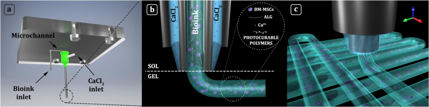
His team – which includes Andrea Barbetta and a group of chemists at Sapienza University of Rome, and specialists in stem-cell research at Oslo University Hospital directed by Jan Brinchmann – is helping to advance cartilage regeneration through the development of 3D-printed biomimetic hydrogel scaffolds. To fabricate these intricate matrix structures for supporting cell growth, the researchers have developed custom apparatus that provides microfluidic control over the dispensing of bioink through co-axial nozzles.
Microchannel control
According to Święszkowski, who has presented the work in the journal Biofabrication, the set-up unlocks a new level of printing accuracy for extrusion-based systems. “Using this apparatus, we can deposit cells with precision beyond the dimension of a single laid fibre,” he explains. “It means that extrusions can contain multiple cell types or biomaterials, which allows us to pattern 3D constructs that more closely mimic the body’s own tissue.”
The technique also makes advances in decoupling printing accuracy and precision from bioink rheology. “We can freely change bioink composition in terms of biopolymer content and/or cellular density, without changing printing conditions such as printing speed, layer thickness, or the distance between fibres,” Święszkowski comments.
This flexibility gives the team more freedom to tailor the structure and composition of the fibre. In the case of cartilage repair, the group has succeeded in bioprinting biomimetic extracellular matrices composed of methacrylated derivatives of gelatin, hyaluronic acid and chondroitin sulphate.
Święszkowski’s team also has projects running that focus on repairing bone, skeletal muscle, tendon and pancreatic tissue. Examining the performance of their fabricated designs, the researchers have shown that three-dimensional constructs bioprinted using the co-axial extrusion system exhibited functional features after culturing in the lab.
In related work, the researchers have formulated and bioprinted a series of biomimetic inks containing human-bone-marrow-derived mesenchymal stem cells (hBM-MSCs), with the aim of examining the influence that each component exerts on cell differentiation. “Interestingly, we found that the composition and the stiffness of the printed bioink plays a key role in the differentiation of hBM-MSCs. We obtain the best results for a bioink composed of gelatin methacrylate and chondroitin sulphate amino ethyl methacrylate,” says Święszkowski.
The group is looking to apply its knowledge to a range of different scenarios, which includes the regeneration of skeletal muscle tissue. Noteworthy preliminary results – attained in collaboration with the group of Cesare Gargioli from University of Rome Tor Vergata – include bioprinting aligned layers of highly-packed hydrogel fibres containing skeletal muscle precursor cells (myoblasts) to create functionally organized and fully differentiated myobundles. “In the future, this 3D bioprinting approach could be used to assemble macroscopic constructs for treating volumetric muscle loss, a severe condition that can arise after injuries, traumas and degenerative disease,” Święszkowski points out.
The results are promising, but there are more challenges to be addressed. “Without any doubt, the major step for me and my team is to bring our system to the next level by improving the current apparatus and building superior structures that can better and more efficiently recreate the complexity and functions of the human body,” Święszkowski comments.
He adds that future progress requires focus not just on the deposition systems, but also on bioink development to expand the choice of natural polymers available for designs. Święszkowski feels that performance gains can be found by focusing on the cellular benefits offered by new blends of matrix materials.
At the same time, he believes that advances in protocols for stem cell differentiation together with progress in understanding key growth-factors and their stimuli will play a major role in delivering further advances in organ healing and tissue repair. “There is still a lot to do in the field of biofabrication and 3D bioprinting,” said Święszkowski, as he looks forward to tackling more challenges in 2018.
Full details of the team’s work can be found in the journal Biofabrication.
This article is one of a series of reports reviewing progress on high-impact research originally published in the IOP Publishing journal Biofabrication.
A huge discrepancy between the observed hyperfine splitting in highly-ionized bismuth-209 atoms and the expected value could be a calculation error rather than evidence for new physics. That is the conclusion of Leonid Skripnikov at St Petersburg State University in Russia and colleagues, who have shown that the magnetic moment of the bismuth-209 nucleus – which is used to calculate the hyperfine splitting — is much smaller than the currently accepted value.
Hyperfine splitting is a small shift in electron energy levels that arises because of the interaction between the dipole magnetic moment of the atomic nucleus and the orbital motion of the electrons. Very precise spectroscopic measurements of hyperfine splitting offer a way of testing quantum electrodynamics (QED). Finding discrepancies between QED and experimental observations could point towards physics beyond the Standard Model of particle physics.
Sole electron
In 2017, Wilfried Nörtershäuser of the Technical University of Darmstadt and colleagues injected bismuth-209 atoms into the Experimental Storage Ring (ESR) at GSI Helmholtz Centre for Heavy Ion Research in Darmstadt. The atoms were stripped of all but one of their electrons. This remaining electron is tightly bound in hydrogen-like orbits that have very large overlaps with the bismuth nucleus. The bismuth-209 nucleus has a huge magnetic moment – and this combined with the close proximity of the electron makes the system ideal for testing QED.
There is an important snag, however, because quantum fluctuations make it extremely difficult to calculate the distribution of magnetization in the nucleus – something called the Bohr-Weisskopf effect. To get around this problem, the team also created lithium-like ions by stripping bismuth-209 atoms of all but three of their electrons. By comparing measurements of the hydrogen-like and lithium-like ions, the team could cancel-out the Bohr-Weisskopf effect and measure the difference between the hyperfine splitting of the hydrogen-like and lithium-like ions.
Whopping deviation
Much to their surprise, the team found that this difference had a whopping 7σ deviation from that predicted by theory, which could be indicative of new physics. But now, new measurements and calculations done by Skripnikov and colleagues (including Nörtershäuser) suggest a more mundane explanation.
Calculating the hyperfine splitting from the experimental data requires an accurate value for the nuclear magnetic moment of bismuth-209. Nörtershäuser along with Darmstadt’s Michael Vogel and colleagues used nuclear magnetic resonance spectroscopy to measure the magnetic moment of the nucleus. This was done by placing an aqueous solution of bismuth nitrate in a powerful superconducting magnet and measuring its radio-frequency spectrum.
An important challenge in making this measurement is accounting for the effect of the bismuth nitrate solution on the local magnetic field that is felt by the bismuth nuclei. This was worked-out by Skripnikov and colleagues, who did sophisticated quantum-mechanical calculations that revealed that the effect on the local field was much greater than expected.
Good agreement
When the new value of the magnetic moment was used to calculate the hyperfine splitting, the result was in good agreement with the original experiment.
“It would be too early to state that this represents the complete solution to the hyperfine puzzle,” says Nörtershäuser, adding “nevertheless, it is for sure a significant part of the solution”. “Further experiments are still needed to achieve complete clarity about the interplay between the atomic nucleus and the shell and, therefore, to verify the theoretical predictions of the nature of quantum mechanics in very strong fields”.


 What if gadolinium-based contrast agents (GBCAs) could be used at just 10% of current dose levels without significantly degrading image quality or contrast enhancement? Artificial intelligence (AI) could make that possible, according to research published in the Journal of Magnetic Resonance Imaging (J. Magn. Reson. Imaging 10.1002/jmri.25970).