Simulations reveal how sharp boundaries endure in soft tissue

Sharp boundaries between different tissues have been modelled by a group of physicists led by Daniel Sussman at Syracuse University in the US. The team used simulations to calculate values of surface tension in cell populations, showing how different cell types maintain soft yet distinctive borders between each other. Their work could make it easier for scientists to model a variety of complex living systems.

Biological processes including embryo development, tumour metastasis and wound healing, all rely on different types of cells becoming compartmentalized with clear boundaries between different cell populations. These systems were previously modelled by treating cells like immiscible fluids such as oil and water. However, living cells can adapt their properties based on their surrounding environments and this makes them very complicated to model as fluids.

Perfectly-fitting polygons

Sussman’s team simplified the process by creating 2D simulations of a population of one type of cell, surrounded by cells of another type. Based on the “vertex model”, the cells in the simulations were modelled as perfectly-fitting polygons, whose shapes were determined by the positions and types of their neighbours. The researchers altered the simulation so that on the borders between different populations, the cells would respond to the different types of neighbouring cell by adapting their shapes to form smooth, distinct boundaries.

To measure the properties of the system, the team determined the surface tension across the “droplet” formed by the central cell population. The surface tension across small fluctuations on the droplet’s surface was also worked-out. They then simulated what would happen if the droplet is squeezed the between two solid plates, which would normally increase the surface tension across droplets of regular fluids.

Low tension

However, Sussman’s team found that the surface tension across the squeezed droplet was, in fact, lower than in the fluctuations. This meant that while bulk tissues can remain soft and squishy under pressure, cells on the tissue boundary will maintain a shape that sustains sharp boundaries between tissues. The researchers’ work is the first to reveal these effects of surface tension and could prove invaluable to simulations of biological tissues in the future.

The simulations are described in Physical Review Letters

Climate change to shift timing of glacial runoff

The distribution of freshwater runoff from glaciers throughout the year may change as the climate warms, predicts a study in Nature Climate Change. During this century, for around half the 56 glaciers examined, annual runoff is expected to increase to a peak then reduce, whilst annual runoff for the remainder seems already to have peaked and is predicted to continue to fall. What’s more, the proportion of the run-off taking place in the melt-season looks set to decrease in more than a third of the basins.

Matthias Huss of the ETH Zürich and the University of Fribourg, Switzerland, together with Regine Hock from the University of Alaska Fairbanks, US, simulated the development of glaciers under climate change for all 56 large drainage basins containing glaciers outside Antarctica and Greenland.

The researchers’ predictions indicate that annual freshwater runoff from glaciers will increase with rising global temperatures, before falling again as the glacier shrinks. Ultimately, each glacier will either disappear or settle at a smaller size but with the same annual runoff as before, having reached a new size equilibrium. Melt-water will no longer contribute to overall runoff as an equivalent amount of water will accumulate in the glacier in winter. If other factors, such as precipitation, have remained unchanged over time then total runoff will return to its initial level.

This annual pattern contrasts with the runoff taking place in summer, during the melt-season. The pattern is similar initially; projected runoff increases with global temperature and reaches a peak. It then falls, stabilizing below its pre-warming level. This is because some of the melt-season runoff represents the melting of a percentage of the glacier itself. As the glacier has reduced in size, this volume is smaller. The annual runoff has returned to its original level thanks to the glacier’s new size equilibrium, yet melt-season runoff has fallen, so a greater proportion of runoff must occur outside the melt-season. This probably results from winter rains and snow, as the glacier is not contributing melt-water and has little effect on winter runoff.

Although there was substantial variation between basins, the researchers came to some key conclusions. More than one third of the basins are set to experience an overall reduction in runoff greater than 10% during at least part of the melt-season by the end of the century, they found. It is important to note that these glacier-containing basins house almost one-third of the global human population. The disruption of glacial runoff could have serious effects on these communities, as well as countless natural habitats downstream from melting glaciers.

Huss and Hock used the Global Glacier Evolution Model (GloGEM) to predict glacier size changes and runoff. The researchers based their calculations on three different greenhouse gas concentration predictions developed by the Intergovernmental Panel on Climate Change (IPCC). RCP2.6 is the closest to the targets set out in the 2015 Paris Climate Agreement, while RCP4.5 and RCP8.5 both predict higher emissions and later peaks in atmospheric greenhouse gas concentration. The simulations ran until the year 2100.

Contact allergy: where are the allergens found?

Nickel remains the primary cause of contact allergy, despite efforts to minimize exposure. Nickel is classified as a hapten, a small molecule that, upon combining with proteins, evokes an immune response. To determine the mechanism involved in contact allergy, a team of scientists from Chalmers University and University of Gothenburg, Sweden, has investigated the penetration and distribution of this hapten through the human skin using imaging mass spectrometry. Their results indicate a new method for investigation of skin distribution of contact allergens, providing an alternative to animal experiments (Contact Dermatitis 78 109).

Exposure to nickel

Imaging mass spectrometry is a valuable analytical tool that uses a beam of primary ions to visualize the spatial distribution of chemical molecules on a surface by their molecular mass. The researchers chose time-of-flight secondary ion mass spectrometry (ToF-SIMS) for this study as it provides high-resolution images.

They exposed samples of human skin (obtained from breast reduction surgery) for 24 h to a nickel sulphate solution; control samples were exposed to deionized water. Both the samples from the tissue exposed to nickel and the control tissue were frozen in liquid nitrogen, sliced and analysed by ToF-SIMS.

Nickel distribution

Analysis of the ToF-SIMS images generated a visualization of the distribution of nickel ions as a function of depth into the sample. The results indicated that the highest intensity of nickel ions was observed in the stratum corneum (the outermost layer of the epidermis, regarded as the major barrier to chemical transfer through the skin). The authors observed a lower density of nickel ions in the upper epidermis, as well as a rapid decrease in the number of nickel ions with skin depth.

Moreover, the authors reported that collagen (one of the most abundant proteins in our body and the building block to the health of our skin) is not found in the stratum corneum but at the interface between the epidermis and the dermis (the inner layer of the skin). Collagen was found there together with lipids with a low mass (such as cholesterol and phosphatidylcholine (PC) headgroup). Surprisingly, these remained unaffected by the nickel sulphate exposure. In contrast, lipids with higher mass were found to have an altered biomolecular composition compared with control tissue, indicating a physiological effect due to nickel sulphate exposure.

Ni+ (red), cholesterol (green) and PC headgroup (blue)

This study is the first to offer information regarding penetration of a hapten in human skin ex vivo and validates ToF-SIMS as a tool to acquire high-resolution images of ion distribution in different layers of the skin. This approach may be expanded for investigation of other skin sensitizers, thus providing new avenues for chemical testing while representing a way to reduce the number of animal experiments.

TRAPPIST-1 exoplanets could harbour significant amounts of water

The chances that several exoplanets in the TRAPPIST-1 system could be habitable have been boosted by new measurements that push the envelope of what exoplanet science can do with today’s telescopes.

The TRAPPIST-1 system is just 39.6 light-years away and comprises seven small worlds that orbit a lone red dwarf star. The inner three worlds were discovered in 2016 by astronomers using the Transiting Planets and Planetesimals Small Telescope (TRAPPIST) at the European Southern Observatory in Chile. The outer four planets were spotted a year later and it is it is possible that all seven worlds could potentially be habitable.

Now, new results have narrowed down the masses of the planets, confirming that they are all likely to be rocky, without the extended atmospheres that miniature versions of Uranus and Neptune would have. Furthermore, five of the planets have densities that suggest a significant amount of water, which is vital for life as we know it.

Transit timing variations

Astronomers led by Simon Grimm of the Centre for Space and Habitability at the University of Bern in Switzerland have produced the most accurate calculations of the planetary masses yet by taking advantage of a phenomenon known as transit timing variations, or TTVs. The seven worlds gravitationally push and pull on each other, resulting in the timing of their transits in front of their star being delayed or advanced by up to 1 h.

In the TRAPPIST-1 system, “the TTV are much stronger and more complicated than in many other systems that have fewer exoplanets,” says Grimm. The challenge of disentangling the data to calculate the planetary masses required new code written to handle 35 different parameters, five for each exoplanet. These are mass; orbital period; eccentricity; the argument of perihelion (the angle between its perihelion position and where the exoplanet’s inclined orbit passes through the ecliptic plane); and the mean anomaly (an angle required to calculate an exoplanet’s location on its elliptical orbit at any given time). The code produced a range of different solutions, from which Grimm’s team determined the configuration that best fits the observational data.

Water worlds

The most massive of the seven worlds is exoplanet “c”, the second from the star, with a mass 1.156 times that of Earth. The least massive is exoplanet d, with less than a third of Earth’s mass. The magnitude of the transits tells astronomers the radii of the exoplanets, and from their radius and mass, their densities can be calculated.

This is where things start to become interesting. Based on their densities, all seven worlds are predominantly rocky, but contain up to 5% water. This is much more water than Earth’s oceans (which amount to just 0.02% of Earth’s mass). However, it remains to be seen whether the TRAPPIST-1 water is present on the surface of the exoplanet in vast, deep oceans, or whether it is as vapour in a dense, steamy atmosphere, or whether it is spread around inside the exoplanet, much like how Earth’s mantle contains the equivalent amount of water as in the oceans.

Based on its temperature, exoplanet e would be the most similar to Earth according to Grimm. This world has 77% of the mass of Earth, but is a little denser, indicating a large iron core and a thin atmosphere, possibly even thinner than Earth’s.

Hubble finds no hydrogen

Meanwhile, new observations by the Hubble Space Telescope support the conclusions of the TTV calculations, confirming the likely terrestrial status of the TRAPPIST-1 exoplanets by ruling out the existence of the extended hydrogen envelopes found in the atmospheres of Uranus and Neptune. Pushing Hubble’s powers of resolution to the limit, a team led by Julien de Wit of the Massachusetts Institute of Technology using Hubble’s Wide Field Camera 3 to make infrared spectroscopic observations did not detect any large, puffy atmospheres around exoplanets d, e, f and g, “leaving many more terrestrial-like possibilities to be explored with future telescopes such as the James Webb Space Telescope,” says Hannah Wakeford, a member of de Wit’s team from the Space Telescope Science Institute in Baltimore.

Hubble had previously searched for hydrogen-rich atmospheres around the innermost exoplanets b and c in 2016, while observations of the outermost world, h, remain inconclusive. The next step is to look in the ultraviolet for hydrogen escaping from the exoplanets’ atmospheres. For the innermost worlds, this would be a sign of a greenhouse effect, whereby the temperature boils the oceans, filling the atmosphere with water vapour, which is then broken down into oxygen and hydrogen by ultraviolet light from the star, allowing the hydrogen to escape into space. It is the same scenario that has taken place on Venus.

Best opportunity

However, Wakeford says that while it is difficult to draw any direct analogues to the Sun’s planets, TRAPPIST-1 “still represents the best opportunity we have for studying Earth-sized worlds outside of our own solar system”.

The research is described in Nature Astronomy and in an upcoming paper in Astronomy and Astrophysics.


Ultrasound stimulates peripheral nerves in vivo

Focused ultrasound (FUS) provides non-invasive, targeted therapy for a wide range of clinical applications. Recently, FUS has proved effective at stimulating or inhibiting neuronal activity in both the central nervous system (CNS) and peripheral nervous system (PNS), offering the potential to replace existing neuromodulation therapies, which are either targeted but invasive, or non-invasive and non-specific.

To date, in vivo studies examining the physiological effects of FUS stimulation have only targeted structures in the CNS. Now, Columbia University researchers have demonstrated the first successful in vivo FUS stimulation of the PNS (Phys. Med. Biol. 63 035011).

“Neurostimulation of the CNS excites (or inhibits) many neurons, while with PNS stimulation, we are targeting either the axon or nerve body of a specific, well understood, nerve branch,” explained first author Matthew Downs. “FUS could be a powerful tool to target multiple nerve types including the vagus nerve, which has the potential to treat diseases such as epilepsy, depression and metabolic disorders.”

It’s also easier to deliver FUS to peripheral nerves, as the ultrasound does not need to pass through skull, which leads to energy loss and scattering. “If we can achieve similar effects by stimulating the peripheral nerves as we do the CNS, then the technique can be more easily implemented,” noted Downs.

Physiological response
To demonstrate FUS stimulation of peripheral nerves in vivo, the researchers stimulated the sciatic nerve in anesthetized mice. They targeted the nerve using ultrasound imaging and stimulated it with a 3.57 MHz transducer. At the same time, they recorded electromyography (EMG) signals via needle electrodes placed into the tibialis anterior muscle, which is activated by sciatic nerve stimulation.

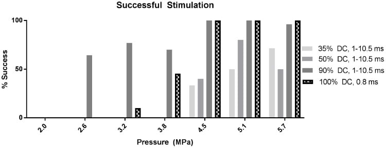
Preliminary experiments revealed a set of FUS parameters that successfully elicited EMG signals and observable muscle contractions. These included a 35-100% duty cycle (DC) and a stimulation duration of 0.8-10.5 ms. An 8 ms stimulation of the sciatic nerve typically produced a single EMG spike, and occasionally a second EMG signal, plus an electromagnetic field (EMF) artefact from the transducer. Reducing the stimulation duration to 0.8 ms at 100% DC (continuous wave) produced single EMG responses with reduced EMF noise.

Stimulation success rates with pressure and pulse length

Varying the duration between 1 and 10.5 ms did not affect the latency (time between stimulation and response) or intensity of the EMG signal, thus the researchers combined results from this range. “From there, we wanted to see if we could achieve stimulation with a shorter duration, so we pushed the software to as fast as it could go, which turned out to be 0.8 ms,” explained Downs. “This was also an attempt to get as close as we could to normal electrical stimulation duration.”

Moving the FUS focal spot away from the sciatic nerve eliminated both observable muscle activation and EMG activity. To further verify that these effects were due to stimulation of the nerve, rather than the surrounding tissue, the researchers clipped the nerve downstream of the FUS target; this transection eliminated the EMG signal.

Comparing ultrasound with electrical nerve stimulation revealed that FUS could elicit comparable EMG spikes. The latency of the electrically stimulated EMG response (average 2.1 ms) was comparable to that of both the 0.8 ms and the 1-10.5 ms FUS stimulation groups. These findings suggest that FUS could serve as an alternative or complimentary treatment for peripheral nerve conditions currently treated with electrical stimulation.

Safety first
To assess the safety of this procedure, the researchers performed an open field test, in which they observed mice in a 30 cm2 box before and three days after FUS stimulation. The motion of the mice did not significantly change after stimulation and was similar to that of a control group – implying that FUS had not damaged the nerve or surrounding tissue.

They also monitored the time mice spent in the box centre and along its walls, to determine anxiety levels. The behaviour of the stimulated group was similar to the control and baseline groups, indicating that the FUS stimulation parameters used to elicit EMG responses are safe. H&E staining revealed no damage to the FUS stimulated nerves, reinforcing the safety of the technique.

Underlying cause
To determine whether FUS elicited a thermal effect, the researchers embedded thermocouples in an ex vivo mouse hind limb adjacent to the sciatic nerve. Stimulation at higher FUS pressures caused a 1.09°C increase in temperature – significantly lower than the 20°C increase shown previously to block nerves from firing.

They also measured the acoustic radiation force generated from the transducer and used this to determine the deformation at the focus. FUS parameters employed in this study generated a large enough displacement (up to 422 µm) to facilitate firing of an action potential that (according to prior studies) will elicit EMG activity. These in vivo findings agree with published ex vivo results stating that FUS excitation of the PNS is a mechanical, not thermal, effect.

The researchers are currently working to develop elastography-based targeting techniques for real-time monitoring of the transducer’s focal area to ensure accurate nerve targeting. They are also investigating whether cavitation is generated during stimulation, to further elucidate the mechanics of peripheral nerve stimulation. “We have also begun preliminary experiments with stimulation of human peripheral nerves using this technique,” Downs told medicalphysicsweb.


3D culture system enables stem cell expansion

Human pluripotent stem cells are cells that can be grown into multiple different types. The differentiation of these stem cells provides a variety of human cell types for use in tissue modelling, cell therapies and even tissue transplantation. However, one of the major limiting factors to the effective use of pluripotent stem cells comes from “scaling-up” of these cells to produce the large number needed for effective studies; with logistical challenges including the expensive techniques required to do this. Additional issues arise from the methods used to culture stem cells, with the majority of protocols relying upon 2D surfaces, which don’t replicate the 3D microenvironment native to cells in the body.

AlgTube

Researchers at the University of Nebraska have developed a potential solution to these issues by creating a cost-effective 3D culture system (named the AlgTube 3D cell culture system) that enables the expansion and differentiation of cells. The AlgTube system is based around a 3D culture platform that allows cell growth within a 3D environment. This offers advantages over traditional 2D culture methods, which fail to replicate the native environment in which stem cells grow and differentiate within the body (Biofabrication 10 025006).

In this system, cells are suspended within alginate hydrogel tubes. This enables interactions between cells encapsulated within the hydrogel, as well as expansion within the hydrogel. The AlgTube system offers a large scope of expansion based on the much-increased area for growth provided by the 3D environment. Hydrogels are solid polymer-based (alginate-based, in this system) biomaterials, which are highly water saturated and porous, and are widely utilized for 3D culture of cells. Alginate, in particular, is affordable and biocompatible, making it perfect for widespread use in hydrogel form.

Improving on cell factories

Whilst “cell factories” (cell culture flasks with large surface areas and multiple plastic layers) enable expansion of stem cells in 2D, their use can be arduous due to their size and they require multiple incubators. AlgTube can provide similar expansion rates to cell factories in a much smaller system, reducing the reliance upon large lab incubators and producing cells that are grown within a physiologically relevant environment.

The research group developed specific spatial configurations that allow for efficient oxygen and nutrient diffusion throughout the 3D system. The system encapsulates cells within a 400 µm diameter, avoiding any physical stress and enabling cell culture medium to easily diffuse to the cells.

The process of cell growth and expansion within AlgTube requires an initial clustering phase – where cells aggregate into clusters – followed by the cell expansion phase, where a large cell mass develops within the device.

The researchers also tested the effect of different initial cell seeding densities on the rates of expansion. They observed similar final cell counts when seeding 1 or 10 million cells, and all cells expressed the important pluripotency marker OCT4. This allows researchers employing the AlgTube system to use their stem cells more sparingly without compromising time or final cell numbers.

Growth kinetics of cells at various seeding densities

The researchers also demonstrated that the AlgTube system can facilitate growth of different stem cells, with induced pluripotent stem cells (iPSCs), mesenchymal stem cells (MSCs) and embryonic stem cells (ESCs) being grown within the system for 10 days without altering their stemness (retention of stem cell pluripotency). These cells could also still be differentiated to the different terminal lineages (ectoderm, mesoderm and endoderm) allowed by pluripotent stem cells.

Future opportunities

The AlgTube system provides a useful tool for producing a large amount of stem cells for a variety of purposes. It also allows for the growth and expansion of cells within a physiologically relevant and 3D microenvironment. This system may enable the easy mass expansion of stem cells for lab groups, without requiring the money and resources of a large pharmaceutical company, thus enabling important studies by university and smaller industrial companies. The authors note that future AlgTube research would benefit from investigating the integration of various human cell types into the system, such as adult stem cells or T-cells.

Pollution boosts risk of lightning

Pollution in the form of aerosols heightens the risk of lightning strikes, say researchers in Israel, Mexico and the US.

The scientists came to the conclusion by correlating the amount of aerosols globally with the record of cloud-to-ground lightning strikes, while excluding weather as a possible influence. Some 70% of the areas studied exhibited more lightning strikes from polluted skies rather than clean skies.

“Significant increase in aerosol concentration is mostly anthropogenic,” said Ilan Koren of the Weizmann Institute of Science, Israel. “Therefore, these results show a global anthropogenic effect on the climate… If we want to predict future climate change correctly, [the aerosol effect on lightning] should be well represented in global climate models.”

Lightning strikes cost lives and damage infrastructure, particularly in the developing world. In South Africa, for instance, scientists have estimated deaths from lightning at 260 per year, while in India the estimated annual death toll is more than 3,000.

Aerosols have long been suspected to raise the likelihood of lightning strikes. When aerosols are present in high concentrations, clouds are believed to take on a deeper, invigorated structure, containing more and bigger ice particles and allowing stronger updraughts. Such a structure is also believed to promote electrical activity, as ice and soft hail “graupel” particles collide and rebound in the presence of supercooled water.

Studies giving evidence of possible links between high aerosol levels and more frequent lightning strikes are not in short supply. According to Koren, however, it has been hard to claim that the link between aerosol and lightning is causal, as it could always be possible that a common factor – weather being the obvious candidate – is influencing the other two. “The same meteorology that favours deeper clouds could induce convergence and bring more aerosols to the column,” he explained.

The study pitched the times and locations of strong lightning events, taken from the World Wide Lightning Location Network, against cloud and aerosol measurements from NASA’s orbiting MODIS instrument. In the first analysis, the researchers showed the link between areas of polluted clouds and areas of significant lightning activity. In the next step, for a few key regions, they sliced the data into strips of similar meteorological conditions. The trend remained, demonstrating that weather could not be a common driving force.

“It is a data-only study that suggests this global trend,” said Koren. “It is important to show how robust the trend is without using models that rely on many assumptions.”

In general, Koren added, the work broadens knowledge of how aerosols affect cloud formation, and could help scientists on their way to a unified aerosol theory that predicts when high aerosol concentrations will yield larger clouds, more rain and more lightning, and when they will have the opposite effect.

“We are now expanding our research to larger scales, to understand how clouds ‘talk’ with their surroundings and how they affect thermodynamics and radiation properties,” he said. “In parallel, we’re developing new approaches to measuring clouds. We think that in the near future more but smaller satellites could provide valuable data on the internal structure of clouds and cloud fields.”

Koren and colleagues published their findings in Environmental Research Letters (ERL).


Shedding light on photochromic responses

Photochromic responses – when exposure to light reversibly changes a material’s absorption properties – have a range of uses from smart windows to data storage. The discovery of photochromic responses in TiO2 embedded with silver nanoparticles greatly expanded the possible applications. Now researchers in France have combined GISAXS and optical transmission measurements to gain a better understanding of how photochromic responses in this nanocomposite take place, which may reveal ways of optimizing them.

Although photochromic behaviour first attracted attention in the late 1880s, like the sepia photographs from that era the response was monochrome. It was not until 2003 that researchers in Japan were able to elicit a multicolour response using mesoporous TiO2 embedded with silver nanoparticles, allowing potential applications in rewritable colour copy paper, electronic paper and high-density multi-wavelength optical memory.

Among those attracted by the potential of the new photochromic material was David Babonneau, CNRS Research Director of the Physics and Properties of Nanostructures at the Université de Poitiers in France, whose team showed that they could also fabricate these nanocomposite films on flexible substrates, opening up further opportunities in high-density multi-wavelength optical data storage, rewritable colour papers, plastic packaging products, and secured credit cards. But making all this potential a reality requires a much better understanding of the mechanisms and limiting factors in the photochromic process, which their latest work now addresses.

On the track of plasmons

Previous studies of the nanocomposite’s photochromic response have indicated the behaviour’s likely origins in the excitation of plasmons – resonant collective oscillations of electrons at the surface of the metal nanoparticles. The optical absorption changes only take place in the presence of oxygen, where visible light leads to redox reactions so that the silver atoms in the nanoparticles oxidize to silver ions. Exciting plasmons in the nanoparticles makes them unstable so that they release these silver ions, resulting in morphological changes in the silver nanoparticle and a change in the optical response. In contrast, exposure to UV radiation, excites electrons in the TiO2 matrix that reduce the silver ions, reversing the whole process. Yet while the initial state of the nanocomposite film, the incident light and environmental parameters seem to have a crucial influence on the process, the role of these factors is little understood.

One approach that has been successful in monitoring the growth of plasmonic nanoparticles is grazing incidence small-angle x-ray scattering (GISAXS), which can reveal changes in the electron density at surfaces and interfaces. Babonneau and colleagues at Université de Poitiers, Université de Lyon, Université Jean Monnet-Saint-Etienne and Université de Grenoble Alpes adopted the same tool to study their TiO2-Ag nanoparticle composites, combined with optical transmission to track spectral changes in response to radiation.

Correlating morphology and photochromic behaviour

With the aid of their real-time measurements, the researchers found a good correlation between the changes in the morphology of the structure and the optical behaviour. Exposing the nanocomposite to visible radiation led to the oxidation of the silver causing the smaller nanoparticles to dissolve, a process that sped up under x-ray radiation. In the absence of these nanoparticles less light is absorbed. The researchers also observed the reversal of the process under UV radiation. However they also noticed that the ionization of the silver atoms to dissolve small nanoparticles outpaced nanoparticle growth, ultimately degrading the material from one cycle to the next.

Babonneau and colleagues conclude, “The study may open up the possibility of exploring the influence of various factors that have an impact on the photochromic transformation process in Ag/TiO2 nanocomposite films (initial state of the film, excitation wavelength and irradiance, environmental conditions, etc), which is mandatory for optimizing their functional properties.”

Full details are reported in Nano Futures.

Going ballistic to pin down Majorana particles

Almost six years ago, Leo Kouwenhoven’s research team glimpsed a particle that is both matter and antimatter, for the first time since it was theoretically predicted in 1937 by Ettore Majorana. Now, thanks to their improved fabrication process, the same collaboration of groups from Delft University of Technology and Eindhoven University of Technology has confirmed their measurements did find the elusive Majorana particle. Their work, published in Nature Nanotechnology, brings Majoranas one step closer to use as robust quantum bits in powerful quantum computers.

The Majoranas with potential for quantum computing are not stand-alone particles, but “quasiparticles” – emerging in special combinations of materials. The Netherlands-based collaboration specializes in combining a semiconductor nanowire with a superconducting film, by partially wrapping a nanowire section with the film to induce an exotic form of superconductivity.

By making an atomically smooth nanowire-superconductor interface, the team reduced electron collisions down to the ‘ballistic’ regime, where electrons can travel the length of the nanowire without scattering. This ideal transport regime maximizes the connection between, and signal of, the Majorana particles at the two nanowire ends. It cleared the way for scientists to exclude alternative explanations for the Majorana signal, which, in their previous nanowire experiment, could have come from the unwanted scattering or disorder.

The signal of an elusive particle

The Majorana particles’ signal lies at the very centre of an energy range usually forbidden for quasiparticles in a superconductor, at the so-called “zero energy”. The zero-energy Majorana can only emerge when the researchers switch on exotic superconductivity in the nanowire, producing the Majorana signature: a “zero-bias peak”. This is exactly the signal the team measured. They then checked the peak occurred at magnetic field strengths and directions predicted by theory, and that it had the characteristic robustness that makes Majoranas so attractive for storing quantum information. Finally, they had collected enough evidence to confirm their glimpsed Majorana a reality, as paper co-author Jouri Bommer concluded: “With our improved Majorana measurements, we can be absolutely certain that the zero-bias peak means that we have actually found Majoranas.”

It looks like a Majorana particle… now can it act like one?

Eighty years on from their theoretical prediction, the dialogue between experiment and theory is what makes Majorana particles an exciting quest for physicists. Even before publication, pioneering theoreticians in the USA were already supporting these new experimental discoveries.

The ultimate test, however, will be when Majoranas are not just found, but used as protected quantum information states. Harnessing and manipulating these exotic particles will require more complex devices, but the Eindhoven Technical University collaborators are already creating nanowire networks that might be the key. The reward would be a quantum leap on the roadmap towards topological quantum computing.

Energy harvesting coiled up in your favourite chair – update from innoLAE 2018

From ferroelectrets and thermoelectrics to photovoltaics and inductive power transfer, Steve Beeby showed attendees of innoLAE 2018 how research at Southampton was making progress towards textiles with energy harvesting properties. While energy scavenged from the environment can come in small packages that are difficult to make use of, Beeby also showed how it was possible to use a coil embedded in your favourite chair to charge your mobile phone.

“Textiles are the most common kind of material we come into contact with,” Beeby told nanotechweb.org following his talk. “They’re found in clothes obviously, but also home interiors, car interiors, bedding – so there’s lots of places that you can put the technology.” Yet while their ubiquity may be convenient, textiles have a number of properties that are far from ideal for fabricating electronic devices.

Overcoming fluff and hot air

“The challenge is textiles are rough and fluffy surfaces for depositing thin films,” Beeby told delegates of innoLAE 2018 in his presentation. He and his team have used a screen-printed interface layer to reduce the roughness of a typical fabric from 150 mm to just a few micrometres. They could then spray on fabric organic solar cell devices that operate with a power conversion efficiency approaching glass-substrate-based counterparts.

Of course fluffiness is not the only challenge as textiles are also generally intolerant of the high processing temperatures used to fabricate conventional energy harvesting devices. In some cases Beeby and his co-workers have managed to reduce the processing temperatures required; in others, such as the thermoelectric nanoparticle-based ink they developed from bismuth telleride powders, they used specialised glass fibres, which can handle the processing temperature of 250 °C required.

Beeby also showed delegates work with fabrics laminated with ferroelectret materials. Ferroelectrets are polymer foams, which can store charge in the foam voids that can be released in pulses under pressure. The resulting piezoelectric behaviour can be useful in energy harvesting. By using fluorinated ethylene propylene (FEP), which holds charge better than polydimethylsiloxane (PDMS), and optimizing the geometry of their device they were able to produce an excellent ferroelectret textile material.

“But the question we always get asked is can I charge my mobile phone with it?” said Beeby. With a system of near-field electromagnetic coupled coils they developed a wireless power transfer system that can . The system works well fitted into furniture upholstery so that you can charge your phone while relaxing in your favourite chair.

Driving forces

Far from curling up in a cosy chair, Beeby’s inspiration to work on electronic textiles came from a love of heavy-metal music in his teens. “Heavy-metal T shirts have a lot of silver and gold type inks on and it made me think why not print conductive materials on textiles and see if you can print circuits onto textiles – and that’s kind of how it started,” he told nanotechweb.org.

He adds that applications in healthcare are likely the driving force behind the current growth in the field. To operate health-monitoring or any other kinds of devices incorporated in garments, a source of power is key.

For more on innoLAE 2018 visit http://www-large-area-electronics.eng.cam.ac.uk/innoLAE2018.

Copyright © 2026 by IOP Publishing Ltd and individual contributors