Drug delivery to the brain has been a longstanding challenge in medicine. One option is focused ultrasound (FUS), which can safely disrupt the blood–brain barrier for drug delivery. But how much of a drug gets into the brain depends heavily on its size. A long-standing assumption is that delivery decreases as drug size increases. But by using a clever spin on MRI physics, researcher Matthew Hoch and colleagues from the University of Virginia have shown that “the smaller the better” might not be entirely correct.
The blood–brain barrier is one of biology’s toughest engineering problems. Composed of a dense, tightly sealed layer of cells lining every blood vessel in the brain, the blood–brain barrier only lets nutrients and oxygen through and protects the central nervous system by blocking bloodborne pathogens. In doing so, however, it also blocks the delivery of drugs to the brain. Brain tumours compound this with their own blood–tumour barrier, further hindering drug delivery. For years, researchers have pondered how therapeutics could be delivered past these checkpoints; the answer may simply lie in the use of sound.
The process begins with the injection of gas-filled microbubbles into the bloodstream, where they circulate freely. Exposure to FUS then expands and contracts the volume of these microbubbles, creating a temporary, localized gap that pries the barrier open just long enough for drugs to slip through. Although the technique works, it is unclear how much of a drug actually comes through. In this new study, the team investigate how the size of the drug influences FUS-mediated drug delivery and introduce a novel technique to measure precisely how much drug is delivered.
The measurement problem
Many neurotherapies, with a range of sizes, could benefit from this drug delivery method. But measuring how much of the drug gets past the blood–brain barrier is challenging with current imaging methods. For example, fluorescence imaging is limited by its semi-quantitative nature, while PET scanning suffers from poor spatial resolution.
Standard MRI methods are not only time-consuming, but, in the form of T1-mapping, are typically limited by the size of commercially available contrast agents, which are small in comparison to larger neurotherapeutics used for immunotherapy and gene delivery. Other standard MRI methods sensitive to the presence of iron (T2-mapping) provide an alternative avenue to measure larger agents, but can again be limited by scan time for 3D visualization. Both approaches also have potential to suffer from low sensitivity at low concentrations.
Hoch and his team solve this measurement problem by introducing quantitative susceptibility mapping (QSM), an MRI technique with diverse applications in neuroimaging including evaluation of haemorrhage, iron deposition and calcification. QSM works by exploiting the distortion of local magnetic fields by different materials, a property called magnetic susceptibility. By injecting iron-based nanoparticles that induce measurable, spatially localized shifts in the MRI signal’s phase, QSM can generate a 3D map of magnetic susceptibility. This can then be used to calculate the actual concentration of iron-based nanoparticles within tissues.
By transforming an MRI scanner into a sensitive nanoparticle detector, the researchers present a novel way to quantify drug delivery down to fractions of a percent of the injected dose.
What did they find?
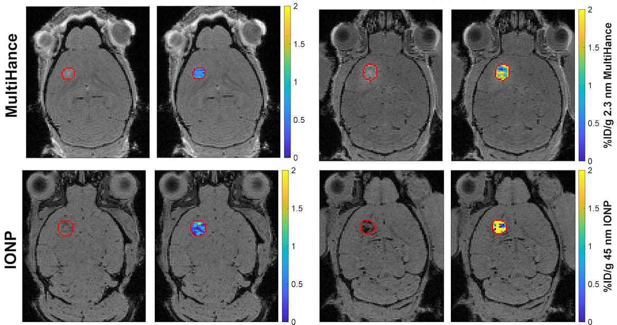
The team tested this approach using four different particles, with a 20-fold range in size. The first was a small gadolinium-based contrast agent, MultiHance (a proxy for small molecule therapeutics), at around 2.3 nm; the other three were iron oxide nanoparticles (IONPs) of 15, 23 and 45 nm, sizes that mirror the dimensions of therapeutic candidates used for immunotherapy and gene therapy.
Using healthy and glioma brain tumour-carrying mice, the researchers used FUS treatment to open the blood–brain barrier and deliver particles of varying sizes. They recorded MRI scans before and after delivery and used QSM to generate a precise measurement of how much of each particle made it into brain tissue.
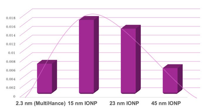
The results were unexpected. Drug delivery followed a bell-shaped curve in healthy mice. From the smallest (2.3 nm) particle to the 15 nm IONP, delivery increased 2.6-fold, then at 23 nm, remained steady. Interestingly, at 45 nm, delivery dropped by 2.5-fold compared with the 23 nm nanoparticle, comparable to that of the smallest agent.
This sweet spot, ranging from 15 to 23 nm, demonstrates that smaller isn’t always better. The researchers suspect that this trend in drug delivery is due to a balance between how easily a particle can squeeze through the barrier (so favouring smaller particles) and how long a particle stays in circulation, giving it more time to travel across the barrier (favouring larger particles).
In the mice with brain tumours, where there is an addition of the tumour’s own leaky, disorganized vasculature, the blood–tumour barrier, drug delivery can differ from that to healthy brain. Specifically, this barrier is thought to make drug delivery more difficult than in healthy tissue due to higher tissue pressures and inconsistent blood flow. But the researchers unexpectedly found that FUS boosted delivery of both small and large agents into tumours, with no real difference to the drug delivery levels seen in healthy brain tissue, despite the added presence of the blood–tumour barrier.
Ultrasound implant helps deliver powerful chemotherapy to brain tumours

“The outcome is exciting because it means that focused ultrasound delivery performance is not expected to diminish in brain tumours,” says senior author Richard J Price in a press statement. “In fact, it may even be enhanced for some types of therapeutics.”
FUS-mediated barrier opening is currently in clinical trials for use in Alzheimer’s disease and glioblastoma, and this research has implications in both drug engineering and treatment planning. By introducing QSM, Hoch and his colleagues have provided a universal, precision technique to quantify contrast agent delivery. Taking existing tools, used for a specific physics measurement problem, and developing them to answer a completely different biological question is no small feat. Here, the researchers present an ingenious intersection of physics and biology, and its potential application in medicine.
The researchers report their findings in Radiology.