Neuroscientists are debating a seemingly simple question: which way does a seizure propagate in the brain? Answering such a question could advance our understanding of epilepsy, but requires the development of cutting-edge microscopy techniques that are far from simple. Researchers from the University of Georgia have now developed an improved light-sheet microscope with the ability to perform fast 3D imaging of zebrafish seizures.
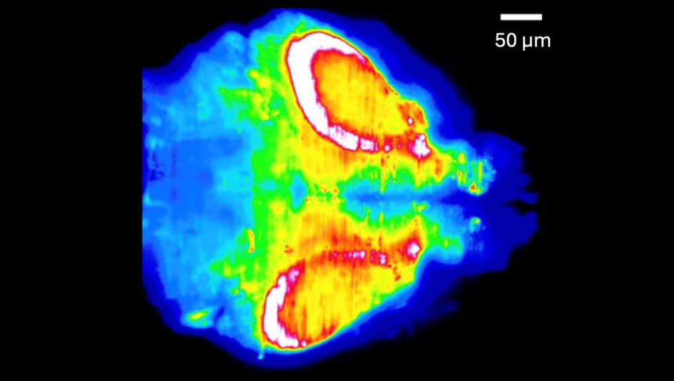
Reporting their findings in Biomedical Optics Express, the researchers found that seizures originated in the hindbrain and propagated forward (anteriorly). This corroborates a previous study examining 3D imaging of zebrafish seizures, but contradicts two earlier studies of 2D imaging (reported in Frontiers in Neural Circuits and eNeuro) that observed propagation in the reverse direction.
“I think the discrepancies about the direction of seizure propagation point to the need for more imaging,” says Peter Kner, an engineering professor who led the research efforts at the University of Georgia. “I believe we still don’t know how probabilistic the behaviour is and what factors are important.”
Adding a third dimension
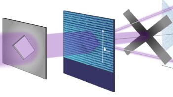
The microscope developed by Kner and his team uses a so-called light sheet — a thin pancake of light that selectively illuminates a single 2D plane of a sample. This technique strongly reduces background noise from other planes that are not illuminated. Fluorescence light from the sample plane passes through a series of lenses, orthogonal to the light sheet, which reimage the sample onto a camera.
“There have been several papers analysing seizure events in zebrafish by looking at 2D imaging. So, the motivation here was to extend that work to 3D imaging”, explains Kner. Compared with other microscopy techniques, such as confocal microscopy, Kner’s team wanted to develop a solution for simple, fast and large field-of-view imaging in all three dimensions.
Light-sheet microscopy works inherently in 2D. To extend it to 3D, the researchers dynamically swept the light sheet through different planes of their sample. To keep the illuminated plane in focus at all times, they synchronously shifted the imaging system’s focal plane using an electrically tunable lens (ETL), which has a focal length that changes with applied current.
While this sounds straightforward in theory, in practice, adjusting the ETL introduces distortions or optical aberrations that degrade image quality. The researchers compensated for wavefront distortions using a deformable mirror whose shape was carefully calibrated at each axial plane imaged.
A wider view of seizures
Using predetermined settings for the deformable mirror, the team imaged volumes of 499 x 499 x 148 µm at a rate of four volumes per second. They also reported a fivefold increase in the area over which imaging remained near-diffraction-limited compared with uncorrected microscope images. These achievements allowed them to image zebrafish seizure propagation in real time under their microscope.
“This work adds pragmatic utility to 3D light-sheet microscopy for fast volumetric dynamics,” says Sixian You, an electrical engineering professor at the Massachusetts Institute of Technology who was not involved in this research. While deformable mirrors have been used to correct ETL aberrations before, she emphasizes that the advance comes from pre-calibration of the corrections; this is what makes the fast, wide field-of-view imaging possible. “I expect the same approach will transfer readily to other scanning modalities that stand to benefit from tunable lenses,” she adds.
Treating epilepsy with physics

After imaging the zebrafish continuously over a 2.5 min period, the researchers found that seizures propagated from the back of the brain to the front, and that the seizures subsided over tens of seconds. The findings add one more data point towards elucidating the statistics of seizure propagation which, if better understood, could aid epilepsy treatments in the future.
Kner’s team hopes to extend this work by next exploring different strains of zebrafish.