A new study showing how tumour cells behave when surrounded by a disordered microenvironment could help cancer researchers by providing a biophysics model describing how cancers spread within the human body.
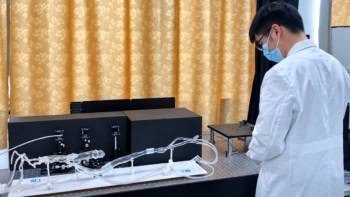
Researchers led by Michael Sixt, head of the Cellular Morphodynamics Group and Edouard Hannezo, leader of the Physical Principles in Biological Systems Group both at the Institute of Science and Technology Austria (ISTA), in collaboration with scientists from the UK’s Francis Crick Institute, have constructed a microfluidic chip experiment and associated computer simulation that shows how heterogeneity (disorder) surrounding a tumour can drive metastasis.
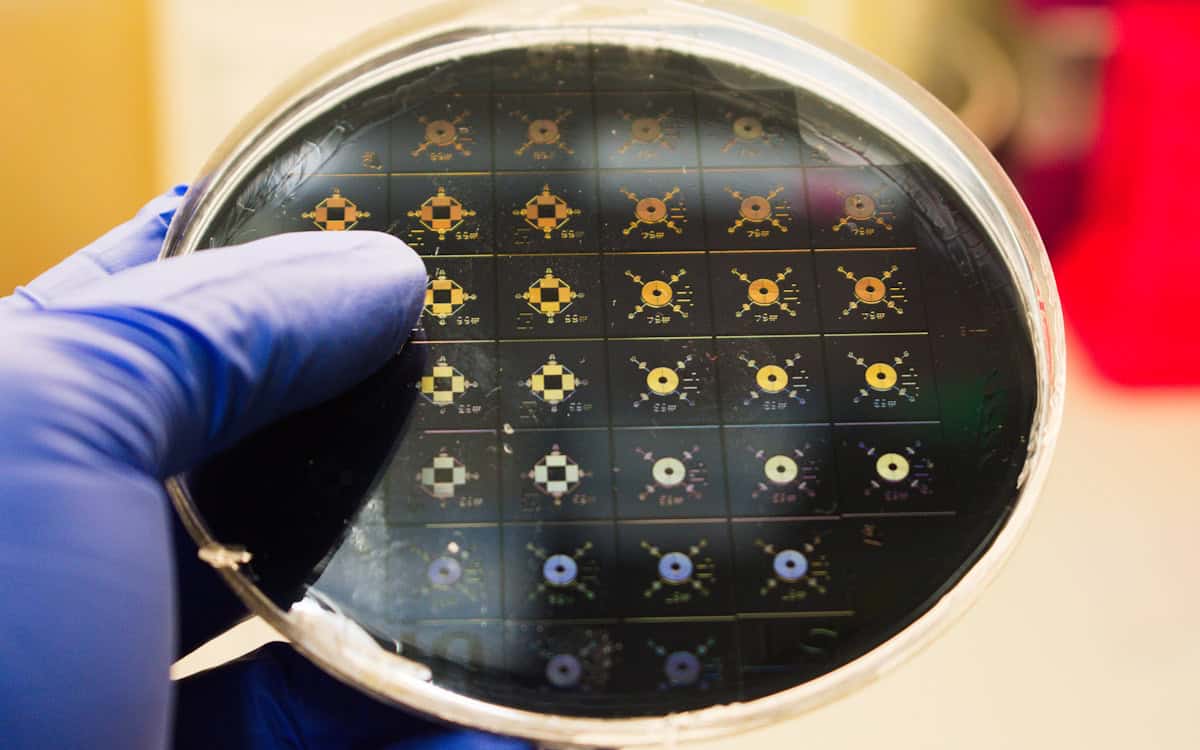
Cancers spread when individual cells detach from the tumour cell collective and invade previously healthy tissue. Whether detachment occurs depends on both genetics and the local microenvironment – including blood vessels, immune cells, signalling molecules and connective tissue. To replicate cancer cells squeezing through pores between the fibres of the tissues they are invading, the researchers created forests of pillars mounted on microfluidic devices for tumour cells to squash past.
As detailed in their recent Science Advances paper, the geometry for some forests was a square lattice of 9 µm-diameter pillars evenly spaced with 9 µm gaps between them, while other forests had their pillars arranged in a disorderly pattern created by moving each pillar from its ordered position by a random angle and distance.
“There were no suitable tools for designing mathematically defined disorder. So I had to write completely new software that can define those patterns, and then translate that into designs that can be manufactured [via standard semiconductor lithography techniques] into microfluidic devices,” explains biotech data science consultant Saren Tasciyan, who designed the microfluidic devices as part of his PhD with the Sixt Group.
The team used a combination of fluorescence imaging and light microscopy to track the behaviour of cancer cells – kept alive for up to eight days thanks to cell culture medium – after they were introduced into the centre of each respective pillar forest. These experiments revealed that the tumour cell collective was more likely to break apart when faced with a heterogeneous environment to navigate compared with a more regular geometry.
In the heterogeneous conditions “we found that the first cells detach, exactly as happens in a cancer metastasis,” says Sixt. The team also observed the pillars increasingly rough up the surface of the spreading cell interface, resulting in finger-like protrusions – from whose tips the cells were more likely to detach.
“When the cell interface moves through this landscape the effects accumulate over time,” continues Sixt, who asked his colleague Hannezo to create computer simulations to verify the experimental results, and study the process over longer periods than the cells could survive.
“Each cell was modelled as a bead that moves through an environment,” says Hannezo group postdoc Zuzana Dunajová, a computational scientist specializing in biological physics, who built the model. These beads are attracted to one another, and move through different geometries that mimic those on the microfluidic devices, she explains.
Cell softening allows cancer cells in rigid tumours to spread
“I kept seeing the same behaviour in my simulations – the beads detached from the collective more often in the disordered environment compared to the ordered. This was very exciting from the physics perspective because it’s not something that we see just in this particular cell type or system, but suggests a more general principle in living, or active systems. The roughness of the cancer-cell invasion actually follows the same kind of universal behaviour as we see in other systems, such as combustion of paper, spreading of fires or drying coffee drops,” enthuses Dunajová.
Next, Sixt – who hopes these insights will help researchers developing new cancer therapies – plans to study whether cell detachment in a disordered microenvironment leads to epigenetic modifications (heritable changes that do not alter the DNA code) and then later on to genetic modifications. “I want to consider whether the detachment process can drive the single cell evolution into something malignant,” Sixt concludes.