Preclinical PET is an ideal research tool for studying small-animal models of disease. To optimize systems for imaging the minute features of mice, developing small-animal PET scanners with high spatial resolution and high sensitivity is a longstanding goal.
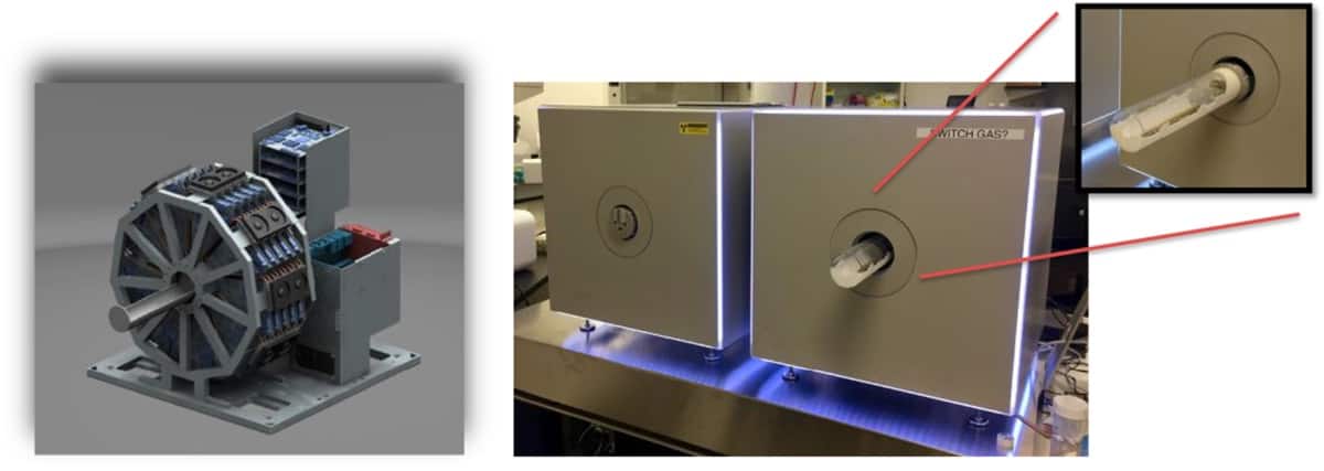
Ghent University spin-off MOLECUBES recently launched a line of dedicated small-animal scanners, including the β-CUBE (PET), X-CUBE (CT) and γ-CUBE (SPECT) systems. The β-CUBE is lightweight, compact (54 cm3) and enables bench-top imaging of both mice and rats. The first β-CUBE and X-CUBE scanners were recently installed in the Small Animal Imaging Facility at the University of Pennsylvania. The team has now reported the findings of a detailed performance evaluation of its β-CUBE (Phys. Med. Biol. 63 155013).
“The main challenge in PET imaging of mice is developing a scanner that offers functionality and practicality for high-throughput imaging, as well as excellent performance,” says first author Srilalan Krishnamoorthy. “The combination of high spatial resolution and high sensitivity is very important – this leads to images that have excellent statistical quality with the ability to achieve quantitatively accurate measurements of radiotracer uptake.”
System specifications
The β-CUBE detector comprises an 8 mm thick monolithic LYSO scintillator coupled to an array of silicon photomultipliers. The monolithic scintillator delivers high intrinsic spatial resolution and enables depth-of-interaction (DOI) measurement. A total of 45 PET detectors arranged in five rings provides a scanner diameter of 7.6 cm and an axial length of 13 cm – suitable for whole-body imaging of both rats and mice.
The PET scanner can be used individually, or in combination with the X-CUBE micro-CT scanner. The animal bed is easily transferred into the X-CUBE, and the 3D PET and CT images are automatically co-registered. The CT acquisitions can be also used to perform all attenuation and scatter corrections necessary to obtain quantitative PET images.
The team first measured the spatial resolution of the β-CUBE using a 22Na point source embedded in an acrylic cube. The source was placed at the scanner centre and stepped radially across the detector field-of-view (FOV). Reconstructing the PET data with a 3D filtered back projection algorithm revealed an excellent spatial resolution of better than 1 mm over the entire FOV.
“The scanner has excellent spatial resolution that is uniform over the imaging FOV, due to its ability to compensate for parallax error in the image reconstruction process,” explains Krishnamoorthy. “This capability is due to the sophisticated monolithic detectors that enable the DOI of the gamma rays within the crystal to be measured.”
Using the same point source, the researchers measured a maximum absolute sensitivity of 12.4% at the scanner centre, using a 50% energy window (255-765 keV). With a 15% energy window (435-588 keV), as is routinely used for animal imaging, they measured an absolute peak sensitivity of 5.7%.
The researchers also used NU-4 standard line-source mouse and rat phantoms to measure scatter fraction (scattered events/scatters plus trues) and noise equivalent count (NEC; trues squared/total counts). They recorded scatter fractions of 11.3% and 15.7%, with the mouse and rat phantoms, respectively. Peak NECs were 300 and 160 kcps in the two phantoms, using the 15% energy window and measured with 900 μCi in the phantoms.
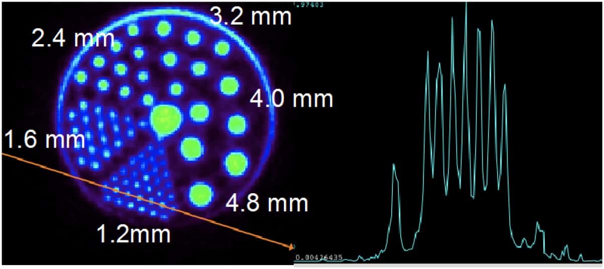
Images of the NU-4 image quality phantom (reconstructed using CT correction of attenuation and scatter) revealed an image uniformity of 7.4% and spill-over ratio of 8%. The researchers measured a contrast recovery of about 70% for 2 mm-diameter rods and full recovery for rods of 3 mm or larger. They also imaged a Micro Derenzo hot rod phantom, and saw that the smallest 1.2 mm rods were clearly visualized and well separated.
Animal investigations
Krishnamoorthy notes that, thanks to an in-house cyclotron and an excellent radiochemistry group at the University of Pennsylvania, the Small Animal Imaging Facility team has used the β-CUBE to perform animal studies with a variety of novel PET radiotracers, in many areas of oncology, neuroscience and cardiology.
In one investigation, they used 18FDG PET to investigate inflammation in osteoarthritis of a rat’s temporomandibular joint (TMJ). They performed a 15-min static PET scan 1-hour post-injection, immediately followed with a CT scan. The co-registered images delineated the small structure of the TMJ from the surrounding jaw, leading to a more accurate measurement of the FDG signal and improved tracking of disease progression.

In a second example, the researchers describe a lung tumour imaging study in mice, using a 15-min static PET scan 1-hour post-injection of 18FDG. They note that the superior spatial resolution and contrast recovery allow better visualization of smaller structures in comparison with their older system, which has 2 mm spatial resolution.
“We are currently working on optimizing our current imaging protocols to fully utilize the MOLECUBES PET and CT scanners,” Krishnamoorthy tells Physics World.


