
Ronan Smith, a postdoctoral research fellow at Adelaide University, has been chosen as the winner of this year’s Physics in Medicine & Biology (PMB) Early Career Researcher Award. The award is presented to the author of the “best paper” in PMB’s 2025 Early Career Researcher Focus Collection, as selected by the journal’s editorial board.
Smith’s research involves implementing X-ray velocimetry (XV), a novel imaging method that uses X-rays to track lung motion during breathing and create 3D maps of local ventilation. In his award-winning paper, Visualising ventilation changes following endobronchial valve placement with x-ray velocimetry functional lung imaging, Smith investigates the potential of XV imaging to detect changes in lung function after insertion of an endobronchial valve (EBV).
EBVs are one-way valves that are placed into the lung to help treat emphysema – a condition that damages air sacs in the lungs, causing air to get trapped inside and making breathing difficult. The EBV, which in some cases can be used in place of surgery, prevents airflow into damaged lung areas so that the rest of the lung can function more effectively.
Successful valve placement causes the targeted area of lung to collapse, which can be imaged using CT. Smith proposed that use of XV functional lung imaging to non-invasively measure regional and local changes in airflow could more accurately assess the clinical impact of EBV placement.
“The lungs are a dynamic organ, their job is to be constantly moving,” he explains. “Because X-ray velocimetry looks at lung motion, it lets us see exactly where the air is or isn’t flowing, so you can instantly see that airflow has changed within the lungs. CT only measures structural changes, which may not necessarily be correlated to changes in lung function.”
In vivo demonstration
To investigate this potential advantage, Smith and colleagues carried out a pilot study on healthy sheep, which have a similar lung size to humans. They performed XV imaging on two anaesthetized and ventilated animals, before and after placing EBVs in their lungs.
The XV scanning process involves recording fluoroscopic videos of individual breaths at various angles around the lung, with anatomic positioning provided by an accompanying breath-hold CT scan. To analyse the data, the researchers used XV LVAS software from 4DMedical, the MedTech company that developed and commercialized the XV technology.
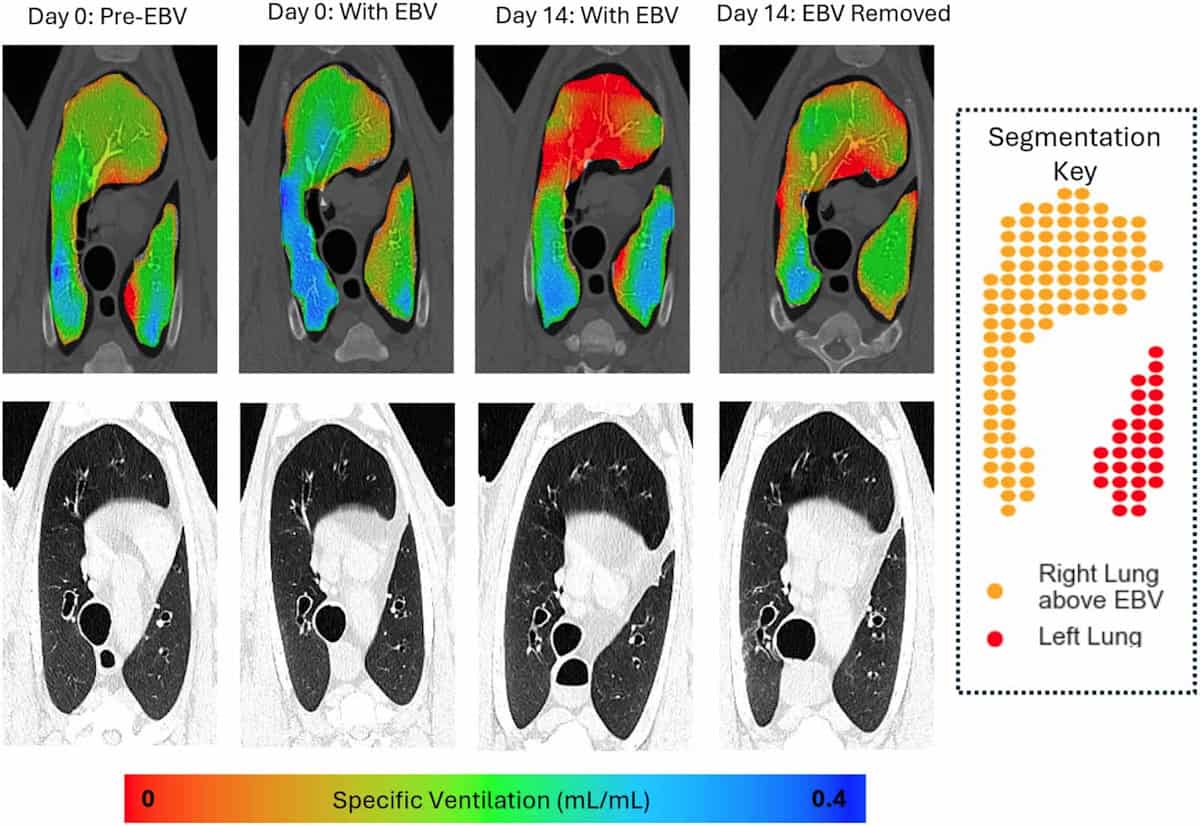
The software correlates motion in the XV videos with CT data to measure the lung’s expansion and contraction during a breath cycle. It creates a 3D map of specific ventilation (the change in voxel volume during a breath, divided by its starting volume) in small voxels throughout the lungs. This map can then be used to calculate mean specific ventilation and ventilation heterogeneity across a given lung region.
As soon as the EBVs were inserted into the animals’ lungs, XV imaging could visualize and quantify a reduction in airflow to areas downstream of the valves. This effect was seen both in regions where collapse was visible in CT scans, as well as those where collapse could not be detected by CT. Ventilation changes were also clearly observed in the remainder of the lungs.
“The main finding of our study was that X-ray velocimetry imaging can detect airflow changes from endobronchial valve placement in the lungs,” says Smith. “Our research could be really important for people [with emphysema], as tools to help with better placement and verification will lead to improved treatment options.”
Future prospects
Since the publication of this paper, Smith has focused on further applications of pre-clinical and clinical XV imaging. “I’ve been working as part of a great interdisciplinary team looking at how lung function changes in a range of diseases, to both understand the diseases, and as an outcome measure when we test treatments,” he says.
Dark-field X-ray imaging reveals potential of nanoparticle-delivered gene therapy
This work includes the world’s first paediatric clinical trial of XV imaging, which is examining the feasibility of using the technology in children with cystic fibrosis. The researchers have imaged around 30 children to date and aim to publish their findings later this year. They are currently planning future studies to see how XV imaging could enhance clinical decision making and improve outcomes for these children, as well as looking at other childhood diseases where it could be of relevance.
“As an early-career researcher, I’m also focussing on developing my own research, looking at another novel X-ray imaging method called dark-field X-ray imaging,” Smith adds.
The perfect award
Smith tells Physics World that he was excited to receive the PMB Early Career Researcher Award, acknowledging the efforts of everyone involved in this “hugely collaborative project”, including clinicians, scientists, 4DMedical and the staff in the preclinical imaging facility where the study was performed.
“As a physicist working in medicine/biology, it feels like the perfect award to get,” he says. “It’s great to see interest in the work we are doing, and fantastic evidence we can use to convince the funding bodies it’s worth continuing this work.”
- The PMB Early Career Researcher Focus Collection 2025 publishes papers from early-career researchers (defined for this collection as a postgraduate student or someone who completed their PhD in 2019 or later) in biomedical physics, with the aim of highlighting research excellence from emerging leaders. The Early Career Researcher Award was introduced to further recognise an outstanding contribution from one of the early-career authors, selected according to the quality of scientific content, number of citations and downloads, and peer review ratings.


