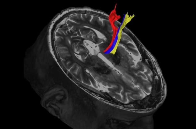
Researchers at the University of Texas Southwestern and the Mayo Clinic in the US have presented the latest developments in the use of MR-guided high-intensity focused ultrasound (HIFU) to treat tremor. Writing in the journal Brain, they describe how these advances are expected to lead to improved clinical efficacy and a reduction of adverse effects.
The first-line treatment to reduce the involuntary trembling or shaking associated with neurologic conditions such as essential tremor or Parkinson’s disease is medication. However, around 30% of patients do not respond well to drugs. This led to the investigation of alternative therapies to alter the connectivity of the thalamus, a symmetric structure composed of grey matter that sits on top of the brain stem and serves as a relay for motor and sensory signals between the body and the rest of the brain.
In the 1990s, deep brain stimulation was developed, in which metal electrodes implanted in the thalamus were stimulated via a battery pack, similarly to a pacemaker. A couple of decades later, advances in MRI allowed real-time guidance of HIFU beams to non-invasively heat and eliminate small sections of the thalamus with millimetre precision.
The main challenge in both of these procedures, however, lies in the correct targeting of the ventral intermediate (VIM) nucleus, a pea-sized region located at the centre of the thalamus that’s involved in the coordination and planning of movement. Precise localization of this structure is important to prevent adverse effects due to incorrect targeting during these procedures, such as speech and swallowing deficits, or sensory and gait abnormalities. While these side effects are usually temporary, they can be permanent in 15 to 20% of cases.

In their paper, Bhavya Shah and colleagues highlight the limitations of current techniques for localizing the VIM nucleus. Established targeting methods either use atlases to identify brain areas from a collection of correctly labelled brain scans or require the use of landmarks on the patient’s brain scans.
We now know that such atlases and landmarks are too simplistic, and that the connections and biology of the brain are too complex and patient-specific to be accurately captured through these methods. Indeed, studies have shown that these approaches can introduce targeting errors of up to 5 mm, causing unacceptable adverse effects.
MRI methods to the rescue
In contrast with traditional localization techniques, the researchers highlight three newly refined MRI techniques that are proving better at delineating the target tissue.
Diffusion tractography seems to be the most promising. It creates precise 3D brain images by taking into account the natural water movement within tissues, which allows identification of not only the VIM nucleus location but also its shape.
Another technique hailed as superior to established targeting methods is quantitative susceptibility mapping, which creates contrast in the image by detecting distortion in the magnetic field caused by substances such as iron or myelin. Finally, the researchers cite fast grey matter acquisition T1 inversion recovery as showing promising results. This technique operates much like a photo negative, turning the brain’s white matter dark and its grey matter white, in order to provide greater detail in the grey matter.
All of these MRI sequences are already approved by the US Food and Drug Administration for guiding HIFU beams during localized ablation of the thalamus. The object of an increasing number of studies, they should improve clinical efficacy and reduce adverse effects from the treatment.


