
Epithelial cancers account for 80% to 90% of all cancer cases in the world, comprising a wide variety of malignancies that begin in the epithelial tissues that form the skin and line the body’s internal passageways and organs. Early diagnosis is critical to improving patient outcomes, but is limited by a heavy reliance on invasive biopsies.
Simultaneous assessment of changes in both the cell nuclei and the microvasculature could potentially improve the accuracy of early cancer and precancer detection, and help guide decisions regarding biopsy and pathology testing. In vivo microscopy (IVM) has shown promise for aiding early diagnosis of epithelial cancers, but has challenges relating to image quality, spatial resolution and field-of-view (FOV).
PrecisionView, an artificial intelligence (AI)-powered, dual-modality endomicroscope in development at Rice University’s Rice360 Institute for Global Health Technologies, addresses these IVM issues. The compact, handheld, easy-to use system enables real-time, wide-area visualization of both subcellular structures and underlying blood vessels, offering higher-quality imaging, a fivefold increase in FOV, and an eightfold increase in spatial resolution over endomicroscopic systems in current clinical use.
The proof-of-concept version of PrecisionView, described in Proceedings of the National Academy of Sciences, is intended for superficial epithelial imaging where scattering-induced distortions remain moderate. And it is specifically designed to be low cost: providing an affordable diagnostic tool for clinics worldwide.
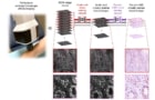
PrecisionView uses simple, off-the-shelf miniaturized optics integrated with a deep learning-based optimization framework. Its AI-supported phase mask design and reconstruction algorithm incorporate dual-modality fluorescence and reflectance imaging to achieve a 5.2 x 3.9 FOV and a 500 µm depth-of-field (DOF) while maintaining cellular-level resolution (4 µm). Images are acquired at approximately 15 frames/s, in 2 s intervals, with image tiles stitched together to form a large-scale, co-registered map of cell nuclei and microvasculature.
Performance assessment
To validate the system, principal investigators Rebecca Richards-Kortum and Ashok Veeraraghavan, and collaborators at the University of Texas MD Anderson Cancer Center, analysed images of ex vivo porcine tongue, human breast and fresh cervical tissue specimens with precancerous lesions.
High-resolution maps of cervical specimens containing squamous intraepithelial lesions (precancer) clearly delineated key anatomic structures, including columnar and squamous epithelium, and the junction between the two epithelial cell types. PrecisionView clearly distinguished abnormal regions from surrounding benign tissue – suggesting that the system may be able to support physicians’ decisions regarding whether or not to conduct a biopsy.
The researchers also performed real-time, in vivo imaging of the oral cavities of healthy volunteers. They report that the system’s large DOF enabled acquisition of high-resolution images of cell nuclei and microvasculature across a large tissue area without requiring refocusing. When the mucosal surface was scanned, the large FOV of each frame, combined with the high video acquisition rate, provided substantial frame-to-frame overlap. This allowed seamless stitching of the dual-modality images to create large-scale, co-registered maps of the oral mucosa, which showed distinct cellular and vascular features.
Meeting a global need
The Rice 360 Institute focuses on designing, implementing and scaling highly effective, low-cost medical technologies for underserved and low-resource communities. The idea for PrecisionView grew out of recognizing a global limitation in access to high-quality diagnostic information.
“In many care settings, especially where conventional pathology resources are limited, clinicians may not have timely access to the cellular- and tissue-level information needed to guide diagnosis and treatment decisions,” says first author Huayu Hou, who worked with co-first author Jimin Wu, to develop the PrecisionView instrument.
Hou explains that although existing clinical endomicroscopy systems can provide valuable microscopic information, their broader use is often limited by cost, complexity, a limited imaging volume and the ability to capture only a narrow range of diagnostic features. Such constraints reflect fundamental design trade-offs in compact clinical optical imaging systems and create significant barriers to practical point-of-care use.
“PrecisionView was motivated by the opportunity to use computational imaging to overcome limitations in existing clinical endomicroscope designs that could not be fully addressed with traditional optical approaches alone, with the goal of bringing high-quality diagnostic information closer to the point-of-care,” he says.
Hou advises that it took about a year to develop the first working prototype, and that several projects are now underway to further enhance both performance and usability for point-of-care applications. “We are working to improve image quality and resolution, so that the system can provide finer cellular details from living systems to real time,” he adds. “We are working to further refine the hardware and workflow to make the device easier to operate in clinical settings.”
Virtual histology may reduce the need for skin biopsies
The team is developing AI-based classification algorithms that can integrate both cellular and vascular information and provide real-time diagnostic guidance. Its long term goal is to create a system that not only visualizes tissue at the cellular level, but also helps clinicians interpret the information efficiently at the point-of-care. “This capability could be especially valuable in low-resource settings, where access to pathology expertise may be limited, and tools that support real-time decision-making could significantly improve clinical usability and impact,” notes Wu.
Clinical validation studies to evaluate PrecisionView’s diagnostic performance across different clinical settings and cancer types are also planned. For instance, a study focusing on head-and-neck cancer detection has already started in collaboration with MD Anderson Cancer Center. Others are being developed for cervical cancer imaging in Mozambique, to evaluate the system’s performance in low-resource settings.


