
Image guided radiotherapy (IGRT) is the state-of-the-art in radiation treatment, and the recent introduction of integrated MRI-linac systems adds potential for real-time tumour tracking during beam delivery. But achieving this requires the ability to quickly and accurately determine the position of the target volume and critical structures from the MR images.
Because radiotherapy uses a divergent beam emanating from a single point, conventional pre-treatment simulation using CT requires the creation of “ray-traced” digital reconstruction radiographs to generate a beam’s-eye-view (BEV) image that represents the path of the treatment beam. Conventional MR image slices, however, have pixels that represent volume elements arranged parallel to each other.
When MRI is used to track structures for real-time radiotherapy guidance, these two coordinate geometries do not match and tracking errors can result, especially for thick image slices. Divergent ray-tracing of MR images is technically possible, but not suitable for real-time guidance due to lengthy 3D acquisition and ray-tracing reconstruction times.
Now, Keith Wachowicz, Brad Murray and B. Gino Fallone from the University of Alberta have developed a theoretical framework that allows – for the first time – direct acquisition of BEV projection images in MRI. They also describe how their concept can be applied to various types of MRI-linac configurations (Phys. Med. Biol. 63 125002).
“The BEV encoding gradients proposed in this work would allow direct acquisition of tracking images in the same geometry as the treatment beam, avoiding any potential for tracking errors due to the geometry mismatch,” explains first author Wachowicz.
Warping fields
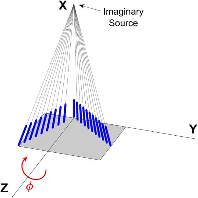
To use MRI to track anatomy in real time, the researchers propose the use of non-conventional gradient field patterns, implemented through hardware additions to a standard scanner architecture. They developed nonlinear encoding gradient fields that allow MR images to be generated in a divergent beam geometry. For MRI-linac systems where the radiation source is fixed relative to the magnet, adding two warping coils to the linear X and Y coils can produce these encoding fields.
For MRI-linac architectures in which the beam source is not fixed to the imaging magnet, the identified warping field pattern will only be appropriate for one source position. In such cases, the researchers showed that a basis set of second-order spherical harmonic functions, together with linear gradients, provides a good approximation of the BEV gradient patterns at any angle.
They propose the use of a set of second-order warping coils fixed to the magnet, employed in various combinations to generate the conditions for divergent imaging as the source rotates. This would require four additional warping coils.
Proof of principle
To test their proposed theory, the researchers used a 3T scanner to image a phantom with nonlinear encoding-gradient field patterns. The phantom comprised gel-filled rods oriented to converge at a single point 100 cm away.
As the derived encoding gradients are not readily available, they approximated the ideal warping field to a second-order field gradient and created this using second-order shim coils. Such coils, however, are not currently designed for rapid switching in tandem with the linear encoding gradients.
“To test the feasibility of this approach in an environment without rapid-switching capability, we had to first find a sequence that maintained as much as possible a constant encoding gradient amplitude during image encoding. The closest match we could find was a short-echo radial acquisition,” Wachowicz explains. “Secondly, we had to manually alter the shim coil currents according to our calculations for each of the 102 radial spokes that we acquired.”
To circumvent hardware limitations, the researchers also created a corresponding virtual phantom. They simulated three images, using: conventional linear gradients; switched linear-encoding gradients and unswitched warping fields (to mimic the experiment); and linear-encoding gradients and warping fields switched in tandem (representing an ideal implementation).
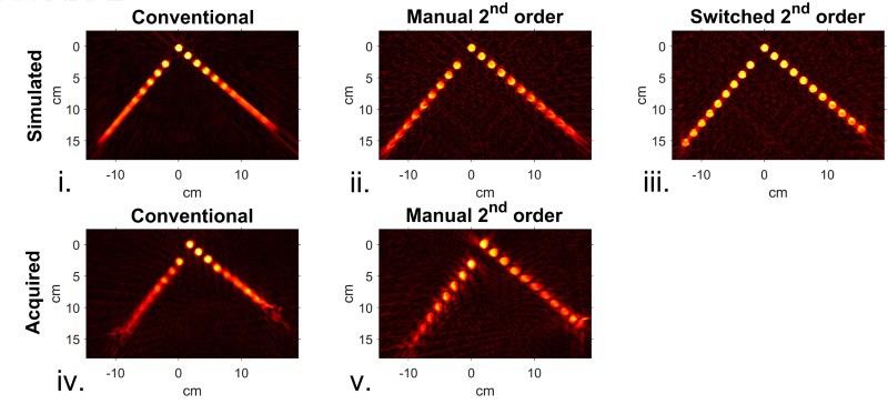
Images of the phantom generated with traditional parallel geometry over its full 12 cm thickness exhibited blurring, as expected. When images were acquired with (unswitched) warping field patterns, much of this blurring was absent. However, the tubes still appeared distorted, particularly those farthest from the isocentre.
The authors believed that a switched set of warping fields – as would be present in any physical implementation of this technique – would remove the bulk of this residual distortion. Simulations with companion fields switched in tandem with the read gradients produced images with all of the tubes clearly discernible, successfully validating their technique.
The team is now planning to move this approach towards clinical application. “We expect to include a set of BEV coils within the Alberta (Edmonton) biplanar linac-MR hybrid design,” says Fallone.


