You might think it would be a tough gig to hold a scientific session at 7 p.m. on a Saturday night anywhere, let alone in San Francisco. But several hundred attendees – including me – turned up at the Moscone Center for a whirlwind tour of the latest breakthroughs in biomedical optics.
The occasion was the Hot Topics session of the BIOS conference, held alongside Photonics West since 2004 to provide a forum for highlighting novel research in biophotonics. Over the years it has become the largest scientific meeting to focus on this increasing important subdiscipline, and the Hot Topics evening slot has become popular among attendees for providing a snapshot some of the most exciting new technologies.
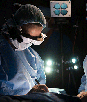
Before the Hot Topics got under way, a keynote address by Samuel Achilefu from Washington University in St Louis offered a brief insight into the use of real-time optical imaging of cancer cells during surgery. Achilefu and his team invented a device that enables surgeons to see cancer cells during an operation, and his focus on producing practical solutions for clinical use was recognized by this year’s Britton Chance Award – given by the SPIE for pioneering contributions to biophotonics techniques and devices.

The motivation for Achilefu is to “eliminate guesswork, prevent local relapse, and allow surgeons to selectively kill cancer cells”. It is also important to ensure that the optical equipment can fit into a crowded operating room, and the solution devised by Achifelu and his team was to engineer a head-mounted display that he calls “cancer vision goggles”. The fluorescence goggle system allows the surgeon to visualize the cancer cells in real time, ensuring that all the diseased cells are removed during the operation. The portability of the cancer goggles also makes them suitable for use in any operating theatre, anywhere in the world.
Among the following Hot Topics presentations, one that captured my interest was by Clare Ewell, a professor of medical physics at University College London. Ewell and her team have been investigating a functional brain imaging technique based on near-infrared spectroscopy (NIRS), and have developed a broadband NIRS system that offers better performance that more common dual-wavelength versions. The wearable system provides high-density optical measurements of cerebral oxygen metabolism, which is important, for example, for investigating cognitive function in brain-damaged patients.
Ewell pointed out that functional NIRS is a particularly useful tool for assessing the brain function of infants and young children. One recent study measured the fNIRS response in babies’ brains, and showed that measurements from babies less than six months old could be used as an early predictor of autism in later childhood.
Ewell is also working the make optical brain imaging available to children who do not have easy access to sophisticated medical equipment. Through the BRIGHT Project, funded by the Bill and Melinda Gates Foundation, fNIRS has been used to investigate the impact of malnutrition on the brains of infants in rural Gambia within their critical first 1000 days of life – which Ewell says is the first time that functional brain imaging has been offered to children in Africa. Her research aims to identify which nutritional and other interventions could protect the infants’ brains and ensure that they reach their full developmental potential.
“One third of the children living in resource-poor settings fail to meet developmental milestones,” Ewell pointed out. “This can impact academic achievements, mental health, and the ability to form and sustain healthy relationships. These children are surviving, but not thriving.”


