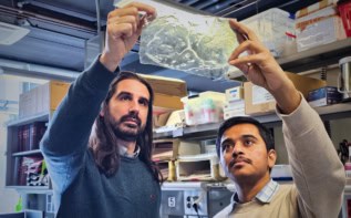
The tooth is the most mineralized tissue in the mammalian body and holds information about the creature it belongs to. One can identify the taxonomy, life history and species simply by examining enamel. However, the origin of enamel thickness differences and how this comes about between species and within populations is still unknown. Now, researchers led by Jukka Jernvall in Finland have developed a mathematical model that simulates enamel secretion from underlying dentine. This model allowed the researchers to reproduce mammalian enamel structures as well as explaining the difference in enamel morphology between species.
Tooth enamel grows from an underlying tissue known as dentine during mammalian development. The interface between these two tissues, the enamel-dentine junction (EDJ), reflects the final shape of our enamel crowns, however, it is not a simple geometric extrapolation of this surface. Hence, the researchers developed a growth model for enamel from the EDJ that assumes enamel growth requires a net influx of a diffusing nutrient substance. By modifying the nutrient concentration, the minimum amount of nutrients for growth and the stiffness of the advancing growth front, the researchers were able to predict the morphology of final enamel structures and compare them to micro-CT scans of tooth samples.
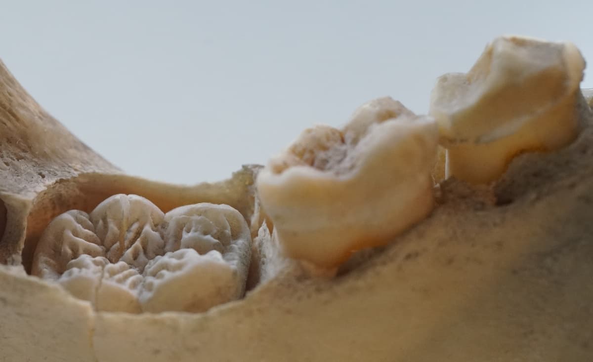
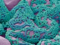
Impressively, the researchers were able to predict the deep narrow furrows present in pig molar enamel, which a simple geometric extrapolation from the EDJ could not. The diffusive nature of enamel growth means that small changes in the EDJ shape correspond to large differences in enamel morphology. Furthermore, the model mimics the incremental lines of enamel growth in pig molars during development, comparable to growth rings in trees. Using tomographic image data from the particle accelerator at the European Synchrotron Radiation Facility, they showed that the model not only reproduces the correct final structure but also the growth mode of the enamel matrix.
Species differentiation by diffusion
Following on from the pig molars, the researchers studied primate molars (including humans). By modifying the orientation of their diffusion-limited model, the researchers could reproduce human enamel morphology with characteristic ripples and larger crowns. Comparing human and orangutan molars, the researchers were able to reproduce the more complex enamel structure of the orangutan using the softer human EDJ simply by modifying the amount of nutrient available. They could also switch the enamels from human-like to orangutan-like for EDJs from either species by modifying the limits of diffusion for the enamel growth.
More than just teeth
This model highlights the necessity of diffusion-mediated matrix deposition for complex enamel development and shows how even simple mechanisms have allowed biology to flourish into complexity. Furthermore, this model allows researchers to study the growth of other diffusion-limited tissues and increases our understanding of enamel matrix formation.
Full details of the research are reported in PLoS Computational Biology