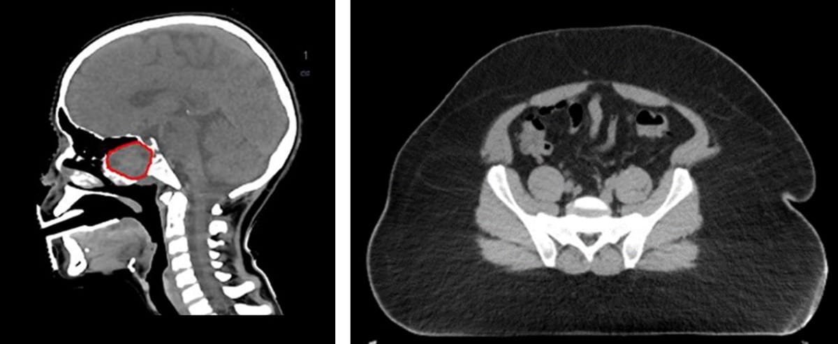
The DirectDensity algorithm from Siemens Healthineers gives radiation oncologists and medical physicists ready access to CT images optimized for both patient contouring and dose calculation

Fundamental transformation doesn’t come easy in radiation therapy given the countervailing forces at play within the clinical workflow. On the one hand, there’s the relentless drive among radiation oncology teams for enhanced efficiency and standardization across treatment planning, management and delivery. At the same time, those multidisciplinary care teams are striving to deliver increasingly personalized radiation treatments tailored to the needs of individual patients and their specific cancer indications.
The trade-offs between standardization and personalization are particularly evident when it comes to CT simulation in the radiotherapy suite – chiefly because of the requirement for patient imaging to service the needs of two very different end-users. Radiation oncologists, for example, seek optimized image quality to support contouring and delineation of the tumour volume as well as adjacent critical structures and organs-at-risk (OARs). Medical physicists, for their part, apply the associated tissue density data in the CT scan to calculate accurate 3D dose distributions for 3D treatment planning.
Clinical impact
Squaring that circle – generating a geometrically accurate and qualitative representation of the patient in tandem with robust dose calculations – remains an ongoing priority for the Cancer Therapy Business Line at Siemens Healthineers. The equipment vendor’s DirectDensity algorithm is a case in point, reconstructing images from single-energy CT acquisitions in which the resulting CT values at any given kV setting can be interpreted to show relative electron or mass density for dose calculations. Currently, radiotherapy treatment planning systems (TPS) require a kV-specific calibration curve to carry out this conversion (i.e. where tube voltage is allowed as an extra degree of freedom during simulation, this implies a time and resource overhead as well as a possible source of error in a busy clinical environment).
As such, the core innovation of the DirectDensity algorithm – a single linear relationship that does not depend on the tube voltage of CT acquisition – is something of a game-changer, unlocking the full potential of CT imaging in radiotherapy by dispensing with the traditional practice of scan protocols at a fixed tube voltage (typically 120 kV)1. By extension, the DirectDensity algorithm also reduces the scope for workflow errors that may be introduced if the medical physicist inadvertently selects the wrong TPS calibration curve.
At the clinical sharp-end, the advanced functionality offered by DirectDensity – the option to vary the tube voltage of the CT scanner while using one calibration curve – opens the way for care teams to design scan protocols that are more personalized to the patient. In the case of bariatric patients, for example, who have a higher X-ray attenuation, the output current of the X-ray tube at lower or conventional kV settings may not be sufficient to produce the required contrast-to-noise ratio. Here, higher X-ray tube voltages might be necessary. For paediatric patients or younger breast cancer patients, who might be unnecessarily exposed to higher radiation doses with a conventional 120 kV scan protocol, the contrast-to-noise ratio in the images could be maintained by using a scan protocol with lower kV setting – thereby lowering the received dose during CT simulation.
Educating the care team
Among the clinical early-adopters of the DirectDensity algorithm are medical physicist Enric Fernandez-Velilla Cepria and his colleagues at Hospital Del Mar in Barcelona, Spain. The clinic’s radiation oncology programme treats around 900 adult cancer patients every year – a wide range of disease indications excluding sarcomas – on a suite of three Varian machines (two TrueBeams and a GammaMedPlus brachytherapy system) and an additional intraoperative radiotherapy system for breast cancer patients.
“We were the first radiotherapy clinic in Spain to deploy the Siemens Healthineers SOMATOM Confidence RT Pro [in 2017], an advanced CT scanner for patient simulation,” explains Fernandez-Velilla. The timing was such that the Hospital Del Mar medical physics team was co-opted to work with SOMATOM Confidence development engineers on road-testing of the CT scanner’s advanced functionality – and specifically the preclinical validation of the DirectDensity algorithm. In this way, the evidence-based evaluation from the Barcelona team informed subsequent iteration of DirectDensity and its user interface ahead of wider clinical roll-out.
“Bear in mind,” adds Fernandez-Velilla, “DirectDensity was so new at the time that our first task here was an education and training process for the multidisciplinary oncology care team. Up to that point, we were used to working at a single CT tube voltage during CT simulation, so we needed to highlight the clinical advantages of imaging at different kV settings.”
The main beneficiaries of DirectDensity, he notes, are Hospital Del Mar’s radiation oncologists, who take advantage of the flexible kV settings of the CT scanner to fine-tune image quality for enhanced delineation and contouring across the clinic’s diverse patient population. When imaging obese patients, for example, the standard 120 kV setting often means too much noise and artefacts on the CT image, so there are improvements to be had by pushing the tube voltage out to 140 kV. “By the same token,” adds Fernandez-Velilla, “we see that CT image quality in head-and-neck patients is enhanced at 80 kV. So that means dose to the patient decreases, also the motion and registration artefacts as there’s no need for a supplementary 120 kV image series.”
Specific reconstructions for specific tasks
Elsewhere, Ghent University Hospital in Belgium is another Siemens Healthineers customer seeing significant clinical upside following deployment of the DirectDensity algorithm into its radiotherapy workflow. Ghent’s radiation oncology programme treats around 2000 patients annually in a facility comprising four linacs (three Elekta linacs and a Varian Clinac iX) and two brachytherapy afterloaders. In April this year, the clinic also completed commissioning and acceptance of Siemens Healthineers’ latest SOMATOM go.Open Pro CT scanner for patient simulation (including the Direct Density algorithm).

“The introduction of DirectDensity means that SOMATOM functionalities such as CARE Dose and CARE kV – which can semi-automatically adapt tube current and, of special interest, tube voltage – can be included in our CT scan protocols,” explains Evelien Bogaert, a medical physicist at Ghent University Hospital and project manager responsible for clinical implementation of the SOMATOM go.Open Pro scanner in the radiotherapy suite. “While the image set reconstructed with the DirectDensity algorithm is used for dose calculation,” she notes, “additionally reconstructed data sets at the same tube voltage optimize acquisition and, with the focus on image quality, aid the task of delineation for Ghent’s radiation oncologists.”
It’s worth noting, though, that the enhanced image quality for patient contouring is not just down to the DirectDensity algorithm, rather a portfolio of software and hardware advances within the SOMATOM go.Open Pro scanner. Key innovations include the high-performance Stellar detector; the use of iterative metal-artefact reduction (iMAR) to suppress beam-hardening artefacts caused by artificial joints, pacemakers, chemotherapy ports or dental implants; and SAFIRE iterative reconstruction algorithms to smooth noise and decrease artefacts at lower doses (thereby increasing lesion conspicuity).
The Ghent team also reports significant advantages for patients imaged with contrast media. “Since iodine contrast appearance is supressed in DirectDensity images for dose calculation, there’s no need for an additional non-contrast CT scan in our clinical workflow,” explains Bogaert. Conversely, enhancement of iodine contrast is the priority when it comes to the specific task of contouring. “The fact that there is improved contrast media visualization at lower tube voltages,” she adds, “allows for a reduction in the aggregate contrast burden to the patient when administering kV-dependent iodine contrast volumes.” In this way, a significant patient cohort in the Ghent oncology programme see benefits, without any loss of contrast enhancement, in their image data sets for target and OAR definition.2
“Ultimately,” concludes Bogaert, “the use of DirectDensity means we profit from optimal image quality for delineation and the specific requirements for dose calculation without having to worry about the potential errors introduced by multiple CT conversion curves – something which prohibited us in the past from exploiting kV optimization for CT simulation of the patient.”
The DirectDensity algorithm3 is available on Siemens Healthineers SOMATOM CT scanners compatible with the software version syngo.CT VA30 (or higher).
- For further information, see the Siemens Healthineers white paper DirectDensity: Technical Principles and Implications for Radiotherapy
In pictures: the DirectDensity algorithm