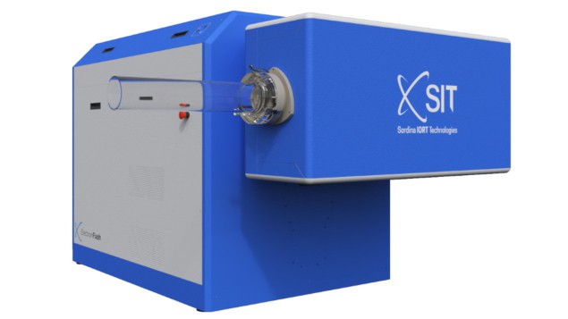
A European research team has used a prototype diamond-based Schottky diode detector to successfully commission an ElectronFlash research accelerator for both conventional and pre-clinical FLASH radiotherapy. The novel detector proved to be a useful tool for fast and reproducible beam characterization, suitable for ultrahigh-dose rates (UH-DR) and ultrahigh dose-per-pulse (UH-DPP) conditions. This is a milestone achievement for its development team, headed up at the University of Rome Tor Vergata, as no commercial real-time active dosimeters for FLASH radiotherapy are currently available.
FLASH radiotherapy is an emerging cancer treatment technique in which target tissues are irradiated using much higher dose rates than with conventional radiation therapy, and consequently, for much shorter irradiation times. This ultrahigh dose rate causes the so-called FLASH effect: a decrease in radiation-induced toxicities to surrounding normal tissues, while maintaining equivalent tumour-kill response.
This emerging technology is being lauded globally as an exciting treatment strategy with potential to change the future of clinical cancer therapy. But there are obstacles to overcome, one of which has been the development of an accurate, efficient-to-use dosimetry system to determine radiation dose in real-time.
Current commercial real-time dosimeters such as ionization chambers and solid-state detectors are not suitable for clinical use, due to recombination, saturation and nonlinearity effects observed in their response. Passive dosimeters such as alanine and GAFchromic films work, but their response may not be generated for hours or even days after an irradiation procedure, making them impractical for daily linac quality assurance.
To overcome these limitations, the team designed the flashDiamond (fD) detector specifically for UH-DR and UH-DPP applications, describing it in a January 2022 article in Medical Physics. Now, principal investigator Gianluca Verona Rinati and colleagues have performed a systematic investigation of fD detector response to pulsed electron beams, validating its response linearity at DPPs of up to about 26 Gy/pulse, instantaneous dose rates of about 5 MGy/s and average dose rates of around 1 kGy/s.
The researchers then used the fD detector to commission an ElectronFlash linac at Sordina Iort Technologies (SIT) in Italy, reporting their findings in Medical Physics.
Dosimetric characterization
To assess the fD prototype, the team first performed absorbed dose calibrations under three different irradiation conditions: 60Co irradiation in reference conditions at the PTW secondary standard laboratory (PTW-Freiburg); UH-DPP electron beams at PTB; and ElectronFlash beams in conventional conditions at SIT.
Encouragingly, the values obtained from calibration procedures at the three facilities agreed well. The sensitivities of an fD prototype obtained under 60Co irradiation, with UH-DPP electron beams and with conventional electron beams were 0.309±0.005, 0.305±0.002 and 0.306±0.005 nC/Gy, respectively. This indicates that there are no differences in the fD prototype response when conventional or UH-DPP electron beams are used, or between 60Co and electron beam irradiation.
The team next investigated the fD response linearity in the UH-DPP range. Varying the DPP between 1.2 and 11.9 Gy revealed that the prototype’s response was linear at least up to the maximum investigated value of 11.9 Gy.
The researchers also compared the fD detector results with those of commercially available dosimeters, including a microDiamond, an Advanced Markus ionization chamber, a silicon diode detector and EBT-XD GAFchromic films. They observed good agreement among percentage depth-dose curves, beam profiles and output factors measured by the fD prototype and the reference detectors, for conventional and (with EBT-XD films) UH-DPP irradiation.
Converted clinical linac delivers FLASH radiotherapy
Finally, the team used the fD detector to commission the ElectronFlash linac, which is capable of operating in both conventional and UH-DPP modalities. The linac is equipped with several cylindrical PMMA applicators, of between 30 and 120 mm in diameter, which are used to vary the DPP. Commissioning was completed by acquiring percentage depth-dose and beam profiles for 7 and 9 MeV pulsed electron beams, using all of the different applicators, and in both conventional and UH-DPP modalities.
The researchers conclude that the fD prototype could prove a valuable tool for commissioning of electron beam linacs for FLASH radiotherapy. They are currently conducting Monte Carlo simulations of both the ElectronFlash linac beams and the fD detector to provide theoretical support to their dosimetric valuations.


