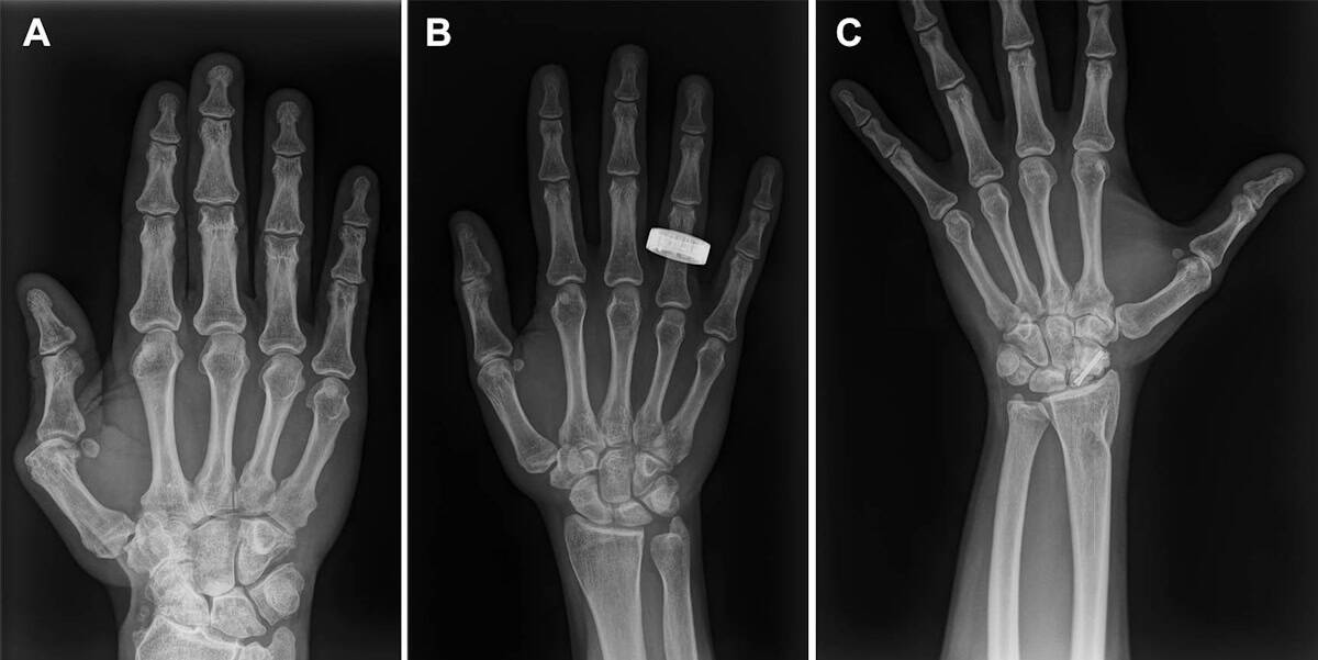
A collaboration of researchers in North America has reported their findings from the first diagnostic X-ray imaging performed in space. X-ray images recorded during an orbital spaceflight exhibited similar overall quality to those taken preflight on Earth, demonstrating the feasibility of adding radiography as a diagnostic tool for crew health.
With humans venturing further into space and spending more time in this harsh environment, the risk of adverse medical events increases. Accurate and timely diagnosis and treatment is a must-have. Currently, the only reliable medical imaging modality available to astronauts in spaceflight is ultrasonography – which is operator dependent, requires substantial training and relies on limited acoustic windows.
X-ray imaging, on the other hand, benefits from low operator dependence, rapid acquisition and typically higher spatial resolution than ultrasound. Digital radiography could also offer superior diagnostic capabilities for many medical conditions of concern during spaceflight, including dental disease, musculoskeletal trauma, inhalational injury, collapsed lung and arthritis.
Traditional X-ray scanning systems are bulky, produce a lot of radiation and are sensitive to motion – making them less than ideal for sending into space. But a new generation of portable X-ray scanners shows potential to survive the spaceflight and be operated by crew members with minimal training.
“It’s been a dream for aerospace medicine to have more than one imaging modality for diagnosing illnesses and injuries in space,” says lead researcher Sheyna Gifford, an assistant professor of aerospace medicine at the Mayo Clinic in Rochester, MN, in a press statement.
Scanning in space
In a study led by co-author David Lerner, the researchers first demonstrated the feasibility of X-ray imaging in simulated microgravity in 2022. During a parabolic flight, they used a commercial off-the-shelf digital radiography system to successfully capture radiographs of a human subject and a phantom during lunar gravity (1/6g), Martian gravity (1/3g) and microgravity.
In this latest project, reported in in Radiology, Gifford and her team investigated the performance of a similar portable digital radiography system aboard the Dragon spacecraft, during the Fram2 spaceflight mission, a 3.5-day polar orbital flight launched on 31 March 2025 on a SpaceX Falcon 9 rocket. The imaging system comprised a commercial off-the-shelf, FDA-cleared, portable, digital X-ray generator (Impact Wireless; MinXray) along with an FDA-cleared flat-panel detector.

Prior to the mission, three crew members received 4 hr of training on the portable X-ray system and acquired preflight X-ray images of the hand, forearm, abdomen, pelvis, chest and a quality control phantom. They then recorded the same anatomic and phantom images whilst in space, and also imaged a smartwatch to investigate the potential for non-destructive testing.
During anatomic imaging, the devices were handheld by the crew members, while the phantom and smartwatch were secured to the detector for imaging. The crew reported that they found it easy to use the X-ray system and follow the imaging protocols.
Back on Earth
To assess the quality of the seven X-ray images acquired by the crew, three independent radiologists evaluated and compared the preflight and in-flight anatomic radiographs, finding no differences in overall image quality, contrast resolution or spatial resolution. For central-body radiographs (chest, abdomen and pelvis), image positioning was worse in-flight than preflight. Despite this, all X-rays achieved good or near-excellent diagnostic quality (rated on a 5-point Likert scale).
Phantom radiographs recorded in space exhibited good spatial and contrast resolution, imaging low-contrast targets down to the smallest 2-mm target and easily visualizing high-contrast meshes ranging from 20 to 60 lines per inch, with 80 lines per inch remaining visible. The smartwatch radiograph clearly showed the watch’s internal components at the submillimetre scale.
The research team found that the X-ray generator had sustained minor re-entry damage, but its internal components and X-ray output were not affected. Similarly, the detector passed visual inspection and quality control tests. The researchers also recorded a set of postflight X-rays (on non-crewmembers) following the same in-flight protocols. These showed no qualitative differences from the preflight or in-flight radiographs.
Study in astronauts could improve health in space and on Earth

“By acquiring the first human and equipment X-rays in space, our study demonstrates the feasibility of in-orbit radiography and expanded diagnostic capabilities for crew health and hardware evaluation,” says Gifford. “Acquiring diagnostically useful X-rays in space is something that anyone can do. Three very talented nonmedical people with four hours of training in one of the harshest environments did it right and did it well.”
Gifford points out that a spaceflight-ready radiography system could also prove indispensable for mission-critical non-medical tasks. “For sustained human presence in space, X-rays are critical not just for crew members but also for other mission components like electronics and spacesuits. The only way to look inside these objects without taking them apart is to X-ray them,” she explains.