PET/CT scanners have become an invaluable imaging tool in clinical oncology because of their ability to measure metabolic activity and evaluate potentially malignant cells. This is not the case for integrated whole-body PET/MRI systems.
Hybrid integrated PET/MRI systems that perform simultaneous PET and MRI acquisitions afford significantly more accurate registration than the sequential scanning of conventional PET/CT scanners. PET/MRI shows soft-tissue contrast better than PET/CT, can yield functional information about perfusion, diffusion and metabolism, and also reduces total radiation dose. However, its adoption has been slower, in great part due to cost.
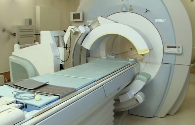
Researchers from the Graduate School of Medicine of Kyoto University have assessed a prototype mobile PET scanner designed to work with both CT and MRI scanners. Their objective is to create a more affordable, more versatile PET scanner.
The team tested two different detector layouts of the flexible PET scanner – called fxPET and manufactured by Shimadzu Corporation. They evaluated image quality, lesion detection rate and parameters such as standardized uptake values (SUVs) and metabolic tumour volume (MTV), compared with those of a conventional PET/CT scanner without time-of-flight capacity (Mol. Imaging Biol. 10.1007/s11307-019-01384-9).
The MR-compatible fxPET scanner, comprised of dual arc-shaped detectors each covering 135°, is a partial-ring system rather than a conventional full-ring scanner, with limited angular coverage. It consists of two detector units that fit beds and patients of various body sizes. The fxPET is designed to fit existing MR systems, allowing it to fuse sequential PET and MR images with minimum misalignment.
The dual arc-shaped detector heads can be arranged in various configurations, including top–bottom and left–right. Each detector unit consists of three rings of 18 detector modules, giving a total of 108 detector blocks. Each detector block comprises four-layer depth-of-interaction crystal blocks of lutetium gadolinium oxyorthosilicate crystals and a 64-channel silicon photomultiplier array.
The arc-shaped detectors have a ring diameter of 778 mm and an axial extent of 150 mm. The spatial resolution of fxPET is estimated to be less than 2.5 mm and the coincidence timing resolution is approximately 500 ps.
Performance comparisons
Because fxPET is a partial-ring scanner, degradation of image quality can occur as a result of incomplete coincident data. Radiologist Masao Watanabe and colleagues conducted a study comparing two layouts: one with the detectors located above and below the patient; and the other with the detectors in the same configuration but closer to the patients to eliminate the gap. They also compared the results with whole-body PET/CT images.
The researchers studied 59 patients with a variety of cancers who underwent whole-body PET/CT scans, followed by fxPET scanning in both layouts. Two radiologists rated each image on a 4-point grading scale. With the three image datasets simultaneously displayed on a diagnostic workstation, Watanabe counted the number of visible lesions for each.
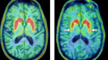
The team found that bringing the detectors closer to the patient improved image quality. The fxPET layout with the detectors closest to the patient identified 172 lesions out of 184 (93.5%), compared with 166 lesions (90.2%) for whole-body PET/CT. The other fxPET layout identified 169 lesions, or 91.8%. The authors also reported that SUVs were larger and MTVs smaller in fxPET than in whole-body PET/CT, especially for lesions smaller than 2 cm.
Co-author Yuji Nakamoto tells Physics World that the researchers have developed a new reconstruction algorithm for the scanner and are currently evaluating image quality, diagnostic performance and quantitative values for images reconstructed with this algorithm. In future, they plan to conduct studies to determine an optimum scanning time for fxPET.
“We believe that the fxPET scanner will be an attractive imaging tool, especially from the standpoint of cost,” the authors say. “In any facilities having a state-of-art MR system, fused PET and MR images can be obtained at low cost, although the scanning is not simultaneous but sequential. Also, because of the wide inner diameter of the fxPET system, it can use the standard radiofrequency coils of MR scanning if a CT attenuation map of them is prepared and incorporated into reconstruction of the fxPET images. For this reason, it is not necessary to prepare dedicated radiofrequency coils for combined PET/MRI. In addition, this system could be installed for other devices, such as CT or radiation therapy equipment.”


