New research by Raymond Goldstein and colleagues at the University of Cambridge has revealed how fluid motion and direct mechanical pressure triggers the emission of bioluminescent light by tiny marine organisms.
Bioluminescence occurs in a wide array of organisms including dinoflagellates, which are single-celled plankton that play a crucial role in aquatic food chains. Despite their global importance, scientists do not have a full understanding of how these organisms emit green-blue light in response to fluid motion in their environments.
Goldstein’s team looked at how the light intensity from dinoflagellates correlates with various mechanical stressors. They did this by exposing the tiny organisms to two types of stimuli mimicking the mechanical attack of a predator and swimming in a densely populated wave.
The team built their study upon previous work by Michael Latz and colleagues at the Scripps Institution of Oceanography in the US. Latz’s team had used macroscopic contracting fl0ws, Couette flow, microfluidic flows, and atomic force microscopy to study dinoflagellates.
“Next logical step”
“Our work is the next logical step that builds on [Latz’s] work, by using micropipette technology to hold individual cells and apply quantified cell wall deformations, all with high-speed imaging,” explains Goldstein.
The bioluminescence of dinoflagellates is unique from many perspectives. The biochemical and cellular cascade leading to a light flash begins when the cell membrane is subject to mechanical stress, which activates a receptor in the membrane. This mechanical signal is translated into an electrical signal by increasing the concentration of calcium ions.
As a result, membranes of inner organelles depolarize and generate an action potential that propagates along the membranes. Ultimately, the pH of the environment decreases, promoting luciferin oxidation catalyzed by luciferase, which is accompanied by a photon release.
Ideal experimental subjects
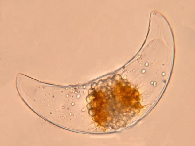
To investigate the underlying biophysical processes, Goldstein and his team held single cells of the dinoflagellate Pyrocystis lunula on a glass micropipette using suction. Measuring 30–1000 μm, they are ideal experimental subjects thanks to their robustness and reduced motility. According to Goldstein, the team often manipulated each cell up to 20 times without any obvious damage.
The cells were mechanically stimulated either by directing a submerged jet of fluid (growth medium) at a single cell or by squeezing a single cell between two pipettes with submicron precision. The cell wall deformation was monitored by direct imaging with a high-speed camera.
The results showed that the cells flash only after the fluid pressure is high enough to induce significant cell wall deformation. The flash occurs and decays within fractions of a second. The bioluminescence intensity eventually decreases, most probably due to exhaustion of the luciferin. Interestingly, bioluminescence tends to occur within the central region around the cell’s nucleus.
For mechanical stimuli, light intensity was low when the deformation was small (whatever the speed), or when the deformation rate was small (whatever the deformation). However, the bioluminescence flash was large when both the deformation and its rate were large. These results suggest that the membrane and calcium channels have viscoelastic properties – which was confirmed by a phenomenological model.
“Welcome contribution”
According to Latz, “this study provides a welcome contribution to our knowledge of mechanosensory responsiveness in the dinoflagellate bioluminescence system. I applaud their use of controlled and quantified modes of mechanical stimulation using sophisticated analytical techniques.”

Turbulence causes swimming algae to congregate in dense patches, say physicists
“One of our challenges is the discrepancy between sensitivity in macroflows and microstimulation, based on the as-yet unknown distribution of mechanosensors within the cell and how their activity is integrated into a whole cell response. As I am now retired, I hope that it will inspire others to continue the work,” Latz adds.
Goldstein adds “Michael Latz has shown, already a number of years ago, that dinoflagellates can be used as local sensors for fluid shear. We are now very interested in the role of dinoflagellates in algal blooms, in extending our understanding of cell wall mechanics, and in force and light distribution over the entire cell body.”
The research is described in Physical Review Letters.


