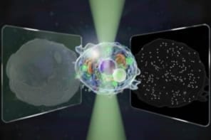
Existing brain imaging techniques are either unable to penetrate the brain or are not of a high enough resolution to follow specific brain processes. Researchers at the Massachusetts Institute of Technology have now created a sensor that can detect neural activity using functional magnetic resonance imaging (fMRI), which is non-invasive.
The new sensor targets calcium ions (Ca2+), the extracellular concentrations of which fluctuate during synaptic activity. These fluctuations can last for tens of seconds and can push Ca2+ concentrations down to as low as 100 µM. Even slower variations are associated with sleep-wake transitions, for example.
Abnormal Ca2+ signals are thought to be implicated in brain disorders, such as epilepsy and Alzheimer’s disease. However, the problem is that most current fMRI techniques are unable to monitor the dynamics of Ca2+ ions over large volumes in brain tissue.
Clustered nanoparticles look darker in MRI
As in standard MRI, fMRI involves using a fixed magnetic field to align the spins of protons inside the water molecules of biological tissue. Radio waves are used to deflect these spins, which then relax back to their original alignment. This relaxation is probed with a radio receiver coil, and the result provides information on the tissue’s composition. In the new calcium-dependent fMRI method, paramagnetic molecules known as “contrast agents” are used to interact with some of the water molecules and change their brightness in scans.
“Our new sensors are bioengineered paramagnetic nanoparticles that cluster together in the presence, but not the absence, of calcium ions,” explains Alan Jasanoff, who led this research effort. “Clustered nanoparticles look darker in MRI than non-clustered ones and this allows us to monitor changes in calcium levels based on the changes in the darkness of MRI scans obtained with the sensors.”
Jasanoff and colleagues engineered their magnetic calcium-responsive nanoparticles using synaptotagmin proteins. These proteins are components of the synaptic machinery that releases neurotransmitters and which naturally responds to changes in Ca2+ concentrations ranging from 0.1 to 1.0 mM. This range is suitable for monitoring extracellular calcium signalling processes in the brain.
Measuring the strength of the contrast agent
The researchers observed the nanoparticles using a technique called dynamic light scattering (DLS). Atomic force microscopy (AFM) further confirmed that the particles aggregate in the presence of Ca2+ ions.
“We injected the nanoprobes into rat brains and showed that the probes produce a MRI response when injected brain tissue is stimulated with chemicals or electrodes,” says Jasanoff. “In one set of experiments, we detected responses to rewarding brain stimuli that emulate the action of certain illegal drugs.”
The researchers say they were easily able to detect the nanoprobes’ responses with MRI by measuring the strength of the contrast agent, or transverse relaxivity, r2. Indeed, they observed a calcium-dependent increase in r2 from 0 mM Ca2+ to 1.2 mM Ca2+.
Towards human trials
“Such functional brain imaging using the new sensors provides us with a more direct measure of neural activation than earlier fMRI methods,” Jasanoff tells nanotechweb.org. “Using the new technique, we should be able to map activity across large areas of the brain with much better precision than current fMRI methods permit. We will also learn how to interpret calcium-related brain imaging signals in terms of the underlying function of specific calcium-related proteins in the brain, including receptors involved in neurotransmitter signals or learning and memory.”
The team, reporting its work in Nature Nanotechnology 10.1038/s41565-018-0092-4, says that it is now busy optimizing its new calcium sensors and the way they are delivered to large regions of the living brain in animals. “In the long term, we are interested in seeing whether we can use the probes in human patients, probably initially in surgical contexts, in which brain injections are feasible.”


