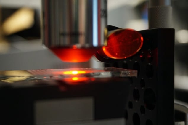
A novel hybrid microscope delivers the same information as standard optical microscopy without the need for detrimental tissue staining, while also providing molecular insight into tissue biopsies. Developed by researchers from the University of Illinois at Urbana-Champaign, the system adds infrared capability to the ubiquitous, standard optical microscope. The new system could deeply impact histopathology, both in the clinics and within research, by offering faster diagnosis, lower cost and wider availability (PNAS 10.1073/pnas.1912400117).
Histopathology is the microscopic study of tissues to spot and identify disease manifestations such as tumours. The gold standard technique requires the addition of dyes or stains to human tissue biopsies. This enables pathologists to see the shapes and patterns of the cells under a microscope and distinguish cancerous tissues from healthy ones. However, even for highly trained readers, such diagnostics can prove tricky and are subjective.
Moreover, the information given by optical microscopes is limited and superficial, as it does not shed any light on the underlying molecular changes driving cancer. Infrared (IR) microscopy, on the other hand, can provide such details by measuring the molecular composition of tissues. But while conventional optical microscopes are widespread and easy to use, IR microscopes are expensive and require the sample to undergo an extensive preparation – making this approach impractical in most settings.
A ready-to-build hybrid microscope
A team led by Rohit Bhargava bypassed the limitations of both techniques by coupling them. The feat was achieved by adding an IR laser and an interference objective to an optical camera, which harnesses the strengths of both modalities.

The hybrid microscope has the same high resolution, large field-of-view and accessibility of an optical system, while its software can use the IR data to compute an image that looks similar to a conventional stained sample. This combination allows researchers to use an all-digital approach to biopsies and derive information about tissue density, scattering, path length or visible absorption that exceeds that offered by standard microscopy.
“We built the hybrid microscope from off-the-shelf components. This is important because it allows others to easily build their own microscope or upgrade an existing microscope,” says first author Martin Schnell, a postdoctoral fellow in Bhargava’s group.
AI helps pathologists
The researchers tested the performance of the hybrid microscope on unstained breast tissue samples and compared the results with conventional techniques. They developed an iterative search algorithm to identify the cell types in each biopsy – such as healthy and malignant cells in the epithelium, stroma, red blood cells and some additional tissues – using the 22 available frequency bands of the IR spectrum. The team subsequently found that only seven bands were needed to obtain accurate classification and five to obtain an area under the curve (a metric assessing a tool’s performance) above 0.90, which could considerably speed up diagnosis.
More work needs to be done on the analysis of the hybrid images. The researchers are now working to optimize machine-learning programs that can measure multiple IR wavelengths, creating images that readily distinguish between multiple cell types, and integrate that data with the detailed optical images to precisely map cancer within a sample.
“It is very intriguing what this additional detail can offer in terms of pathology diagnoses,” Bhargava says. “This could help speed up the wait for results, reduce costs of reagents and people to stain tissue, and provide an ‘all-digital’ solution for cancer pathology.”


