A team of US-based researchers has demonstrated how cutting-edge catheter technology can help improve the accuracy of radiofrequency ablation (RFA), often used to treat cardiac arrhythmias. So, what technology does the new catheter use — and how exactly does it work? And what are the ongoing prospects of using it in clinical settings in the future?
Real-time tissue imaging
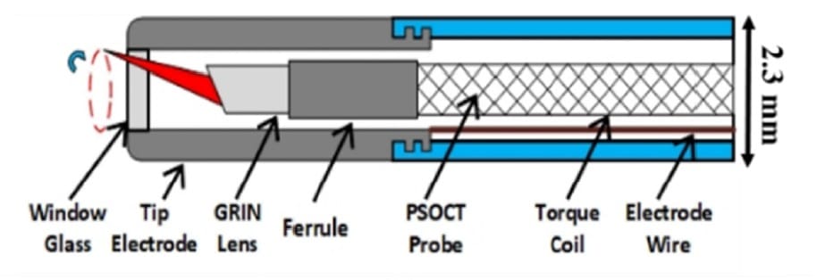
Although RFA is often used to treat cardiac arrhythmias, there is currently no way to directly monitor the formation of the ablated lesion. To increase the accuracy of this therapy, the researchers propose a novel combination of RFA with polarization-sensitive optical coherence tomography (PSOCT) to create an innovative catheter device that is capable of providing real-time, high-resolution tissue imaging.
The team demonstrated the feasibility of such an integrated RFA/PSOCT catheter by constructing and testing a prototype (Biomed. Opt. Express 10.1364/BOE.9.006400).
“We showed that the prototype can ablate normally and can image the tissue in order to confirm good catheter contact with the heart wall, and monitor that the tissue is ablated as the RF energy is delivered,” says co-author Andrew Rollins from Case Western Reserve University. Rollins prepared the paper in partnership with fellow academics at the university, as well as clinicians based at Rainbow Babies and Children’s Hospital and University Hospitals Case Medical Center.
“This validation was needed before tests can move forward to testing in large animals, and eventually to helping to treat human patients,” Rollins adds.
An RFA catheter is a 2 m-long cable that a doctor threads through a patient’s veins to their heart. The device, which contains an electrode that delivers radiofrequency energy to burn spots on the heart wall, is used to treat arrhythmias like atrial fibrillation (AF). It does this by ablating tissue in the correct spots to stop the AF or keep it from travelling.
“PSOCT is like super-high-resolution ultrasound using infrared light,” says Rollins. “The integrated catheter has PSOCT at the tip so the doctor can see the tissue to be ablated, and hopefully do it better and safer.”
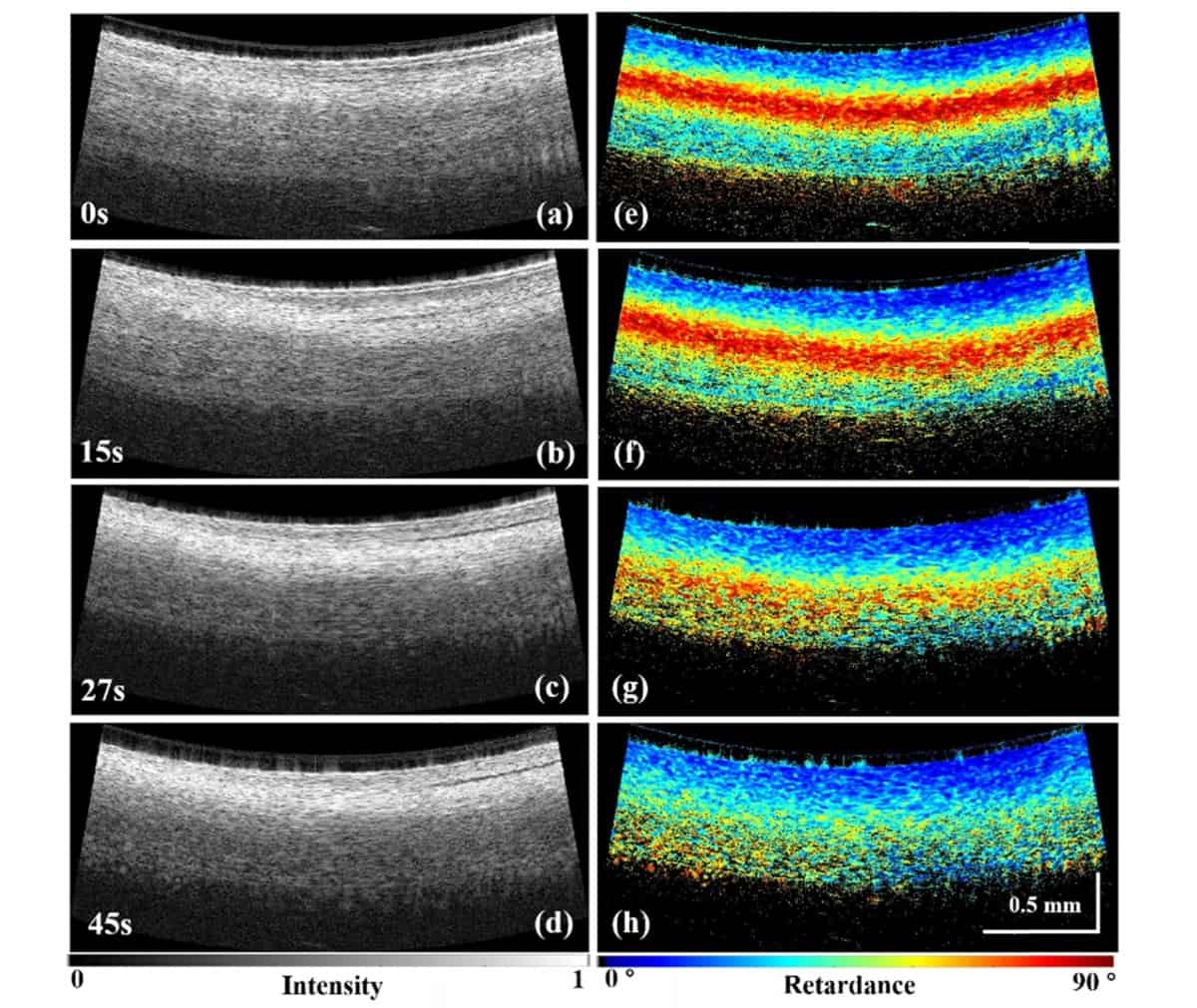
According to Rollins, one of the main potential advantages of the new device is that it makes RF ablation safer and more effective. He also points out its capacity to improve effectiveness by directly detecting whether the ablation is complete, and by helping determine where to ablate.
“It can improve safety by detecting signs of over-treatment before a complication, for example a steam pop, occurs,” he says.
Continuous improvement
Commenting on the prospects for use of the new catheter in clinical settings in the coming years, Rollins highlights the fact that the process of getting new medical technology into the hands of doctors — including the necessary steps of regulatory approval and commercialization — is long and expensive, especially for a high-stakes scenario such as cardiac catheter ablation.
“We are on the path, but there is a long way to go,” he tells Physics World. “Our next step is to demonstrate that an integrated catheter can work as intended in large animals. Then we can test the catheter in animals with disease to determine whether the technology improves the treatment. Along the way, we need to continue to improve the catheter design and develop data analysis methods and user interfaces to make the technology useful in the real-time clinical setting.”