
In its first clinical trial, a novel PET radiotracer developed in China outperformed the traditional 18F-FDG tracer in identifying primary malignant melanoma and nodal metastases. The new PET probe – 18F-PFPN – detected 365 metastases that were missed in 18F-FDG PET imaging. Investigations in healthy volunteers also confirmed that the new PET tracer is safe and well tolerated.
Currently, 18F-FDG PET is used to stage patients with advanced malignant melanoma and to monitor the effects of cancer treatments. 18F-FDG PET, however, lacks the sensitivity to diagnose early-stage disease and is unable to identify small metastases (less than 1 cm) to the lung, liver and brain.
The new radiotracer is designed to target melanin, which exists in most melanomas. Principal investigator Xiaoli Lan, from Union Hospital, Tongji Medical College of Huazhong University of Science and Technology, explains that the 18F-PFPN tracer was based on a melanin-targeted nicotinamide probe (18F-FPN) that the researchers had previously developed, optimized to have a higher tumour-to-normal liver ratio and radiochemical yield. It is characterized by negligible accumulation in the liver and rapid renal clearance, enabling its safe use in clinical imaging studies.
Safety assessment
Lan and colleagues initially investigated the biodistribution, pharmacokinetics, radiation dosimetry and safety of 18F-PFPN in five healthy volunteers. The tracer was safe and well-tolerated; none of the volunteers exhibited any changes in vital signs or experienced adverse reactions to the tracer.
The researchers performed serial whole-body PET scans on the volunteers 30, 60, 120 and 240 min after injection of 18F-PFPN, and calculated the tracer uptake in the gallbladder, urinary bladder, stomach and liver at each time point. To investigate the tracer pharmacokinetics, they determined the radioactive counts of blood, plasma and urine samples at different time points, observing a rapid renal clearance. The bladder wall showed the highest dose activity, followed by the kidneys. The total effective dose was 2.01 × 10⁻² mSv/MBq.
Follow-up exams did not reveal any abnormal changes in liver and kidney function and none of the volunteers reported any subsequent problems.
Tracer comparison in patients
The team also examined nine patients with suspected malignant melanoma and 12 with confirmed malignant melanoma. All patients received both 18F-FDG and 18F-PFPN PET scans, spanning the brain down to the bottom of the foot. The resulting images were interpreted by two experienced nuclear medicine specialists. For patient-based analysis, they identified either the primary tumour or the single lesion showing the highest tracer uptake at each metastatic site. For each site, the experts also conducted lesion-based analysis, either on all lesions (for up to 10) or on the 10 with the highest tracer uptake.
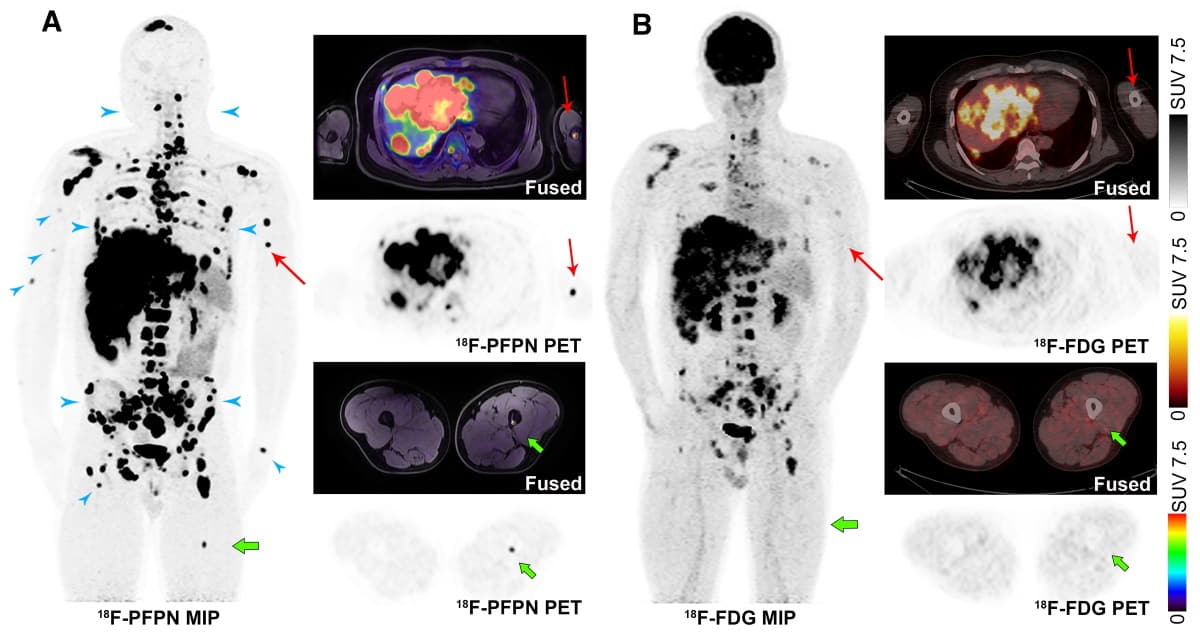
In the patient-based analysis, both types of tracer identified all eight primary tumours and performed similarly with all types of metastases, with 18F-FDG images identifying 35 metastases and 18F-PFPN images identifying 39. The team note that 18F-PFPN uptake was higher than 18F-FDG for both primary tumours and nodal metastases.
In the lesion-based analysis, 18F-PFPN significantly outperformed 18F-FDG, identifying 100% of all lesions. Specifically, 18F-PFPN identified 394 bone metastases compared with 18F-FDG’s 151 (100% versus 38.32%); 141 liver metastases compared with 49 (100% versus 34.75%); 124 lymph node metastases compared with 98 (100% versus 79.03%); and 33 metastases to other sites compared with 29 (100% vs 87.88%). In total, 18F-PFPN PET detected 365 metastases that were missed when using 18F-FDG PET.
The team also applied a visual scoring system based on the number of lesions identified in each patient. Here, 18F-PFPN also outperformed 18F-FDG for detection of distant metastases to the liver, bone, lymph nodes and other distant sites.
“We found that 18F-PFPN PET was capable of identifying early T-stage lesions (for example, T2b),” comments Lan. “We will continue to enrol more patients with early stage to further confirm the feasibility, which may help the patients’ clinical management. Early surgical excision of localized malignant melanoma portends favourable outcomes, and surgical strategies vary on different T-stages. In this scenario, both prompt diagnosis and accurate disease staging are paramount to reduce mortality.”
“We aim at exploring the diagnostic value of 18F-PFPN in patients with clinically suspected or confirmed malignant melanoma,” she adds. Although 18F-PFPN helps to distinguish between pigmented and non-pigmented lesions, our planned research will not focus on the evaluation of non-pigmented lesions.”
The study is described in the Journal of Nuclear Medicine.


