Neutron scattering experiments have allowed researchers in the US to map out the precise positions of hydrogen atoms in a coronavirus protein for the first time. The approach, taken by Andrey Kovalevsky and colleagues at Oak Ridge National Laboratory, reveals key clues of how binding occurs between specific enzymes and protein chains involved in the replication of the virus. The team’s discoveries could lead to advanced computational designs of targeted drugs.
Within the SARS-CoV-2 virus (which causes COVID-19) the information required for replication is encoded on two overlapping chains of protein molecules. To use this information, the virus must first break these chains down into individual functional proteins. This requires a specific enzyme called the main protease – which interacts with protein chains at a specific group of atoms called the “active site”. Currently, researchers are aiming to reduce the activity of these enzymes by developing inhibitor drugs that tightly bind to their active sites – preventing further reactions.
Developing these inhibitors requires a detailed knowledge about the locations of hydrogen atoms within the main protease enzyme because these atoms define the nature of the hydrogen bonds between enzymes and protein chains. Normally, the locations of atoms in a molecule can be determined using X-ray crystallography. That technique, however, is not sensitive to hydrogen so the team used neutron scattering instead. Another benefit of neutron scattering is that unlike X-ray techniques, it does not cause any radiation damage to samples.
Key players
“Half of the atoms in proteins are hydrogen. Those atoms are key players in enzymatic function and are essential to how drugs bind,” explains Kovalevsky. “If we don’t know where those hydrogens are and how the electrical charges are distributed inside the protein, we can’t design effective inhibitors for the enzyme.”
How physics is helping in the war against COVID-19
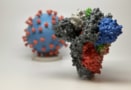
Using instruments at ORNL’s Spallation Neutron Source, the researchers determined the precise structure of the virus’s main protease enzyme, and compared the result to structures obtained through X-ray crystallography. Whereas the locations of hydrogen atoms could only be inferred from the X-ray data, neutron scattering allowed Kovalevsky and colleagues to determine their positions to subatomic resolution. This enabled them to identify electrical charges, as well as intricate networks of hydrogen bonds across the enzyme’s active site.
The team is now the first to determine the exact structure of a coronavirus protein. Their findings represent a crucial advance in our understanding of how the SARS-CoV-2 virus replicates, and will now provide critical information for the computational design of inhibitor drugs that are specifically tailored for targeting the electrostatic environment of the main protease enzyme. If created, such drugs could soon become a key aspect of global efforts to contain the spread of the virus.
The research is described in The Journal of Biological Chemistry.