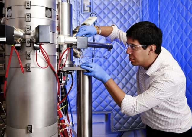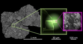
Researchers at the Oak Ridge National Laboratory in the US and Uppsala University in Sweden have developed a new electron-microscopy technique that can detect magnetism at the atomic scale. The technique exploits distortions – or “aberrations” – in how the microscope’s electron beam is focussed, and could be used could be used to study magnetic domains in devices such as computer hard-disk drives.
Electron microscopes focus beams of electrons in much the same way that optical microscopes focus light. Just like their optical counterparts, the lenses in an electron microscope are not perfect, and this results in distortions in microscope images. These distortions can be minimized using an aberration-correction system, and now Oak Ridge’s Juan Carlos Idrobo and colleagues have used such a system to introduce a specific aberration to their electron beam to make it sensitive to tiny magnetic domains in a material.
Electron energy loss
The new technique is based on an effect called “electron energy-loss magnetic circular dichroism” (EMCD), whereby a magnetic material will absorb energy at different rates from electron beams with different values of orbital angular momenta. Beginning in 2006, physicists in Austria and Germany have shown that EMCD can be used in electron microscopes to image magnetic domains as small as 1–2 nm. While this is tiny, it is still much larger than individual atoms in a solid, which tend to be about 0.1 nm in size.
This latest work improves the spatial resolution of EMCD by using the fact that an electron beam with a specific type of aberration will interact with a magnetic material in much the same way as an electron beam carrying orbital angular momentum. Idrobo and colleagues used their microscope’s aberration-correction system to create an electron beam with an aberration called four-fold astigmatism. They fired the beam at a sample of lanthanum manganese arsenic oxide (LaMnAsO) and used a technique called electron energy-loss spectroscopy (EELS) to analyse how the electron beam lost energy as it passed through the sample.
By scanning the beam across the sample, they were able to build up an image of the checkerboard antiferromagnetic ordering in the material. Specifically, they were able to see that the direction of the magnetic moments of neighbouring manganese atoms alternated from pointing up to pointing down.
Highly distorted
“Four-fold astigmatism is a type of aberration present in electron lenses,” explains Idrobo. “Imagine a wine glass full of water. If you place the glass at a certain distance from an object, you can see how the glass behaves as a magnifying lens. However, you will also notice that the magnification is better at the centre of the glass and becomes highly distorted at the edges of it. This is spherical aberration.
“If the glass were not spherical but instead wider or taller, then you would see the effects of two-fold astigmatism because of light having different foci in the vertical and horizontal directions of the glass. Four-fold astigmatism occurs in a lens that has four different directions; each with focusing planes rotated 45° with respect to each other.” The reason why the researchers used four-fold astigmatism to measure the magnetic ordering in LaMnAsO is because this material has four-fold crystal symmetry.
Popular instrument
Idrobo says that the team’s accomplishment is important for two reasons. The first is that it shows that, rather than always being a bad thing, aberrations in electron beams can be used to perform useful measurements. The second reason is that the method they developed for controlling the electron beam is easy to implement in aberration-corrected scanning transmission electron microscopes. “Since most modern materials-science characterization laboratories use this kind of instrument, studying magnetism in materials at high spatial resolutions will now be [with]in the reach of a large number of scientists,” he adds.
The team, reporting its work in Advanced Structural and Chemical Imaging, says that it is now trying to see what other physical phenomena, besides magnetism, it can measure using these aberrated probes. “We have some ideas of what can be done and are working really hard to see how far we can go. So stay tuned!” Idrobo says.
- A version of this article first appeared on nanotechweb.org


