When you’re sick, you may reach for a thermometer to take your body’s temperature. But what if you need to take your brain’s temperature?
Events like ischemic stroke can increase brain temperature by several degrees Celsius and can lead to brain malfunction. These large changes are linked to worse patient outcomes and recovery and don’t always correlate with body temperature.
A team of researchers in the United States is working to help doctors understand and monitor brain temperature in sickness and health by creating personalized temperature maps of the brain using magnetic resonance (MR) thermometry and a new biophysical model. Results of the researchers’ proof-of-concept study are published in Communications Physics.
“You cannot assign a single temperature to a brain”
The brain is a highly sensitive and finely tuned organ that requires a sizeable fraction of the body’s energy to function and produces heat in doing so. How well a patient’s brain recovers after injury and returns to normal is critical.
“We’ve known for decades that brain temperature is important for [patient] recovery,” says Candace Fleischer, from the Emory University School of Medicine and Georgia Institute of Technology. But, she adds, “there’s no clinical standard for measuring brain temperature”.
In addition, brain temperature varies throughout the brain as blood flow and metabolic demands change. This means that “you cannot assign a single temperature to a brain,” explains Andrei Fedorov from Georgia Tech.
Today, doctors may attempt to measure brain temperature directly by implanting a probe called a thermocouple in the brain. But because this technique is invasive and gives a measurement of brain temperature only at a single point, it isn’t suitable for every patient, particularly those who are not in critical care. Furthermore, doctors might be able to make more informed monitoring and treatment decisions if they can use a non-invasive technique that provides them with a precise, three-dimensional map of temperatures throughout a patient’s brain.
Going back to basics
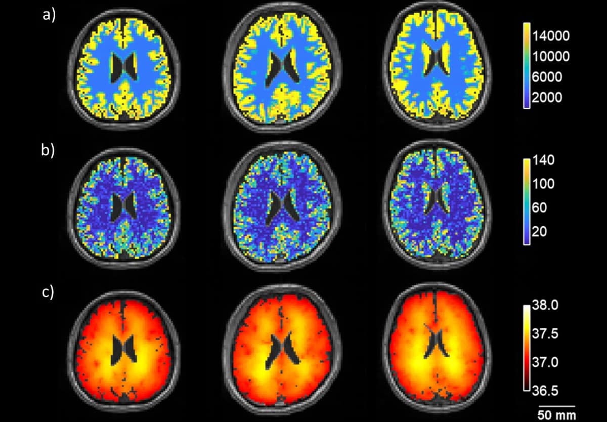
In their study, the researchers collected structural MR brain images from three healthy volunteers and developed a biophysical model to predict brain temperature. After a couple of hours of data analysis and simulations, the model computes a three-dimensional distribution of brain temperature that the researchers visualize as three-dimensional brain temperature maps.
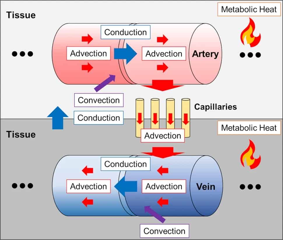
The model itself relies on two fundamental principles of physics – conservation of energy and conservation of mass – and incorporates all the ways in which heat is generated through metabolism and dissipated via blood flow throughout the brain (conduction, convection and advection). These features of the model allowed the researchers to predict brain temperature without using any empirical information about how the brain behaves under individual conditions.
To validate their brain temperature predictions, the researchers collected temperature measurements using whole-brain MR thermometry, compared these measurements to the model’s predictions, and found that they agreed.
Marrying fundamental science with clinical applications
Fleischer and Fedorov, along with their graduate student Dongsuk Sung and research engineer Peter Kottke, are pursuing several projects that expand upon this study in collaboration with Emory clinicians. They are validating their model in a large group of healthy people, incorporating the behaviour of local blood flow to predict how brain temperature responds to changes in blood supply to different brain regions, and creating temperature maps after injury.
“By combining fundamental physics, experimental measures of brain temperature and direct clinical application, model development and accurate brain temperature predictions in patients are propelled forward,” Fleischer says.
Though their technique is limited to individuals who can receive an MRI, the researchers are confident in its utility.
“All of the complexity [in the brain’s functioning] results in a very unique personal temperature map that actually tells us a lot about what may be happening to us, what may have happened in the past, or what may happen if we somehow perturb us as a human,” Fedorov says.


